Objective Differential Characteristics of Cystic Biliary Atresia and Choledochal Cysts in Neonates and Young Infants: Sonographic Findings
Lu-Yao Zhou, Bu-Yun Guan, Le Li, Zuo-Feng Xu, Chang-Ping Dai, Wei Wang, Hui-Min Xia, and Xiao-Yan Xie. JUM June 1, 2012 vol. 31 no. 6 833-841
Objectives— To prospectively evaluate the objective
differential characteristics between cystic biliary atresia and choledochal
cysts on sonography among neonates and young infants.
Methods— Twenty-three patients who had sonographic findings of a portal cyst and
a final diagnosis were included. Their final diagnoses were cystic biliary
atresia in 12 patients and choledochal cysts in 11. All of them underwent
detailed sonographic scanning. Data for cystic biliary atresia and choledochal
cyst groups were compared by the χ2 test for categorical
variables and an unpaired t test for continuous variables.
Results— The triangular cord sign was detected in 11 patients in the atresia
group but in none in the cyst group (P < .001). Nine of 11 patients
in the cyst group had dilatation of intrahepatic bile ducts, whereas none in
the atresia group had that feature (P < .001). Sonography also showed
sludge deposits in the cysts in 6 of 11 patients in the cyst group, whereas
none in the atresia group had sludge deposits (P = .005). The mean width
and length of the cysts in the cyst group were significantly larger than those
in the atresia group (P< .05 for both). The mean hepatic artery
diameter was significantly larger in the atresia group than in the cyst group (P
< .001). The difference in gallbladder abnormalities between the atresia (n
= 11) and cyst (n = 0) groups was also significant (P < .001). When
all specific sonographic features were used, all patients were correctly
classified into the atresia and cyst groups.
Conclusions— The triangular cord sign, intrahepatic bile duct
dilatation, and echoic cysts might be regarded as objective sonographic
features for differentiating cystic biliary atresia and choledochal cysts.
Other sonographic features might be very supportive.
Biliary
atresia is a destructive inflammatory obliterative cholangiopathy of neonates
and infants that affects varying lengths of both intrahepatic and extrahepatic
bile ducts.1
It is an uncommon disorder with a prevalence of about 1 per 5000 in East Asian
countries.2
No analogous pathologic process exists in older children or adults. Cystic
biliary atresia is a subtype of biliary atresia with portal cysts; it has an
occurrence rate of 8% to 11%3–5
among patients with biliary atresia. Because of its specific appearance, cystic
biliary atresia is the only type of biliary atresia that can be detected
antenatally with sonography.6
A choledochal cyst in a neonate or young infant is another uncommon entity with
an appearance similar to that of cystic biliary atresia on medical imaging.
Both are well-known causes of jaundice in neonates and young infants,5
with a cyst at the porta hepatis.
However,
cystic biliary atresia and choledochal cysts are two entities with dramatically
different management approaches and prognoses.3,7
Patients with cystic biliary atresia can have a better prognosis with timely
and early portoenterostomy.8,9
If they are left untreated, progressive liver cirrhosis leads to death by 2
years of age. Some patients with cystic biliary atresia do not have jaundice
relief even after early portoenterostomy. On the other hand, a choledochal cyst
is a curable choledochal malformation that can have an excellent prognosis with
resection of the cyst and hepaticojejunostomy.5,6
In addition, although there is an opinion that a surgical delay might increase
the risk of liver fibrosis in patients with choledochal cysts,3
the optimal time for surgery of choledochal cysts has not been decided yet.
Hence, it is very important to differentiate cystic biliary atresia from a
choledochal cyst when a portal cyst is found in a neonate or young infant. Sonography usually is recommended as the initial tool for evaluating a neonate with jaundice. Some literature has reported that sonography can play an important role in differentiating cystic biliary atresia from a choledochal cyst by characterizing the appearance of the cyst and gallbladder.5,10,11 However, the published literature is sparse, with small sample sizes. Furthermore, the sonographic technique for diagnosis of biliary atresia is still progressing. New sonographic features have been identified, such as the triangular cord sign and hepatic subcapsular flow, which are very effective in identifying biliary atresia. If sonographic features such as those could also be observed in patients with cystic biliary atresia, then there might be new differential characteristics between cystic iliary atresia and choledochal cysts on sonography.
This
study aimed to review the clinical and sonographic features of jaundiced
neonates and infants younger than 6 months with a final diagnosis of cystic
biliary atresia or a choledochal cyst and to assess the differential
characteristics between them.
Materials and Methods
A study
evaluating the value of sonography for biliary atresia screening among neonates
with pathologic jaundice was initiated in July 2009. The study was approved by
our Institutional Clinical Research Ethics Committee, and informed parental
consent was obtained for every case. Between July 2009 and March, 2011, 263
neonates and infants younger than 6 months who had pathologic jaundice
prospectively underwent sonographic screening to rule out biliary atresia.
Twenty-six of them showed portal cysts on sonography. Of these, 2 patients who
were suspected of having cystic biliary atresia and 1 patient who was suspected
of having a choledochal cyst did not undergo surgery in our hospital and were
lost to follow-up. As a result, 23 patients who had sonographic findings of a
portal cyst and a final diagnosis were included.
One
pediatric radiologist with 3 years of experience in pediatric sonography
performed all sonographic examinations. Sonography was performed with an
SSA-660A scanner (Toshiba Medical Systems Co, Ltd, Tokyo, Japan) incorporating
a 3.5-MHz curvilinear transducer and a 12-MHz linear array transducer and an
EUB-7000HV scanner (Hitachi Medical Corporation, Tokyo, Japan) incorporating a
2- to 5-MHz curvilinear transducer and a 6- to 13-MHz linear array transducer.
Immediately after detailed scanning, the diagnosis of cystic biliary atresia or
a choledochal cyst was made by the pediatric radiologist, who was blinded to
the clinical findings for every patient. The patients were not fed for at least
4 hours before the sonographic examinations. During the examinations, the
patients were allowed to feed to keep quiet. The presence and thickness of a
triangular cord in the porta hepatis and the diameters of the portal vein and
hepatic artery were recorded in each case. A triangular cord was defined as a
thickness of the echogenic anterior wall of the right portal vein just proximal
to the right portal vein bifurcation site of greater than 4 mm.12
The diameter of the hepatic artery was measured at the level of the right
proximal hepatic artery running parallel to the right portal vein.13
The maximum width and length of the gallbladder and the cyst were also
measured. The presence or absence of dilatation of the intrahepatic bile ducts,
the presence or absence of sludge in the cyst, a communication between the
gallbladder and the cyst, hepatomegaly, splenomegaly, ascites, and the
echogenicity of liver parenchyma were noted during the examinations. The liver
was considered enlarged if the right lobe extended below the right kidney.
Splenomegaly was defined as a spleen of greater than 6 cm.14
The
gallbladder was classified as normal, atretic, or irregularly elongated. A
normal gallbladder was defined as one that was 15 mm or greater in length and
globular or ovoid without wall abnormalities. An atretic gallbladder was
defined as one that was less than 15 mm in maximum length. An irregularly
elongated gallbladder was defined as one that was 15 mm or greater in length
and had an abnormal wall with an irregularly compromised lumen. An atretic or
irregularly elongated gallbladder was regarded as an abnormal gallbladder.
Patients
with an abnormal gallbladder or the presence of the triangular cord sign on
sonography were considered to have cystic biliary atresia. Those with a normal
gallbladder and absence of the triangular cord sign were considered to have
choledochal cysts. Dilatation of the intrahepatic bile duct and sludge in a
large cyst were supportive features of choledochal cysts. Dilatation of the
hepatic artery and a small cyst without sludge were supportive features of
cystic biliary atresia.
Cystic
biliary atresia was confirmed by surgical cholangiography and pathologic
examination in 11 patients and with liver biopsy in 1. Choledochal cysts were
confirmed by surgery in 11 patients. Eleven patients with cystic biliary
atresia underwent excision of the extrahepatic bile duct, including the cyst,
and Roux-en-Y portoenterostomy. The other patient who did not have surgery
because he was older than 4 months was confirmed to have biliary atresia by
liver biopsy. All 11 patients with choledochal cysts underwent cyst excision
and hepaticojejunostomy or choledochojejunostomy.
The
sonographic findings were analyzed with a focus on the presence or absence of a
triangular cord, the size of the cyst, the presence or absence of sludge in the
cyst, dilatation of the intrahepatic bile ducts and hepatic artery, portal vein
diameters, and the sizes and morphologic characteristics of the gallbladder,
spleen, and liver. Cyst sizes in cystic biliary atresia between those with a
communication between the gallbladder and the cyst and those without were
compared. Clinical data such as age, sex, and serum bilirubin level were also
recorded and analyzed. Statistical analysis of the two groups was performed
with the χ2 test and Fisher exact test for counting data and a t test for
measurement data. P < .05 was considered statistically significant.
The sensitivity, specificity, and positive and negative predictive values for
gallbladder size, gallbladder abnormalities, the triangular cord sign, and
sludge in the cyst for predicting cystic biliary atresia were calculated.
Results
Final
diagnoses were cystic biliary atresia in 12 patients and choledochal cysts in
11. The ages of patients with cystic biliary atresia and choledochal cysts at
the time of presentation ranged from 23 to 150 (mean ± SD, 76.4 ± 41.1) and 10
to 120 (60.9 ± 36.4) days, respectively. There were 6 boys and 6 girls in the
atresia group and 4 boys and 7 girls in the cyst group. There were no
significant differences in age (P = .351) and sex (P = .68)
between the atresia and cyst groups.
The
total bilirubin levels in the atresia and cyst groups ranged from 123.2 to
308.0 (208.4 ± 57.4) and 44.9 to 269.4 (150.6 ± 79.7) mmol/L, respectively (P
= .113). The direct bilirubin levels in the atresia cyst groups ranged from
93.6 to 226.5 (161.0 ± 39.6) and 34.2 to 190.9 (112.3 ± 56.9) mmol/L (P
= .062). There were significant differences in the frequency of several sonographic features between the atresia and cyst groups (Table 1). The triangular cord sign was detected in 11 patients (91.7%) in the atresia group (Figure 1) but in none in the cyst group (P < .001). Intrahepatic bile duct dilatation was detected in 9 of 11 patients (81.8%) in the cyst group, whereas none had that feature in the atresia group (P < .001).

Table 1.
Comparison
of Sonographic Features Between Cystic Biliary Atresia and Choledochal Cysts in
Neonates and Young Infants
Sonography
showed a cystic structure in the porta hepatis in all patients in both groups.
The sizes (maximum width × maximum length) of the cysts on sonography ranged
from 4 × 3 to 47 × 24 mm in the atresia group (n = 12) and 20 × 7 to 130 × 80
mm in the cyst group (n = 11). Specifically, it was observed that 1 patient
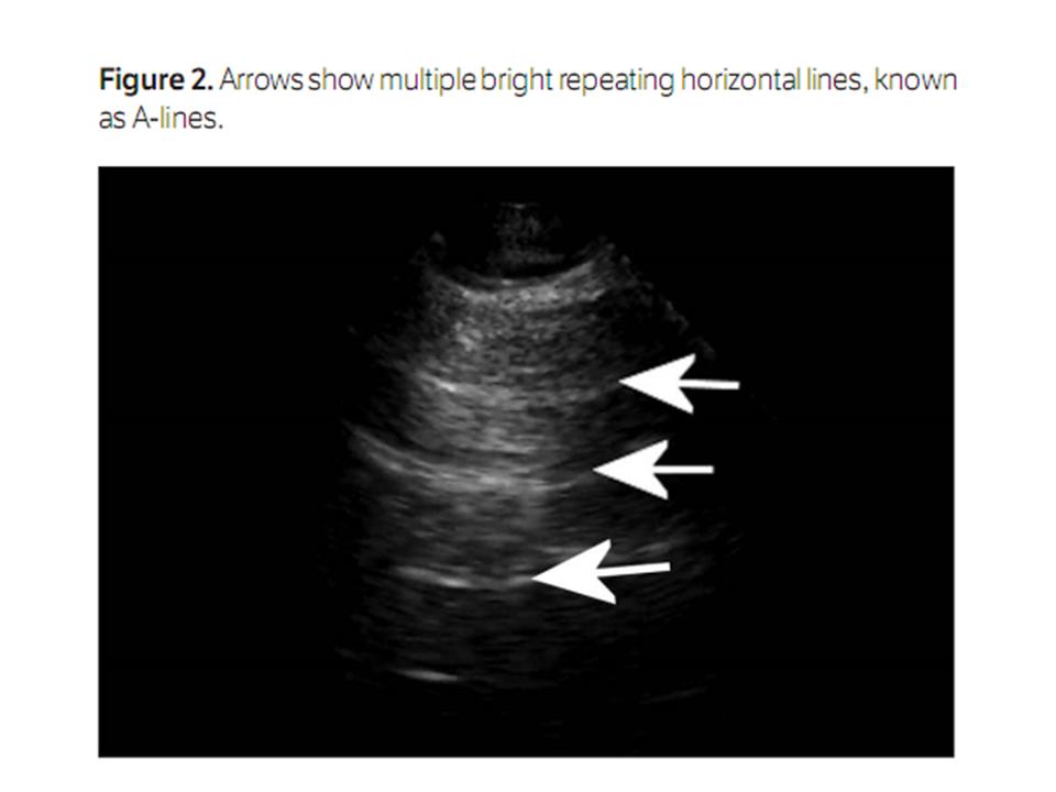
with later-stage biliary atresia had 4 cysts (Figure 2)
in the porta hepatis, with diameters ranging from 7 to 10 mm; only the largest
cyst was taken into account. The mean width and length of the cysts in the cyst
group (62.2 ± 39.9 and 41.1 ± 30.7 mm, respectively) were significantly larger
than those in the atresia group (16.2 ± 13.2 and 8.9 ± 6.5 mm; P = .002;
P = .001). However, the width to length ratios between the two groups
were not significantly different (0.64 ± 0.22 versus 0.63 ± 0.20; P =
.934). Sludge deposits in cysts were found in 6 of 11 patients (54.5%) in the
cyst group (Figure 3),
whereas none in the cystic atresia group had sludge deposits (P = .005).
On
sonography, the mean hepatic artery diameter in the atresia group was
significantly larger than that in the cyst group (2.4 ± 0.4 versus 1.7 ± 0.2
mm; P < .001), whereas no significant difference was found for the
portal vein diameter between the two groups (4.1 ± 0.8 versus 3.8 ± 0.6 mm; P
= .373). 
Figure 1.
Cystic
biliary atresia in a 51-day-old girl. A, Cyst (cursors) at the porta hepatis with a size of 46.6 × 23.5 mm. B, Irregularly elongated gallbladder (cursors) with
a length of 42.5 mm and a width of 7.8 mm. C, Triangular cord sign (arrows) at the anterior
wall of the right portal vein with a thickness of 7.0 mm. D, Enlarged hepatic artery (cursors) with a
diameter of 2.4 mm. CY indicates cyst.

Figure 2.
Later-stage
cystic biliary atresia in a 123-day-old boy. A, Four cysts (arrows) at the porta hepatis with
diameters ranging from 7 to 10 mm. Arrowheads indicate an atretic gallbladder
with a length of 9 mm. B, Triangular cord sign
(arrows) at the anterior wall of the right portal vein with a thickness of 8.0
mm. C, High-frequency image
showing fluids around the liver (arrows) and a coarse liver echo texture. D, Color Doppler image showing reopening of the
umbilical vein (arrow).
In the
atresia group, sonography showed atretic gallbladders in 2 patients (16.7%),
irregularly elongated gallbladders (Figure 1)
in 9 (75%), and a relatively normal gallbladder in 1 (8.3%). In the cyst group,
all 11 patients (100%) were observed to have normal gallbladders. The
morphologic characteristics of the gallbladders between the two groups were
significantly different (P < .001). Two patients in the atresia group
had gallbladder lengths of less than 15 mm, whereas all patients in the cyst
group had gallbladder lengths of greater than 15 mm. However, the mean
gallbladder length and the gallbladder width to length ratio were not
significantly different in the atresia group (26.4 ± 11.9 and 0.25 ± 0.10 mm)
compared with the cyst group (32.7 ± 10.2 and 0.27 ± 0.07 mm; P = .189; P=
.476). On the contrary, the mean gallbladder width in the atresia group (5.6 ±
2.0 mm) was significantly smaller than that in the cyst group (8.5 ± 2.5 mm; P
= .005).
Sensitivity,
specificity, and positive and negative predictive values for gallbladder size,
gallbladder abnormalities, the triangular cord sign, and sludge in the cyst for
predicting cystic biliary atresia are shown in Table 2.
When all specific sonographic features were taken into account, all patients
were correctly classified into the atresia and cyst groups, for an overall
accuracy rate of 100%.
Six of
12 patients (50%) in the atresia group were observed to have a communication
between the gallbladder and portal cyst on sonography, which was later
confirmed by intraoperative cholangiography. The cyst sizes were different
between those who had a communication between the gallbladder and cyst and
those who did not. The length of those with a communication (n = 6) ranged from
11 to 47 (25.5 ± 13.1) mm, whereas the length of those without a communication
(n = 6) ranged from 4 to 10 (6.8 ± 2.3) mm (P = .006).
Two
patients in the atresia group had an abnormal coarse liver echo texture and a
reopened umbilical vein (Figure 2)
on sonography. One of them also had ascites. All patients in the cyst group had
a normal liver echo texture, and no ascites was detected. Discussion
Sonography has been considered very helpful in making a preoperative differential diagnosis between cystic biliary atresia and choledochal cysts.5,10,15,16 Patients with larger cysts, dilated intrahepatic ducts, and normal gallbladders are more likely to have choledochal cysts. Inversely, those with smaller cysts, nondilated intrahepatic ducts, and abnormal gallbladders are more likely to have cystic biliary atresia. However, debates still exist as to this point of view. Some authors have reported cystic biliary atresia with large cysts or with dilated intrahepatic ducts.5,9,16 In addition, it is hard to draw a clear line between a large cyst and a small cyst. Some authors proposed that the shape of the cyst might play a role in distinguishing cystic biliary atresia from a choledochal cyst5,10 because the cysts in cystic biliary atresia were more likely to be teardrop shaped, whereas the choledochal cysts were more likely to be fusiform. However, we thought it was a little too subjective to differentiate cystic biliary atresia from choledochal cysts by judging the shape of the cysts. Some more specific and objective sonographic features for differentiating the diagnosis are urgently needed.
The state of the art in diagnosis of biliary atresia by sonography has been updated in recent years. Apart from gallbladder abnormalities, other sonographic features have been identified as useful in identifying biliary atresia, such as the triangular cord sign, dilatation of the hepatic artery, and hepatic subcapsular flow. The triangular cord sign was considered an objective criterion for identifying biliary atresia on sonography.12 Although the sensitivity of this sign for identifying biliary atresia varies, it is widely considered a specific sonographic marker of the disease, with specificity ranging from 96% to 100%.12,17– 20 Enlargement of the hepatic artery in infants with biliary atresia was also reported.13,19,21 With a critical diameter of 1.5 mm, the sensitivity and specificity of an enlarged hepatic artery for identifying biliary atresia were 92% and 87%, respectively.13 Hepatic subcapsular flow, defined as vascular structures contiguous with the liver capsular surface on color Doppler sonograms, was found to have sensitivity of 100% and specificity of 86% for identifying biliary atresia.21 A combination of all of these features could improve the accuracy of sonography for diagnosis of biliary atresia to 98% to 100%.17–19 However, few studies in the literature have assessed the value of these sonographic features for differentiating cystic biliary atresia from choledochal cysts.

Table 2.
Specific
Sonographic Features as Predictors of Cystic Biliary Atresia
In this
study, we found that the triangular cord sign and a dilated hepatic artery are
very useful for differentiating cystic biliary atresia from noncystic biliary
atresia. The triangular cord sign was detected in 11 of 12 patients (91.7%) in
the atresia group, whereas no patient in the cyst group had this sign. The
sensitivity and specificity of the triangular cord sign were 91.7% and 100%,
respectively. Although the triangular cord sign was not observed in 5 cases of
cystic biliary atresia10
because of the negative effect of the cyst at the porta hepatis, we thought
that the triangular cord sign could be detected by an experienced operator
performing careful scanning. However, it should be noted that not every case of
cystic biliary atresia might show the triangular cord sign. The incidence of
the triangular cord sign among patients with cystic biliary atresia needs to be
further assessed with more cases. We also showed that the caliber of the
hepatic artery is significantly larger in patients with cystic biliary atresia,
which is the same as that in patients with isolated biliary atresia. We think
that the reason for this finding is the same as for that in isolated biliary
atresia, even if the latter is still not known clearly.22
Although there is still no standard critical value for determining a larger
hepatic artery from normal, we think that a prominent hepatic artery can be
supportive in differentiating cystic biliary atresia from choledochal cysts.
Nevertheless, we have to confess that measurement of the hepatic artery in
patients with larger cysts at the porta hepatis was very difficult.

Figure 3.
Choledochal
cyst in a 74-day-old boy. A, Cyst at the porta hepatis
with a size of 52.1 × 50.0 mm. At the bottom of the cyst, sludge (arrow) without
a posterior acoustic shadow is shown. B, Normal gallbladder (cursors) above the cyst with a size of 22.3 × 4.5
mm. C, Dilated intrahepatic bile
duct (arrow) with a diameter of 1.7 mm. D, Hepatic artery (arrow) with a diameter of 1.8
mm. CY indicates cyst.
We
confirmed that an abnormal gallbladder is very critical for distinguishing
between cystic biliary atresia and choledochal cysts on sonography. Although
some authors reported that the gallbladder length in biliary atresia is usually
smaller than that in nonbiliary atresia,19,20
when it comes to cystic biliary atresia and choledochal cysts, this conclusion
might not be applicable.10
In this study, the occurrence of only 2 gallbladders (16.7%) in the cystic
biliary atresia group with a length of less than 15 mm was dramatically smaller
than that in isolated biliary atresia. Furthermore, the mean gallbladder length
in the atresia group was not significantly smaller than that in the cyst group
in this study. We think that it is the shape and not the size of the
gallbladder that is useful for differentiating cystic from noncystic biliary
atresia. As reported in the literature,5,10
the shape of the gallbladder in patients with cystic biliary atresia was much
more likely to be irregularly elongated in our study, with a frequency of 75%
(9 of 12).
The
mean cyst size in the atresia group was smaller than that in the cyst group in
our study. This result was the same as what most authors have noted.5,10,11
However, a cyst in a patient with cystic biliary atresia with a diameter of 80
mm reported by Caponcelli et al9
is clearly an exception to this outcome. Kim et al5
noted that it is useful to differentiate cystic biliary atresia from a
choledochal cyst by the shape of the cyst. However, it is a little too
subjective to define the shape as a teardrop, fusiform, or ovoid. In this
study, we compared the cyst length to width ratios between the cyst and atresia
groups, and the difference was not significant, which we consider a supportive
point for our opinion. In this study, 6 patients (54.5%) with choledochal cysts had sludge deposits, whereas no cyst with sludge was found in the patients with cystic biliary atresia. Similarly, Casaccia et al15 reported that echoic cysts were strongly suggestive of choledochal cysts. This finding could be explained by the pathologic changes that occur in these two entities. A choledochal cyst is an abnormality in which only the distant extrahepatic bile duct might be obstructive, whereas the proximal bile duct is patent. With the bile continuing to be secreted and flowing into the cyst, some components in the bile deposit and become sludge. On the contrary, biliary atresia is usually an entity with obliteration of the hepatic bile duct or the most proximal part of the extrahepatic bile duct.1 Hence, the bile flow cannot reach the cyst, and the fluid in the cyst does not contain the necessary components to form sludge. The white cystic fluid in cystic biliary atresia found during surgery23 might support this explanation. In addition, the reason why the intrahepatic bile ducts of choledochal cysts are more likely to be dilated than those of cystic biliary atresia was also explained. In our study, the incidence of dilated intrahepatic bile ducts in the cyst group was 81.8% (9 of 11), which was comparable to incidence rates of 47.7% to 100% noted by other authors.5,10
In this study, we also noted that one 123-day-old patient with later-stage biliary atresia had multiple cysts at the porta hepatis. We speculated that these might have been secondary intrahepatic biliary cysts caused by the ongoing process of hilar bile duct obstruction. Generally, multiple intrahepatic biliary cysts occur more often in postoperative biliary atresia. However, a few cases of cystic biliary atresia with multiple intrahepatic biliary cysts found before surgery have been reported.24
The main limitation of our study was that we did not correlate the triangular cord sign with the location of the cystic biliary atresia because of a lack of related information. However, we think that there might be a correlation between the presentation of the triangular cord sign and the location of the biliary atresia. Another limitation of our study was that the sample was very small. Large clinical trials are required to further test our findings.
In
conclusion, with the use of a combination of all specific sonographic features,
all 23 patients in our study were correctly classified as having cystic biliary
atresia or choledochal cysts. Although measurements of the cyst size and
hepatic artery diameter could be supportive points for differentiating cystic
biliary atresia from choledochal cysts, we think that the triangular cord sign,
gallbladder abnormalities, intrahepatic bile duct dilatation, and echoic cysts
might be more valuable sonographic features for differentiating cystic biliary
atresia from choledochal cysts. Furthermore, the triangular cord sign,
intrahepatic bile duct dilatation, and echoic cysts might be regarded as
objective sonographic features for distinguishing between cystic biliary
atresia and choledochal cysts because they can be detected objectively on
sonography.
© 2012
by the American Institute of Ultrasound in Medicine.


+W534-9,+Fig.+2.jpg)