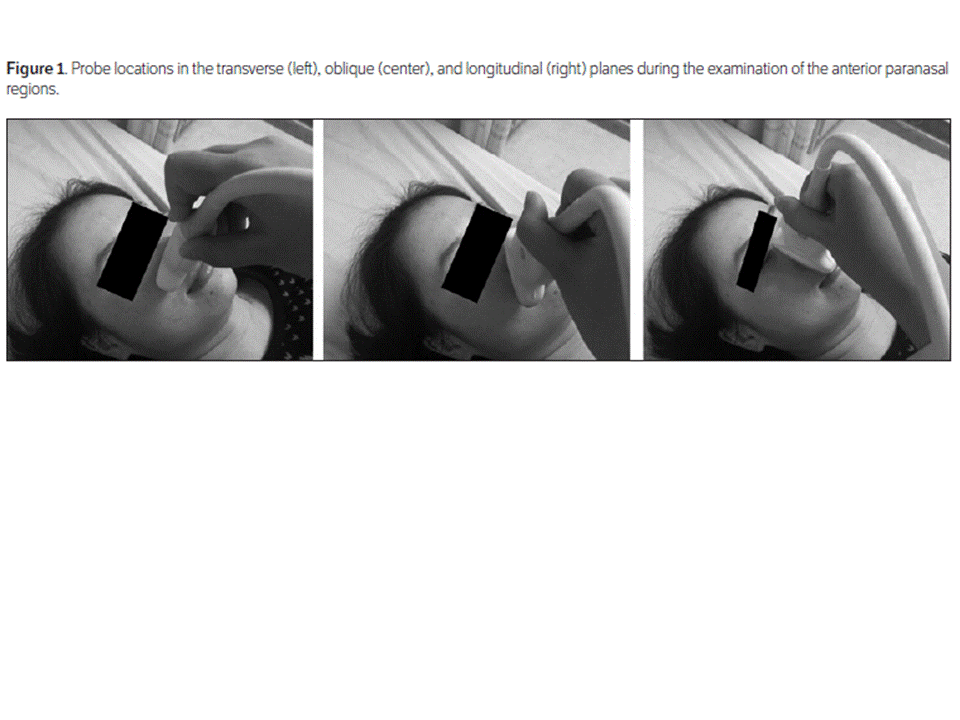
GASTRIC ULTRASOUND: ANTRAL GASTRITIS and H.pylori.
Sonography can be used effectively to evaluate the stomach and duodenum. A mucosal
thickness greater than 4 mm in the gastric antrum may suggest the presence of
gastritis. Marked transmural gastric wall thickening is typical of gastritis,
with documented resolution after appropriate therapy. Loss of the normal
multilaminar gut signature at the posterior wall of the gastric antrum is another
useful sonographic characteristic of inflammation.
The mucosa often seems to be
hyperenhancing adjacent to the hypoattenuation of edematous submucosa.
Antral
gastritis displays a spectrum of findings on double-contrast upper
gastrointestinal tract examinations, including thickened rugal folds, erosions,
mucosal nodularity, and antral striae. Wall thickening that is focal,
eccentric, and enhancing can resemble a malignant tumor, and further steps must
be taken with this possibility kept in mind.

Our study is unique and original, since we suggest that antral
wall and mucosal layer thicknesses as well as the mucosal layer-to-antral wall
thickness ratio may be predictive parameters for detection of antral gastritis
and H pylori infection on sonography. Cutoff values have not yet been
established, and further controlled studies are necessary for validation and
standardization of these parameters.
Inflammation and structural changes in the gastric mucosa
seem to be more prominent in the presence of H pylori. Therefore, careful
investigation for detection of H pylori and effective treatment for its
eradication should be mainstays of the management strategy.
This study recommends that sonography of the gastric
antrum can be beneficial for patients with presumed antral gastritis. If
thickening of the antral walls and mucosal layers is detected, antral gastritis
and H pylori infection must be kept in mind, and further diagnostic and therapeutic
steps should be taken accordingly. Even though the sonographic appearance of
antral thickening can rarely be found in healthy individuals, this
characteristic appearance should strongly suggest gastric disease,
necessitating an upper gastrointestinal series.
There are several noninvasive methods for evaluating the
severity of inflammation in the stomach and the presence or absence of H pylori
infection, such as a serum pepsinogen test, including pepsinogen I/II,
pepsinogen II, and immunoglobulin G antibody for H pylori. In this study, we
found that sonography of the stomach may be quite useful for noninvasive
evaluation of the antrum. Radiologists should be aware of the usefulness of
these specific criteria in the evaluation of gastric wall thickening on sonography
to better differentiate gastritis or a normal stomach from malignant or
potentially malignant lesions that warrant further diagnostic evaluation.
Our study had several limitations. Since we only measured
the gastrointestinal wall in white participants in Gumushane, Turkey, the
reference values may not be transferrable to populations with other diets or
ethnicities.
In addition, gastrointestinal wall thickness differs with
the ages, heights, weights, sexes, and smoking habits of patients. Some history
details and factors that may influence the outcome may not haven been
completely documented. Another limitation of our study was the lack of absolute
proof of antral gastritis in all cases, as no perfect reference standard exists
for this diagnosis. Furthermore, the wall thickness in vivo can be influenced
by muscular contraction, especially in the gastric antrum. The correlation
between the wall thickness evaluated by sonography and the real thickness of
the stomach as determined by macroscopic features (or histologic sections) of
resected stomach may well be different. However, a single 20-mg dose of butylscopolamine
was administered via an intramuscular route before the sonographic examinations
in all patients in our study. In addition, the degree of atrophic change (or degree
of inflammatory cell infiltration) that may influence the degree of wall
thickness in H pylori–positive patients was not considered in this study. Due
to these restrictions, associations should be interpreted with caution. Further
prospective and controlled studies with larger patient populations are
warranted for making more accurate interpretations of the sonographic findings
of antral gastritis and H pylori infection of the stomach. In terms of methods,
we have used fasting guidelines and intramuscular administration of a
pharmacologic agent.
Since most patients present with generalized abdominal symptoms,
and routine use of this protocol would be impractical in most busy ultrasound
practices, a fluid-filled antrum can be alternatively used for measuring the
wall layers.
The main limitation of this study was the relatively small
number of patients. Furthermore, the format of expressing average measurement
data for the 3 groups may not allow for determination of threshold values and
true-positive and -negative rates. Because of these restrictions, associations
should be interpreted with caution. However, this article raises awareness of
sonographic clues and will lead to further studies of this method.
In conclusion, our results suggest that antral gastritis caused
by H pylori infection is associated with characteristic features such as
thickening of the antral walls and mucosal layers on sonography. Thus, these
clues may be useful for diagnosis of gastritis cases that require further investigation
and may avoid at least some unnecessary interventions and measures.