Tổng số lượt xem trang

Thứ Hai, 7 tháng 9, 2020

What's best for lactating women: US vs Mammography?

By Theresa Pablos, AuntMinnie staff writer


September 3, 2020 -- Ultrasound may be the ideal imaging modality for evaluating palpable breast masses in lactating women, according to the results of a September 1 study in Radiology. For breastfeeding patients, targeted ultrasound identified just as many cancers as mammography with fewer false-positive findings.

The results are especially notable since more women in the U.S. are delaying childbearing into their 30s and 40s -- ages when mammography is typically considered the ideal first-line imaging modality for symptomatic patients. The findings support guidelines recommending the use of ultrasound for breastfeeding women of all ages.

"These findings suggest that sensitivity of mammography is slightly limited and inferior to that of [ultrasound] during pregnancy and lactation because of increased breast density," wrote the authors, led by Dr. Maggie Chung from the department of radiology and biomedical imaging at the University of California, San Francisco.

Ultrasound is often recommended as a first-line imaging modality for symptomatic, lactating women, but the evidence to support this recommendation for patients ages 30 and older is "near nonexistent," the authors noted. This lack of evidence prompted the team to conduct their own investigation.

The retrospective study included 167 lactating women with palpable breast masses who underwent ultrasound diagnostic evaluation at an academic institution between January 2000 and July 2017. The women were not currently pregnant and had no known malignancies at the time of evaluation. Their ages ranged from 17 to 52, with a median age of 35.

While all 167 women underwent ultrasound evaluation, 59% also underwent mammography. Mammography was performed at the discretion of the interpreting radiologist because the institution did not have a specific imaging policy for breastfeeding women -- a reflection of larger uncertainty around ideal imaging modalities for mothers in their 30s and 40s.

Targeted ultrasound scans identified a sonographic correlate to the palpable lump for 66% of women. A little more than half of these women had findings categorized as benign or probably benign, with cysts, inflammation or infectious changes, and galactoceles representing the most common benign findings.

An additional 53 women were recommended for biopsy due to a BI-RADS 4 or 5 lesion on the targeted ultrasound scan. Of these women, five were diagnosed with invasive ductal carcinoma. Mammography identified seven additional suspicious lesions but no additional cancers.

On its own, targeted ultrasound achieved a 100% sensitivity and 67% specificity. When combined with mammography, the modality maintained a 100% sensitivity rate, but the specificity rate dropped to 61%.
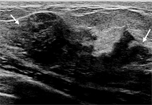
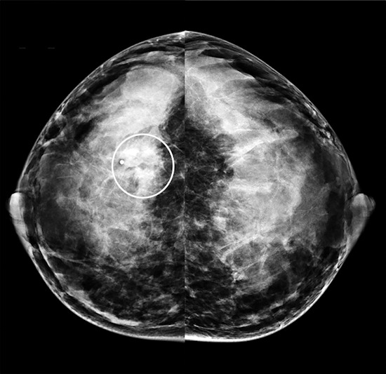
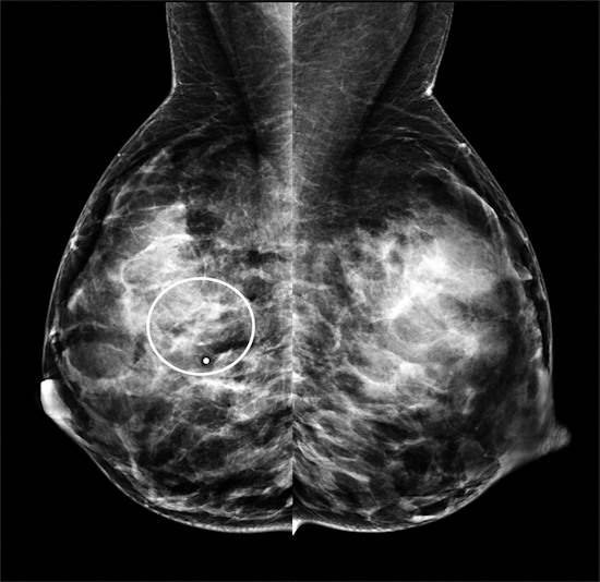
Imaging from a 40-year-old breastfeeding woman with a palpable lump in the right breast who was diagnosed with invasive ductal carcinoma
Imaging from a 40-year-old breastfeeding woman with a palpable lump in the right breast who was diagnosed with invasive ductal carcinoma. This targeted gray-scale ultrasound image shows a 2.4-cm hypoechoic irregular solid mass with indistinct margins (arrows) at the site of the palpable abnormality. All images courtesy of the RSNA.
Craniocaudal mammogram from the same 40-year-old patient with invasive ductal carcinoma
Craniocaudal mammogram from the same 40-year-old patient with invasive ductal carcinoma. The patient's mammogram images show a subtle, obscured mass at the site of the palpable abnormality (region of interest) with some associated amorphous calcifications.
Mediolateral oblique mammogram from the same 40-year-old patient with invasive ductal carcinoma
Mediolateral oblique mammogram from the same 40-year-old patient with invasive ductal carcinoma. Both mammograms depict heterogeneously dense breast tissue.

The authors attributed mammography's lower specificity due to the modality's struggle depicting cancers in dense breast tissues during pregnancy and lactation. Notably, 98% of the patients in the study had dense breast tissue, including 72% of whom had extremely dense breast tissue.

The authors cautioned that selection bias could have even overstated the performance of mammography in their study. Still, they noted mammography may benefit lactating women ages 40 or older, which only comprised a small subset of their study population.

"Mammography may be of greater benefit in lactating women age 40 years or older in whom the incidence of breast cancer is higher," they wrote. "More data are needed to confirm the best imaging algorithm in this small subset of patients.

Thứ Ba, 1 tháng 9, 2020

Blood pressure monitor trades compression for US


By Theresa Pablos, AuntMinnie staff writer

September 1, 2020 -- Researchers from Japan have developed a blood pressure monitor that utilizes a tiny ultrasound probe instead of compression to monitor systolic and diastolic changes. The science behind the prototype was presented as a virtual poster at the 2020 European Society of Cardiology (ESC) Conference. Lead author Dr. Kazunori Uemura, PhD, and his team from the National Cerebral and Cardiovascular Center in Japan designed the ultrasound-based monitor to unobtrusively track blood pressure on-the-go. Once calibrated, the ultrasound-based device produced blood pressure readings comparable to conventional technology in dogs.

"This method reliably tracks [blood pressure] changes without occlusive cuff inflation," the authors wrote in their poster abstract.

Monitoring blood pressure out-of-the-office and at regular intervals is important for hypertension diagnosis and management, but current compression-based blood pressure monitors can interfere with the daily lives of patients. Uemura and colleagues saw ultrasound as a potential alternative to compression-based technology.

The cuff they created utilizes a tiny ultrasound probe about half the size of a box of matches. The probe sits snugly in between a patient's skin and the larger blood pressure cuff.

Upon mild cuff inflation, the ultrasound transducer's 65 piezoelectric elements measure changes to an artery situated underneath the probe. Uemura and colleagues use these measurements to calculate estimated diastolic and systolic blood pressure.


(A) Schematic illustration of the cuff and ultrasound probe wrapped around the upper arm and positioned over the brachial artery. (B) Longitudinal cross section illustration of the ultrasound probe and inflated cuff. When the cuff is inflated, the device tracks the ultrasonic dimension of the brachial artery. Image courtesy of Dr. Kazunori Uemura, PhD.
The authors tested the cuff's accuracy for blood pressure monitoring on the right femoral artery of six anesthetized dogs. Measurements taken with the ultrasound-based device correlated strongly with compression-based readings, according to coefficient of determination (R2) analysis.

With just one-time calibration, the diastolic blood pressure readings had a bias of 3.9 ± 7.9 mmHg. This measurement fell within the acceptable bias range of < 5 ± < 8 mmHg set by the Association for the Advancement of Medical Instrumentation (AAMI).

However, with one-time calibration, the systolic blood pressure readings had a bias of 2.6 ± 18.9 mmHg, outside of the AAMI acceptable range. To improve the systolic readings, the authors added machine learning and a support vector algorithm to the initial, one-time calibration. The new analysis yielded systolic measurements with a bias of 0.7 ± 6.9 mmHg, meeting the AAMI guidelines.

"Once calibrated, this method measures [diastolic blood pressure] accurately," the authors wrote. "With the aid of machine learning, precision in [systolic blood pressure] prediction was greatly improved to acceptable levels."

This type of ultrasound-based technology is a long way from use in the clinic, and the research team still has yet to test the device's accuracy on people. But if the cuff holds up in further testing, the combination of ultrasound and machine learning could lead to better mobile blood pressure monitoring.

"This method with machine learning approach has potential for stress-free [blood pressure] measurement in ambulatory [blood pressure] monitoring," the authors concluded.