Tổng số lượt xem trang

Thứ Sáu, 15 tháng 4, 2022

U S offers alternative to MRI for bone stress


By Kate Madden Yee, AuntMinnie.com staff writer

March 24, 2022 -- Ultrasound appears to be an effective point-of-care alternative to MRI for the evaluation of bone stress injury, according to a study published March 22 in the Journal of Ultrasound in Medicine.

MRI is typically used to assess bone injuries, but the study results suggest that clinicians may have a more easily accessible tool for this application, wrote a team led by Dr. Isaac Syrop of Columbia University in New York City and Dr. Yaeko Fukushima, PhD, of Kansai Medical University in Osaka, Japan.

"There are many advantages to ultrasound imaging over MRI, including its dynamic practicality, which provides the treating clinician with an opportunity to evaluate local soft tissue sites in real-time," the group wrote. "Ultrasound imaging takes significantly less time than MRI to perform ... and can be done as part of the clinical evaluation."

Bone stress injuries are common in young adult athletes, and can negatively affect performance over the long term, the investigators noted. It's crucial to diagnose these injuries early so that athletes can be treated effectively and return to play.

X-ray and CT aren't very effective for early diagnosis of bone stress injuries and expose patients to radiation; since the 1980s, MRI has been used as the diagnostic standard. But MRI is expensive and time-consuming.

"There is good reason to believe that the more affordable and accessible diagnostic option of musculoskeletal ultrasound imaging may help to address the shortcomings of MRI," the authors noted.

The researchers sought to compare the sensitivity and specificity of ultrasound to MRI in the diagnosis of bone stress injury through a study that included 37 young adult athletes presenting in an academic sports medicine clinic between 2016 and 2020 with suspected lower extremity bone stress injury. Sports included everything from crew, field hockey, gymnastics, and running to soccer, tennis, track, and volleyball. All patients underwent MRI and ultrasound exams.

Of the 37 study participants, 30 (81%) had bone stress injuries. The most common injuries were in the metatarsal bones (54%) and the tibia (32%).

Ultrasound scored relatively high across a range of performance measures relative to the gold standard of diagnosis on MRI scans, the group found.

Performance of ultrasound for diagnosing bone stress injuries compared with baseline assessment MRI
MeasurePerformance
Sensitivity80%
Specificity71%
Positive predictive value92%
Negative predictive value45%

"In summary, ultrasound imaging may be a point-of-care tool for the current practicing sports medicine provider to combine with their clinical evaluation in the diagnosis of bone stress injuries of the lower extremity," the team concluded.
Thứ Sáu, 18 tháng 3, 2022

5 rules for diagnosing fetal problems on ultrasound

 By Amerigo Allegretto, AuntMinnie.com staff writer


March 16, 2022 -- So, you perform a fetal ultrasound scan and find something that might indicate an ectopic pregnancy. But how can you be sure? A presentation at the 2022 American Institute of Ultrasound in Medicine (AIUM) annual meeting gave the answer.

In his talk, Dr. Peter Doubilet, PhD, from Brigham and Women's Hospital in Boston listed five rules for ultrasonographers and clinicians to consider when diagnosing fetal problems in the first trimester when women present with abnormal vaginal bleeding, which in turn influences treatment decisions.

"The key principle for ultrasound in early pregnancy loss is to virtually eliminate false positives," Doubilet said.

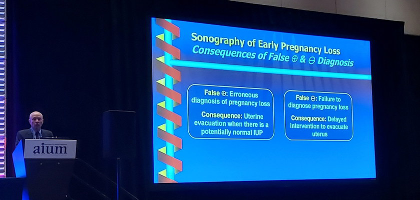
Peter Doubilet talked about ultrasound
Dr. Peter Doubilet, PhD, talked about ultrasound's use in determining whether a pregnancy is ectopic, as well as the consequences of false-positive and false-negative readings.

Ultrasound is used for patients who present with positive symptoms of pregnancy, with the gestational sac typically being the first object seen about five weeks into a pregnancy. However, abnormal vaginal bleeding is a sign of early pregnancy loss or ectopic pregnancy. Doubilet said that about 25% of early, clinically recognized pregnancies end in miscarriage.

"And those are only the clinically recognized ones, and then there are others that aren't recognized that end in miscarriage," he said.

However, false-positive and false-negative results have consequences, with Doubilet saying that the former is "way more consequential." This is because treatments following false-positive readings for ectopic pregnancies, such as methotrexate, can inadvertently cause miscarriages, stillbirths, or birth defects for healthy embryos.

Doubilet's five rules to consider for fetal ultrasound include the following:

  1. If a mass is extraovarian, it's "almost certainly" an ectopic pregnancy. If the mass is inside the ovary, it's a corpus luteum.
  2. If you're unsure about masses in or beside the ovary, press with a transvaginal transducer. If the mass moves with the ovary, it's most likely a corpus luteum. If it moves separately from the ovary, it's an ectopic pregnancy.
  3. A gestational sac located in the cervix is likely a cervical ectopic pregnancy, especially if it's well-formed and has a fetal heartbeat. It is likely also a miscarriage in progress if it is a flattened sac without a heartbeat. If you can't tell and the patient is stable, leave the scan for one or two days. The ectopic pregnancy will still be there while the miscarriage in progress will have passed.
  4. If you have an eccentric gestational sac in the uterus, it's likely to be an interstitial ectopic pregnancy. This also applies if it bulges the uterine contour and has no visible surrounding myometrium. If you're unsure, 3D ultrasound can help provide the correct diagnosis.
  5. If you see a gestational sac in the uterus on ultrasound, a separate adnexal mass is likely to be a tubal ectopic, heterotopic pregnancy. This also applies if it has a heartbeat or a yolk sac in the adnexal mass in addition to the intrauterine pregnancy. The mass is likely to be a corpus luteum if there is no heartbeat or yolk sac. Heterotopic pregnancies are "very rare" while corpus luteums are common.