
A 27-year-old woman was admitted to the obstetric emergency department
for abdominal pain without bleeding at 8 weeks’ gestation. She had a previous
uneventful pregnancy delivered vaginally at term, and an early scan performed 2
weeks before the symptoms occurred revealed an ongoing intrauterine pregnancy.
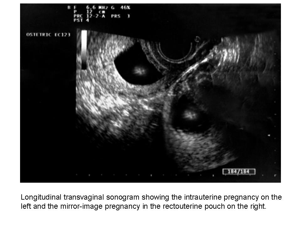
The patient underwent transvaginal sonography (MyLab 25; Esaote SpA,
Florence, Italy), which showed an intrauterine pregnancy with a live embryo
measuring 29 mm, consistent with a gestational age of 8 weeks 5 days. A second
gestational sac with irregular and undefined boundaries containing an active
embryo measuring 17 mm was depicted in the rectouterine pouch (Figure 1). Both
ovaries were visualized as normal, and no pelvic free fluid collection was
noticed during the scan. These findings were consistent with a diagnosis of
heterotopic pregnancy.
Heterotopic pregnancy refers to the rare occurrence of both
intrauterine and ectopic pregnancies usually located in one fallopian tube,
cervix, or, more rarely, abdomen. Assisted reproduction techniques, tubal
surgery, pelvic inflammatory disease, and the use of intrauterine devices
represent the most common risk factors.1
Sonography is the mainstay for diagnosis of heterotopic pregnancy,
allowing for the detection of two gestational sacs located inside and outside
the uterus, respectively, and blood collection in the pelvis. In addition,
clinical symptoms such as pain and genital tract bleeding can help in achieving
the diagnosis.1
In this case, the patient had no risk factors for this condition, with
a previous uneventful pregnancy delivered at term and no history of pelvic
surgery or disease. To confirm the diagnosis, the woman was asked to fill her
bladder, and transabdominal sonography was performed. Interestingly, only the
intrauterine gestational sac was found, with a normal appearance of the
rectouterine pouch and no ectopic pregnancy detected.
Không có nhận xét nào :
Đăng nhận xét