Tổng số lượt xem trang

Thứ Ba, 28 tháng 1, 2014

FACTORS CORRELATING with ARFI ELASTOGRAPHY in CHRONIC HEPATITIS C


Abstract

AIM: To investigate the factors other than fibrosis stage correlating with acoustic radiation force impulse (ARFI) elastograpy in chronic hepatitis C.
METHODS: ARFI elastograpy was performed in 108 consecutive patients with chronic hepatitis C who underwent a liver biopsy. The proportion of fibrosis area in the biopsy specimens was measured by computer-assisted morphometric image analysis.
RESULTS: ARFI correlated significantly with fibrosis stage (β  = 0.1865, P  < 0.0001) and hyaluronic acid levels ( β = 0.0008, P = 0.0039) in all patients by multiple regression analysis. Fibrosis area correlated significantly with ARFI by Spearman’s rank correlation testbut not by multiple regression analysis. ARFI correlatedsignificantly with body mass index (BMI) ( β = -0.0334, P = 0.0001) in F0 or F1, with γ-glutamyltranspeptidase levels ( β = 0.0048, P = 0.0012) in F2, and with fibrosis stage (β  = 0.2921, P  = 0.0044) and hyaluronic acid levels ( β = 0.0012, P = 0.0025) in F3 or F4. The ARFIcutoff value was 1.28 m/s for  F ≥ 2, 1.44 m/s for  F ≥ 3, and 1.73 m/s for  F4.
CONCLUSION:
ARFI correlated with fibrosis stage and hyaluronic acid but not with inflammation. ARFI was affected by BMI, γ-glutamyltranspeptidase, and hyaluronic acid in each fibrosis stage. 

© 2014 Baishideng Publishing Group Co., Limited. All rights reserved

Core tip: The assessment of liver fibrosis stage is important to estimate prognosis and to identify the patients requiring antiviral treatment in chronic hepatitis C. Liver biopsy is a gold standard for assessing fibrosis, but is invasive. Thus methods for noninvasively assessing fibrosis have been developed. Liver stiffness measurement (LSM) by Fibroscan and acoustic radiation force impulse correlate with fibrosis stage. However, LSM may be affected by factors other than fibrosis, such as edema, steatosis, and inflammation.

DISCUSSION

The assessment of fibrosis stage is important to estimate prognosis and to identify the patients requiring antiviral treatment in chronic hepatitis C. A lot of noninvasive methods to assess liver fibrosis stage other than liver biopsy are available, for example, ARFI, TE, real-timeelastography [23], and algorithm of serum fibrosis markers such as FibroTest [24] and APRI [25]. They provide good performances in estimation of fibrosis stage, while there are problems such as influence of inflammation. In the present study, factors other than fibrosis stage that affect ARFI were investigated in patients with chronic hepatitis C.
The present study confirmed findings reported previously that ARFI correlates with fibrosis stage [10-13,26,27].
The ARFI cutoff values for different fibrosis stages were 1.28 m/s for F ≥ 1, 1.28 m/s for F ≥ 2, 1.44 m/s for F ≥ 3 and 1.73 m/s for F4. This result suggests that distinguishing between F0 and F1 is impossible, as the cutoff value for F ≥ 1 and that for F ≥ 2 are the same. However, Sporea et al [26] reported that the cutoff value is 1.19 m/s for F ≥ 1, 1.33 m/s for F ≥ 2, 1.43 m/s for F ≥ 3, and 1.55 m/s for F4 [26]. Rizzo et al [13] reported that the cutoff value is 1.3 m/s for F ≥ 2, 1.7 m/s for F ≥ 3 and 2.0 m/s for F4 [13]. Thus, discrepancies are apparent among the cutoff values reported in different studies. The discrepancies are probably attributed to the difference in the population studied. Further studies should be conducted to establish standard ARFI cutoff values for staging fibrosis.
In the present study, AST, ALT and inflammatory grade were correlated with ARFI in the univariate analysis that included all patients, but were not selected as factors independently correlating with ARFI in the multiple regression analysis. In addition, inflammatory factors did not correlate with ARFI when patients with different fibrosis stages were analyzed separately. These results suggest that inflammatory activity does not affect ARFI in patients with chronic hepatitis C. Rizzo  et al [13] also reported that ARFI is not associated with ALT, BMI, Metavir grade, or liver steatosis, whereas TE is significantly correlated with ALT[13]. Bota et al [10] reported that discordance of at least two fibrosis stages between ARFI and histologic assessment were associated with female sex, interquartile range interval (IQR) ≥ 30%, high AST and high ALT in univariate analysis,while, in multivariate analysis, the female gender and IQR ≥ 30% (P = 0.004) were associated with the discordances. In contrast, Yoon et al [12] reported that the optimum ARFI cutoff values are 1.13 m/s for F ≥ 2 and 1.98 m/s for F4, whereas these values decreased to 1.09 m/s for F ≥ 2 and 1.81 m/s for F4 when patients with normal ALT levelswere selected. Chen et al [11] reported that ALT, ActiTest A score, Metavir activity (A) grade, Metavir F stage, BMI, and platelet count are independently associated with ARFI and suggested that a 100 IU/L increase in serum ALT levels augmented ARFI by approximately 0.155 m/s. In the present study, only 25 patients had ALT levels of 100 IU/L or higher. The low ALT levels among the patients studied may be a reason why ALT was not correlated with ARFI.


A multiple linear regression analysis in our previous study on TE selected fibrosis area, ALT levels, γ-GTP levels, prothrombin time, and hyaluronic acid levels as factors correlating with TE[21]. Many studies on TE have reported that LSM is affected by ALT levels. Franquelli et al [28]  reported that TE fibrosis staging is overestimated by necroinflammatory activity and steatosis. Coco et al [7] found that LSM is higher in patients with an elevated ALT than in those with either spontaneous biochemical remission or after antiviral therapy. Thus, it is probable that ALT or inflammatory activity affects TE. However, it is still unclear whether they also affect ARFI. Further studies are needed to clarify factors that affect ARFI other than fibrosis stage. ARFI was significantly correlated with BMI in the 31 patients with stage F0 or F1; the higher the BMI, the lower the ARFI. However, ARFI was not associated with steatosis grade. Motosugi et al [29] reported that fat deposition in the liver does not affect ARFI. Thus, the negativecorrelation between BMI and ARFI could not be attributed to steatosis, which accompanies higher BMI [30].
Actually, BMI and steatosis grade were not correlated in patients with stage F0 or F1 in the present study (data not shown). The mechanism of the association between higher BMI and lower ARFI is unclear. Because a higher BMI is associated with lower ARFI, and may cause anunderestimation of fibrosis staging, careful attention should be paid to BMI during ARFI staging of fibrosis in patients with stage F0 or F1 disease.
ARFI significantly correlated with γ-GTP levels in patients with F2 and with fibrosis stage and hyaluronic acid levels in patients with stage F3 or F4. γ-GTP[24,31] and hyaluronic acid [32,33]  levels have been regarded as the most informative fibrosis markers. Thus, it is reasonable that γ-GTP and hyaluronic acid levels independently correlated with ARFI. Isgro  et al [20]  showed that the collagen proportional area has a better relationship with TE and with hepaticvenous pressure gradient compared with Ishak stage. In the present study, fibrosis area was correlated significantly with fibrosis stage, but only fibrosis stage and hyaluronic acid levels were selected as factors independently correlating with ARFI. Our previous study demonstrated a better correlation of TE with fibrosis stage than with fibrosis area in patients with chronic hepatitis C[21]. The Metavir stages represent categories of increasing fibrosis severity based on a combination of location and quantity of scarring as well as whether the fibrous tissue forms septa, bridges, or nodules. Fibrosis area represents only the quantity of fibrosis in liver tissues. Our results indicate that not only the quantity of fibrosis but also other histological factors such as patterns of fibrosis also affect ARFI.
The present study demonstrated that ARFI correlated with fibrosis stage but was not associated with inflammation. BMI negatively correlated with ARFI in the patients with stage F0 or F1. γ-GTP and hyaluronic acid levels were positively correlated in those with stage F2 and in those with F3 or F4, respectively. Thus, careful attention should be paid to BMI, γ-GTP levels, and hyaluronic acid levels when estimating fibrosis stage by ARFI. Fibrosis stage showed a better correlation with ARFI than fibrosis area, indicating that not only the quantity of fibrosis but also other factors such as patterns of fibrosis also affect ARFI. Since the number of the patients studied is small,further studies are needed to confirm the conclusion of the present study.

Thứ Hai, 27 tháng 1, 2014

NHÂN CA HYDROCELE of CANAL of NUCK @ MEDIC CENTER


Bệnh nhân đã được mổ cấp cứu vào mùng 25 Tết tại bệnh viện Chợ Rẫy, chẩn đoán sau mổ là nang ống Nuck xuất huyết và tái tạo thành bẹn theo phương pháp Shouldice.
Ca tràn dịch ống Nuck khác, với chẩn đoán lâm sàng là thoát vị bẹn P, bệnh nhân không mổ vì không đau và vì đã biết từ khi còn bé.
 Cả 2 ca đều có cấu trúc dạng ống ngay trên gai mu và đầu trên có hình dấu phẩy đặc trưng, là nơi nối tiếp với phúc mạc, đoạn đầu của processus vaginalis.


Sơ đồ cho thấy cơ chế thành lập hydrocele ở nam (T) và nữ (P) là phần processus vaginalis của phúc mạc không xẹp dính lại sau năm tuổi đầu tiên. 


Thứ Hai, 20 tháng 1, 2014

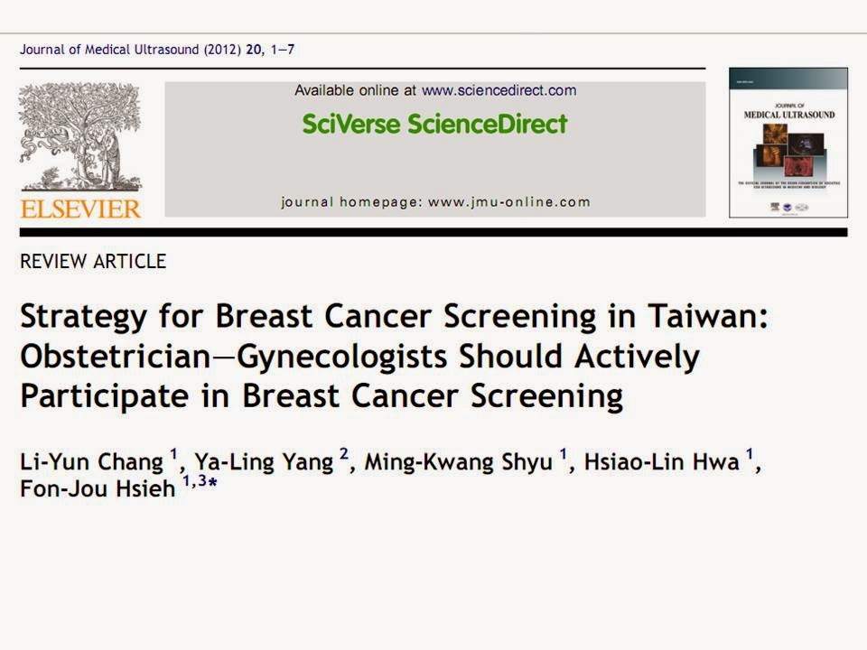
STRATEGY for BREAST CANCER SCREENING in TAIWAN


Breast cancer (BC) has become a global disease among women. Cost-effective strategies in reducing mortality caused by BC are highly desirable. Here, we suggest a working program based on an overall evaluation of the shortcomings and advantages of the current strategy for BC screening in Taiwan. From 1995 to 2002, cervical cancer was the most frequent cancer in women in Taiwan. However, invasive BC has moved from the second-most to the most frequent cancer since 2003. The incidence of BC increased by 14.69% between 2003 and 2008. In the same time interval, the incidence of cervical cancer decreased by 4.59%. Age analysis for BC incidence showed that 11% were at a relatively young age (30e39 years) and the peak incidence was in the group aged 40e49 years. Furthermore, only 31% of patients were at stage I according to data for newly diagnosed patients at National Taiwan University Hospital for 2004-2009. With the increase in pregnancy at an advanced maternal age, obstetrician egynecologists should be alert to the possibility of coexisting BC and pregnancy. To facilitate early detection of BC, a campaign for “Three points examined together” {uterine cervix and 2 breasts} should be implemented. Obstetrician-gynecologists should perform breast examination including palpation and ultrasound examination at the same time as the annual Pap smear. Mammography should be performed every 2-3 years or when indicated. If suspicious lesions are found, patients should be referred to a breast imaging laboratory, where a definitive diagnosis can be established. For confirmed cases of BC, appropriate surgery, chemotherapy and radiotherapy should be mammography or ultrasound provided. In short, active participation of obstetrician-gynecologists is a must in the campaign against BC in Taiwan.

Mammography or ultrasound

Although far from being a perfect tool, mammography has been the mainstay of BC screening. Mammography can detect microcalcification in breast lesions for stage 0 BC patients. However, it involves ionizing radiation. It is not applicable in women under 40 years of age in general. It has to be performed in rooms shielded with lead. Many studies have documented that the morphological view of breast tissue in Asian women is denser than that in Caucasian women. The pain and discomfort caused by compression during the mammography have deterred many women from BC screening programs. In fact, the acceptance rate for mammography in Taiwan has been as low as 17%. In addition, microcalcification in breast lesion is difficult to be detected by mammography for Asian compared to Caucasian women.
By contrast, ultrasound is not ionizing in nature. It is widely acceptable and available in Taiwan and it is performed in an OB/GYN clinic setting where Pap smears are performed. Thus, it is possible for “Three points examined together” to become more than a slogan. When a suspicious breast lesion is found, an experienced physician can undertake fine needle aspiration or core biopsy to make a definitive diagnosis right on site.
Taken together, these considerations indicate that the best strategy for BC detection might be to perform both mammography and ultrasound examinations without an additional charge at each visit. For women not at high risk of BC, mammography can be performed every 2-3 years, while breast ultrasound is performed annually together with Pap smears.
If we can convince 2000 obstetricianegynecologists in Taiwan to actively participate in BC detection, we believe that the BC mortality rate in Taiwan could be markedly reduced within a few years via early detection and management of disease. A good example is the maternal serum screening campaign for Down syndrome (Fig. 4) [19]. With the right strategy and a collaborative team consisting of patients, obstetrician-gynecologists and other medical care providers, the Down syndrome live birth rate decreased by 70% in 3 years. It was estimated that at least 200 families in Taiwan each year would benefit from this campaign.


Framework for a BC screening campaign in Taiwan

We suggest that the following framework should be established without delay in the fight against BC, the most frequent female cancer in Taiwan:
(1) First line. Physical breast examination should be performed followed by breast ultrasound examination at the time of annual Pap smears. Mammography can be performed every 2-3 years in the radiology department. Whether cases have family history of BC or not should be carefully scrutinized. If a patient has a family history of BC, extra caution should be exercised.
(2) Second line. Breast imaging laboratories should be established in regional hospitals and medical centers and operated by experienced radiologists or breast physicians. When a suspicious breast lesion is found during first-line screening, the patient should be referred to this breast imaging laboratory, where an expert can  use breast ultrasound, mammography, or MRI coupled with aspiration cytology or core biopsy to establish a definite diagnosis.
(3) Third line. Breast surgeons should be available to perform adequate surgery for confirmed BC cases.
(4) Fourth line. High-quality pathological diagnosis, radiotherapy, chemotherapy, and genetic counseling should be provided.

Experience in NTUH
A breast imaging laboratory was established in NTUH in 2003 and has been functioning effectively since then. The laboratory has actively engaged in training for breast ultrasound imaging. Physicians from surgery, obstetrics and gynecology, and family medicine can receive 3-month fellowship training in the laboratory. A 1-week introductory course on breast ultrasound is also available for members in Taiwan Society of Obstetricians and Gynecologists. The breast imaging laboratory at NTUH is a successful model that other hospitals in Taiwan can duplicate. We hope that this type of breast imaging laboratory will become available in an increasing number of hospitals all over Taiwan.

Conclusion
The incidence of female BC in Taiwan increased by 14.69% from 2003 to 2008 and some 8136 women were newly diagnosed with BC in 2008. The incidence of female BC at NTUH increased by 62.02% in 2009 compared with 2004 (Fig. 5). The peak incidence was in the group aged 40-49 years and 11% of cases occurred in the group aged 30-39 years. The percentage of early BC detected annually is pproximately 30%, a figure much lower than that in Western countries.
The following steps are suggested to boost early detection of BC in Taiwan:
(1) The health authority should implement a policy of “Three points examined together” by establishing a workable scheme. Thus, Pap smears and BC detection can be performed at the same site during the same visit.
(2) Active participation of obstetricianegynecologists is absolutely necessary for a successful BC detection campaign.
(3) The Taiwan Society of Obstetricians and Gynecologists and all teaching hospitals should gear up in establishing an educational program in breast medicine for residents and attending physicians to provide adequate care for breast disorders.
(4) Facilities to provide Pap smears and breast examinations simultaneously should be available at as many OB/GYN clinics and hospitals as possible.
(5) Easily accessible breast imaging laboratories should be set up at regional hospitals and medical centers to provide a definite diagnosis for patients with suspicious lesions found in first-line clinics.
(6) The health authority and relevant medical societies should work together to run quality assurance programs to familiarize doctors with various breast examinations.
(7) Active public campaigns should be implemented to raise public awareness of BC detection strategies.

Over the years, obstetricians and gynecologists in Taiwan have used Pap smear screening and screening of maternal serum for Down syndrome to great effect. This has resulted in marked reductions in cervical cancer cases and live births affected by Down syndrome. If a widespread screening program is set up involving first-line clinics and a good breast imaging laboratory, we may soon witness another miracle in BC screening in terms of early detection of (pre) cancerous lesions.

Thứ Tư, 15 tháng 1, 2014

ULTRASOUND NEWS on AuntMinnie


AJR: Using ultrasound 1st for appendicitis saves money


January 14, 2014 -- Using ultrasound as a first-line evaluation tool for patients suspected of having appendicitis -- and sending equivocal cases on to CT -- reduces radiation dose and saves healthcare dollars compared with a CT-only protocol, according to a new study in the January American Journal of Roentgenology.


Researchers from Thomas Jefferson University conducted a comparative effectiveness research study to compare the costs of an ultrasound-CT protocol versus a CT-only protocol for assessing appendicitis. They found that ultrasound is effective enough to be used as a first-line evaluation tool, even though it's slightly less accurate than CT.

What really tips the scales in favor of ultrasound is its lack of radiation dose. When the downstream costs of CT radiation dose are included, using ultrasound first results in savings of more than $330 million, according to study authors Laurence Parker, PhD, Dr. Levon Nazarian, and colleagues (AJR, January 2014, Vol. 202:1, pp. 124-135).

Advantages of ultrasound

Ultrasound's advantages include its relatively low cost, wide availability, and lack of radiation; its perceived disadvantage is that it is more dependent on the operator's skill than other imaging tests, the authors wrote. CT is highly accurate for detecting acute appendicitis, but it is expensive and exposes patients to radiation.


"The reason we did this study is that there are quite a few articles in the literature that compare how good ultrasound and CT are for the diagnosis of appendicitis, but we felt that none of them had an algorithmic approach that addressed the ramifications of the two technologies in terms of cost," Nazarian told AuntMinnie.com. "Just to compare CT to ultrasound for this purpose and to say that CT is more accurate than ultrasound, while true, only gives part of the story."

For their study, the researchers used information from the U.S. Centers for Medicare and Medicaid Services (CMS), national hospital discharge surveys, and U.S. Census Bureau life tables.

The group proposed the following imaging protocol: Patients presenting in the emergency department with right lower quadrant abdominal pain would receive an ultrasound first. If the ultrasound was positive, the patient would be sent for an appendectomy because ultrasound has a high positive predictive value for appendicitis.

However, if the ultrasound findings were negative or uncertain, patients would undergo a CT exam. If the CT findings were positive, they would have an appendectomy; if negative, no further treatment would be performed.

The group performed a meta-analysis of ultrasound's performance versus CT for detecting acute appendicitis.

Ultrasound vs. CT for appendicitis
Ultrasound
CT
Mean sensitivity
87.5%
93.4%
Mean specificity
92.7%
95.3%
Positive predictive value
91%
92.5%
Negative predictive value
89.8%
95.9%

Meanwhile, the analysis of CMS files showed that CT was used for almost exactly two examinations per patient (one abdominal and one pelvic) and ultrasound was rarely used. The cost of the CT-only imaging protocol was $547 per patient, whereas the cost of a limited ultrasound study was $88 per patient. For the total U.S. population, the cost savings of an ultrasound-first protocol minus the cost of extra surgeries and extra surgical deaths would be $24.9 million per year, according to the authors.

High costs of radiation

Not only does the study underscore the cost savings an ultrasound-first protocol could provide, it emphasizes the surprisingly high downstream costs of radiation exposure in diagnostic imaging.

"The ultrasound-CT protocol we have proposed saves $24.9 million over the CT-only protocol when the costs of the tests and excess surgeries and mortalities from using a less sensitive test are considered," they wrote. "Radiation exposure savings are more than an order of magnitude greater -- $339.5 million."

Appendicitis evaluation represents another diagnostic imaging problem for which ultrasound could provide substantial cost savings but is underutilized, and when current radiation exposure models are applied to appendicitis evaluation, the number of excess cancer deaths resulting from current practice is striking -- approximately 50 full lives per year, the group wrote.

But clinicians should not necessarily stop using CT to evaluate appendicitis, Parker said.

"It's not that we're taking CT out of it," he said. "But if we can do an ultrasound first, we can avoid enough CTs to gain significant cost savings and avoid radiation exposure. It's interesting that a slightly less accurate technique is actually superior when you look at the big picture."

Related Reading


Copyright © 2014 AuntMinnie.com

Study shows preabortion ultrasound doesn't often change decisions


January 14, 2014 -- Although many abortion opponents believe that women who view ultrasound scans of their fetuses will cancel their decision to pursue abortions, a new study in Obstetrics and Gynecology suggests the impact of the scans may be limited.

In an analysis of medical records of more than 15,000 women seeking abortion care at Planned Parenthood Los Angeles (PPLA), the researchers found that 98.4% of women who voluntarily elected to view preabortion ultrasound images went on to terminate the pregnancy, compared with 99% of women who did not view the images.

However, viewing ultrasound images was significantly associated with a decision not to terminate the pregnancy in the 7.4% of women who had medium or low certainty in their decision to have an abortion.

"Voluntarily viewing the ultrasound image may contribute to a small proportion of women with medium or low decision certainty deciding to continue the pregnancy; such viewing does not alter decisions of the large majority of women who are certain that abortion is the right decision," wrote a team led by Dr. Mary Gatter, medical director of PPLA.

Small but statistically significant difference

Preabortion ultrasound has become a political lightning rod in the U.S. as a number of states have implemented laws requiring pregnant women to view an ultrasound image before going ahead with the decision to terminate a fetus. However, whether such laws actually affect the decision to go ahead with an abortion hasn't been heavily studied.

Seeking to add to the small body of research, a group from PPLA and the University of California, San Francisco (UCSF) analyzed deidentified records for 15,575 women who sought abortion care at PPLA in 2011 (Obstetrics and Gynecology, January 2014, Vol. 123:1, pp. 81-87).

A previous analysis of this dataset had found that ultrasound images were viewed 42.5% of the time. Of these women, 98.4% went on to terminate their pregnancy, a difference of 0.6% (p < 0.001) from the 99% termination rate among those who did not view the images.

Among the group of patients who viewed the images, 95.2% of women with a medium or low decision certainty proceeded to an abortion, compared with 97.5% of women who had a high degree of certainty.

In contrast to two existing studies in the literature that did not show a link between ultrasound viewing and termination rates, the current study found that voluntary viewing was associated with the decision of some women to continue their pregnancy.

"However, the effect was very small -- and should be considered with caution -- and limited to the 7% of patients with medium or low decision certainty," they wrote. "This population may not have been substantially present in prior studies drawing on much smaller samples of patients."

The authors emphasized that the role of ultrasound viewing in abortion care needs to be viewed in context. Other factors, such as gestational age, had stronger effects on the likelihood of whether a woman continued a pregnancy, according to the researchers. The study showed an increase in the odds of continuing a pregnancy that was associated with each gestational age category after nine weeks.

"The importance of gestational age for women deciding to continue the pregnancy suggests that it is the information the ultrasound scan renders -- i.e., gestational dating -- rather than the image that influences women's decision-making," the authors wrote.

The results cannot be generalized to women's experience of ultrasound viewing in settings where it is mandatory, "although given the very high percentage of women proceeding with abortion after viewing the ultrasound image, it is unlikely that mandatory viewing would substantially affect the number of abortions performed," the authors noted.

"It may, however, affect patient satisfaction and health outcomes, which research shows are enhanced when patients feel control over decisions related to their care," they wrote."Mandating that women view their ultrasound images may have negative psychological and physical effects even on women who wish to view."

Voluntary viewing

As for the clinical implications of their findings, the researchers said that women should be offered the opportunity to voluntarily view their ultrasound images before abortion. Mandatory viewing should be avoided, however, as less than half of women want to view the images, according to the group.

"Second, healthcare providers engaged in ultrasound viewing should be sensitive to how patients react to their images but avoid making assumptions about the effect of viewing on patient decision-making," they wrote. "Patients with low decisional certainty about the abortion decision may need more time and support in reaching a decision about whether abortion is the correct decision for them."

Related Reading


Copyright © 2014 AuntMinnie.com