Tổng số lượt xem trang
Thứ Sáu, 3 tháng 7, 2015
ONSD and RAISED INTRACRANIAL PRESSURE
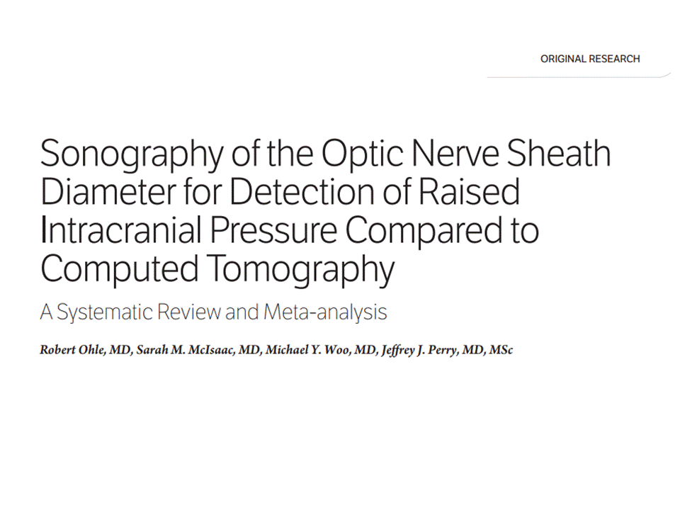
Objectives—The diagnosis of raised intracranial pressure (ICP) is important in many critically ill patients. The optic nerve sheath is contiguous with the subarachnoid space; thus, an increase in ICP results in a corresponding increase in the optic nerve sheath diameter. The objective of this study was to assess the diagnostic accuracy of sonography of the optic nerve sheath diameter compared to computed tomography (CT) for predicting raised ICP.
Methods—We searched PubMed, EMBASE, and the Cochrane database from 1986 to August 2013 and performed hand searches. Two independent reviewers extracted data. Study quality was assessed by using the Quality Assessment of Diagnostic Accuracy Studies (QUADAS) tool. We calculated κ agreement for study selection and evaluated clinical and quality homogeneity before the meta-analysis.
Results—From 1214 studies, we selected 45 for full review. Twelve studies with 478 participants were included (κ = 0.89). Ocular sonography yielded sensitivity of 95.6% (95% confidence interval [CI], 87.7%–98.5%), specificity of 92.3% (95% CI, 77.9%–98.4%), a positive likelihood ratio of 12.5 (95% CI, 4.16–37.5), and a negative likelihood ratio of 0.05 (95% CI, 0.02–0.14). Average quality according to the QUADAS tool was 7.4 of 11. There was moderate to high heterogeneity based on the prediction ellipse area and variance logit of sensitivity (2.1754) and specificity (2.6720).
Conclusions—Ocular sonography shows good diagnostic test accuracy for detecting raised ICP compared to CT: specifically, high sensitivity for ruling out raised ICP in a low-risk group and high specificity for ruling in raised ICP in a high-risk group. This noninvasive point-of-care method could lead to rapid interventions for raised ICP, assist centers without CT, and monitor patients during transport or as part of a protocol to reduce CT use.
VTI and VTQ on BREAST TUMORS
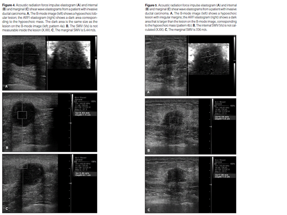
Objectives—Breast cancer is the second leading cause of death from cancer in women, and early detection is the key to successful treatment. Unfortunately, even with technological advances, the specificity of imaging modalities is still low. Therefore, we evaluated the value of a newly developed noninvasive technique, acoustic radiation force impulse imaging, for differentiating benign versus malignant breast lesions.
Methods—We prospectively examined 141 breast lesions in 122 patients. All lesions were classified according to the American College of Radiology Breast Imaging Reporting and Data System (BI-RADS) for mammography, BI-RADS for sonography, and Virtual Touch tissue imaging (VTI; Siemens Medical Solutions, Mountain View, CA) pattern. Internal and marginal shear wave velocity (SWV) values for the lesions were noted. The sensitivity, specificity, accuracy, and positive and negative predictive values for VTI and Virtual Touch tissue quantification (VTQ; Siemens Medical Solutions) were calculated.
Results—The marginal SWV values were statistically higher in malignant lesions (mean ± SD, 5.41 ± 1.37 m/s) than benign lesions (2.91 ± 0.88 m/s; P under .001). When the SWV cutoff level was set at 4.07 m/s, and the higher of the internal and marginal values was adopted, the combination of VTI and VTQ showed 95.1% sensitivity, 99.0% specificity, and 97.8% accuracy.
Conclusions—Breast Imaging Reporting and Data System category 4 lesions are the main focus of research for early detection of breast cancer. Unfortunately, BI-RADS category 4 assessment covers a wide range of likelihood of malignancy (2%–95%). This wide range reflects the necessity for a more specific imaging modality. The combination of VTI and VTQ could increase the diagnostic performance of conventional sonography.
Đăng ký:
Bài đăng
(
Atom
)


