Tổng số lượt xem trang

Thứ Tư, 2 tháng 5, 2018

Automated 3D US effective for diagnosing hip dysplasia


By Kate Madden Yee, AuntMinnie.com staff writer
April 30, 2018 -- Automated 3D ultrasound is just as effective as 2D ultrasound for diagnosing developmental dysplasia of the hip (DDH) in infants: In fact, it reduces the number of studies characterized as borderline by more than two-thirds, according to research published online April 24 in Radiology.
The study's findings suggest that automated 3D ultrasound could serve as an even more effective tool for diagnosing this condition -- which, if untreated, can cause long-term damage, wrote the team led by Dornoosh Zonoobi, PhD, from the University of Alberta in Edmonton, Canada.
"Three-dimensional ultrasound has potential advantages in feasibility in a screening setting for hip dysplasia because the 3D indexes of dysplasia are calculated automatically from surface models generated with minimal user input, or potentially completely automatically calculated by using deep-learning tools," the researchers wrote.
Developmental hip dysplasia in infants is associated with premature osteoarthritis later in life, and it is the cause of 30% of hip arthroplasties in patients younger than 60. The condition is usually treated in infants with a harness, and a prompt and accurate diagnosis reduces its negative long-term effects. 2D ultrasound has long been used to identify DDH, but it has limitations, including operator variability and overdiagnosis.
3D ultrasound overcomes these limitations because it offers a more complete view of hip geometry than 2D ultrasound and also because it is automated, Zonoobi and colleagues wrote. The modality was first suggested for the diagnosis of DDH in the 1990s, but only recently has transducer technology evolved enough to make the use of 3D ultrasound for this application feasible.
For their study, Zonoobi and colleagues added 3D ultrasound to conventional 2D ultrasound exams of 1,728 infants (mean age, 67 days) to evaluate the children for DDH; the exams were performed between January 2013 and December 2016. Custom software automatically calculated measures such as 3D posterior and anterior alpha angle and osculating circle radius. Of the infants imaged, 1,347 were normal, 140 were borderline for the condition, and 241 were dysplastic.
The researchers found that 3D ultrasound helped correctly categorize 97.5% of the dysplastic and 99.4% of the normal hips, and no dysplastic hips were categorized as normal. 3D ultrasound provided a correct diagnosis in 69.3% of cases categorized as borderline at initial 2D ultrasound. The modality also reduced the number of borderline diagnoses to 39, compared with 140 with 2D ultrasound.
The study results justify generalized implementation of 3D ultrasound for DDH diagnosis in clinical settings, Dr. Diego Jaramillo of Nicklaus Children's Hospital in Miami wrote in an editorial that accompanied the study.

Thứ Sáu, 27 tháng 4, 2018

Spinal Pain in Space and US


By Kate Madden Yee, AuntMinnie.com staff writer
April 26, 2018 -- Astronauts armed with a compact ultrasound system successfully performed scans on each other while on the International Space Station. The scans were part of a study to assess spinal changes during long-term spaceflight that could lead to back pain, researchers wrote in the April issue of the Journal of Ultrasound in Medicine.

A group from Henry Ford Hospital worked with NASA to train astronauts on the International Space Station to use ultrasound for imaging the spines of their colleagues during flight. The researchers found that it was feasible to teach these novice users to use ultrasound effectively for this purpose. In addition, the data collected could help in the development of countermeasures to protect astronauts' spines during spaceflight, as well as the creation of protocols for treating injury once the astronauts have returned.
"Focused ultrasound monitoring of the spine for longitudinal changes during long-duration spaceflight may influence additional strategies or nutrition/drug therapies to reduce disk degeneration," lead author Kathleen Garcia and colleagues wrote. "[Our] study demonstrates a potential role for ultrasound in evaluating spinal integrity and alterations in the extreme environment of space."
Aches and pains
Starting with the Apollo program and continuing into the International Space Station era, moderate to severe back pain has been a common medical complaint among astronauts, corresponding author Dr. Scott Dulchavsky, PhD, told AuntMinnie.com.
"When there's no gravity, the spine loosens, making it less stable and putting stress on muscles and ligaments," he said. "The spine can actually elongate by as much as three inches, and that puts astronauts at higher risk of problems when they return."
MRI and CT are the clinical standards for spinal imaging, but they aren't available in space. Ultrasound can be carried on space vehicles thanks to its compact size, but a framework for imaging spinal structures in space hasn't been clearly formulated, Garcia's team wrote.
Dr. Scott Dulchavsky, PhD
Dr. Scott Dulchavsky, PhD, from Henry Ford Hospital.
To address this problem, the researchers developed an ultrasound protocol for spaceflight, and they investigated whether astronauts on the International Space Station could effectively perform ultrasound assessments of the lumbar and cervical regions of the spine. Seven astronauts participated in the study and served as both ultrasound operators and research subjects; two additional crew members were trained as backup operators. The exams were read remotely, and the researchers then compared these in-flight results with preflight and postflight MRI and ultrasound exams (J Ultrasound Med, April 2018, Vol. 37:4, pp. 987-999).
The astronauts were trained six months before their mission via an online program that included a review of spinal anatomy, procedure demonstrations, equipment setup orientation, and a software review, as well as a one-hour, hands-on session during which they alternated between patient and operator roles. The exams were conducted with GE Healthcare's Vivid q device, a laptop-sized ultrasound scanner. The astronauts were assisted remotely by experts at NASA's Lyndon B. Johnson Space Center in Houston.
When the astronauts underwent the exams, they were placed supine on a medical restraint system on board the space station. To evaluate the effects of a lack of gravity on the spine over time, each study participant had three in-flight ultrasounds: one at day 30, one at day 90, and one at day 150.
The astronauts easily obtained high-quality images of the lumbar and cervical vertebrae, the researchers found. Overall success rates for image acquisition were 95% in the lumbar spine and 90% in the cervical spine. In addition, there was "no appreciable difference in success rates for either image acquisition or image quality between expert operators and astronaut crew members in the lumbar and cervical regions," they wrote.
The study findings fill in a data gap, according to Garcia and colleagues.
"Given the previous void of in-flight spinal imaging capabilities in space, to our knowledge, this study represents the first attempt to monitor microgravity-associated acute changes to the spine while they are occurring," they wrote.
Greater purpose
One of the benefits of this kind of research is that the findings can influence healthcare on Earth, according to Dulchavsky.
"By putting smart people into constrained environments like space, we can find solutions to health problems that can be used beyond the space station," he said. "Our work here found not only that nonphysicians can be trained to effectively use imaging devices, but it also pointed to further research on exercise and dietary regimens that could help keep the spine healthy in patients on Earth."
As the U.S. sets its sights on sending astronauts on longer missions -- such as to Mars -- understanding how the human body is affected by space is crucial, Garcia and colleagues wrote.
"As the duration of space missions continues to increase, [ultrasound's utility] will only gain importance in monitoring crew health and diagnosing disorders," the group concluded. "Further investigations should be performed to corroborate this imaging technique and to create a larger database related to in-flight spinal disorders during long-duration spaceflights.

Chủ Nhật, 22 tháng 4, 2018

PROTOCOLS for SHOCK

LINK DOWNLOAD
PROTOCOLS FOR SHOCK


Shock is a major morbidity in emergency and critical care and is also one of the important prognostic factors affecting  in-hospital mortality [1]. Timely diagnosis and treatment of shock reduce the length of stay (LOS) and mortality rate at the emergency department (ED). The early use of the point of care ultrasound (POCUS) can reduce the diagnostic time as well as increase the accuracy of diagnosis [2]. The first protocol Undifferentiated hypotension protocol (UHP) was release on 2001, [3]; and more than 15 subsequent protocols were developed [4].

Future and discussion
Most currently available protocols are focused on diagnosis.Blanco et al. suggested that an ultrasound scan should also assess the efficacy of treatment [5]. For example, the use of velocity time integral (VTI) of LVOT to measure the responses to fluid and inotropic agent to help further management.
Point of care ultrasound is a powerful tool in emergency setting. The emergency ultrasound procedures for shock can reduce the LOS at the emergency department.
Therefore, POCUS is an essential skill for personnels in the emergency department and critical care units.

PROTOCOLS for SHOCK

Thứ Bảy, 21 tháng 4, 2018

CẮT TÚI MẬT NỘI SOI: TIÊU CHUẨN SIÊU ÂM TIÊN LƯỢNG KHÓ


Abstract

Purpose: Laparoscopic cholecystectomy (LC) has become the treatment of choice for cholelithiasis. Still some patients required conversion to open cholecystectomy (OC). Our aim was to develop a standardized Ultrasound based scoring system for preoperative prediction of difficult LC.

 Methods and materials: Ultrasound findings of 300 patients who underwent LC were reviewed retrospectively. Four parameters (time taken, biliary leakage, duct or arterial injury, and conversion) were analyzed to classify LC as easy or difficult. The following ultrasound findings were analyzed: GB wall thickness, pericholecystic collection, distended GB, impacted stones, multiple stones, CBD diameter and liver size. Out of seven parameters, four were statistically significant in our study. A score of 2 was assigned for the presence of each significant finding and a score of 1 was assigned for the remaining parameters to a total score of 11. A cut-off value of 5 was taken to predict easy and difficult LC.
 Results: 66 out of 83 cases of difficult LC and 199 out of 217 cases of easy LC were correctly predicted on the basis of scoring system. A score of >5 had sensitivity 80.7% and specificity 91.7% for correctly identifying difficult LC. Prediction came true in 78.8% difficult and 92.6% easy cases. US findings of GB wall thickness, distended GB, impacted stones and dilated CBD were found statistically significant.
Conclusion: This indigenous scoring system is effective in predicting conversion risk of LC to OC. Patients having high risk may be informed and scheduled appropriately and decision to convert to OC in case of anticipated difficulty may be taken earlier.

ª 2017, Elsevier Taiwan LLC and the Chinese Taipei Society of Ultrasound in Medicine. This is an open access article under the CC BY-NC-ND license (http://creativecommons.org/licenses/ by-nc-nd/4.0/).

TI-RADS REDUCES BIOPSY

Thyroid Imaging Reporting and Data System Reduces Biopsies

Apr 18, 2018
Criteria from the American College of Radiology (ACR) Thyroid Imaging Reporting and Data System (TI-RADS) offers a meaningful reduction in the number of thyroid nodules recommended for biopsy, according to a study published in the journal Radiology
Researchers from several states performed a retrospective study to compare the biopsy rate and diagnostic accuracy before and after applying ACR TI-RADS criteria for thyroid nodule evaluation. Eight radiologists with three to 32 years of experience in thyroid ultrasonography were asked to review the ultrasound features of 100 thyroid nodules that were cytologically proven and/or pathologically proven. Nodules evaluated in five US categories and biopsy recommendations were provided based on the radiologists’ practice patterns without knowledge of ACR TI-RADS criteria. Three other expert radiologists were reference standard readers for the imaging findings. ACR TI-RADS criteria were retrospectively applied to the features assigned by the eight radiologists to produce biopsy recommendations. Comparison was made for biopsy rate, sensitivity, specificity, and accuracy.
The results showed 15 of the 100 nodules (15 percent) were malignant. The mean number of nodules recommended for biopsy by the eight radiologists was 80 ± 16 (standard deviation) based on their own practice patterns and 57 ± 11 with retrospective application of ACR TI-RADS criteria.
Without ACR TI-RADS criteria:
Sensitivity  95 percent
Specificity  20 percent
Accuracy  28 percent
With ACR TI-RADS criteria:
Sensitivity 92 percent
Specificity 44 percent
Accuracy 52 percent
Expert consensus:
Sensitivity 87 percent
Specificity 51 percent
Accuracy 56 percent

The researchers noted that although fewer malignancies were recommended for biopsy with ACR TI-RADS criteria, the majority met the criteria for follow-up US. Only three of 120 (2.5 percent) malignancy encounters required no follow-up or biopsy. Expert consensus recommended biopsy in 55 of 100 nodules with ACR TI-RADS criteria.
Not only did the ACR TI-RADS criteria offer a meaningful reduction in the number of thyroid nodules recommended for biopsy, the researchers wrote, they significantly improve the accuracy of recommendations for nodule management.

Thứ Tư, 18 tháng 4, 2018

SUBCUTANEOUS FAT MEASUREMENT by US


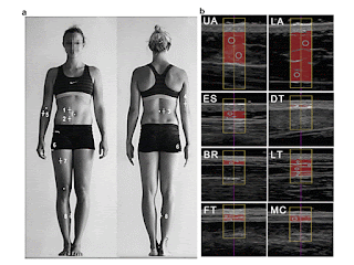
Abstract—A recently standardized ultrasound technique for measuring subcutaneous adipose tissue (SAT) was applied to normal-weight, overweight and obese persons. Eight measurement sites were used: upper abdomen, lower abdomen, erector spinae, distal triceps, brachioradialis, lateral thigh, front thigh and medial calf. Fat compression was avoided. Fat patterning in 38 participants (body mass index: 18.6–40.3 kgm22 ; SAT thickness sums from eight sites: 12–245 mm) was evaluated using a software specifically designed for semi-automatic multiple thickness measurements in SAT (sound speed: 1450 m/s) that also quantifies embedded fibrous structures. With respect to ultrasound intra-observer results, the correlation coefficient r 5 0.999 (p < 0.01), standard error of the estimate 5 1.1 mm and 95% of measurements were within ±2.2 mm. For the normal-weight subgroup, the median measurement deviation was 0.43 mm (1.1% of mean thickness), and for the obese/overweight subgroup it was 0.89 mm (0.5%). The eight sites used here are suggested to represent inter-individual differences in SAT patterning. High measurement accuracy and reliability can be obtained in all groups, from lean to overweight and obese, provided that measurers are trained appropriately.
Fig. 1. B-Mode ultrasound measurement of uncompressed subcutaneous fat. (a) Sites for ultrasound measurement of subcutaneous adipose tissue (SAT) patterning: upper abdomen (1, UA), lower abdomen (2, LA), erector spinae (3, ES), distal triceps (4, DT), brachioradialis (5, BR), lateral thigh (6, LT), front thigh (7, FT) and medial calf (8, MC). Body height was used as the reference length for all distances. A detailed description of the standardized marking and of the ultrasound measurement technique can be found in M€uller et al. (2016). (b) Ultrasound images and evaluations of SAT thicknesses. Participant A: male, body mass index 5 25.5, body mass 5 80.7 kg, stature 5 1.78 m. (c) Survey plot of SAT patterning according to (b). The columns represent the mean values of the semi-automatic multiple thickness measurements for the eight ultrasound sites. The mean thickness value of the SAT thickness in a given ultrasound image (within the region of interest) is termed dINCL (gray) when fibrous structures are included and dEXCL (black) when fibrous structures are subtracted. Sum of the eight thicknesses DINCL 5 44.3 mm (DEXCL 5 39.8 mm). (d) Survey plot of a participant B with similar body mass index. Body mass index 5 25.4 kgm22 ; body mass 5 75.5 kg, stature 5 1.72 m. The SAT thickness sum was 86.9 mm (77.9 mm), 96% higher than in participant A.


(E-mail: wolfram.mueller@medunigraz.at) 2016 The Authors. Published by Elsevier Inc. on behalf of World Federation for Ultrasound in Medicine & Biology. This is an open access article under the CC BY-NC-ND license (http://creativecommons.org/licenses/by-nc-nd/4.0/).

Key Words: Body composition, Subcutaneous adipose tissue, Overweight, Obesity, Ultrasound measurement precision.

How can unnecessary ABUS recalls be avoided?


By Kate Madden Yee, AuntMinnie.com staff writer
April 16, 2018 -- Automated breast ultrasound (ABUS) can yield lower recall rates than handheld ultrasound, but unnecessary recalls still occur -- and it's important to find ways to reduce them, according to research presented at the recent American Institute of Ultrasound in Medicine (AIUM) annual meeting in New York City.

Why? Because having fewer recalls leads to fewer unnecessary biopsies and also less-stressed patients, lead researcher Dr. Beverly Hashimoto of Virginia Mason Medical Center in Seattle told AuntMinnie.com via email.
Dr. Beverly Hashimoto
Dr. Beverly Hashimoto of Virginia Mason Medical Center.
"One of the weaknesses of screening breast ultrasound is that it results in a high number of women being called back for additional imaging," Hashimoto said. "When we started our automated breast ultrasound program, we wanted to determine if there were lessons we could learn from reviewing our ultrasound callbacks to reduce the number of patients recalled in the future."
Reducing recalls
ABUS offers benefits over handheld ultrasound for breast cancer screening, in part because it reduces the modality's dependency on operator skill. But the technology doesn't completely eliminate screening downsides such as unnecessary recalls, study presenter Elissa Picozzi from the University of Washington told session attendees.
She and her colleagues examined 18 months of their experience with screening ABUS. All patients included in the study were asymptomatic and had heterogeneous or extremely dense breast tissue. The researchers focused their attention on patients who were called back for additional imaging due to a category 0 ABUS screening assessment ("incomplete").
Elissa Picozzi
Elissa Picozzi from the University of Washington.
At Virginia Mason, patients called back for a possible ABUS abnormality undergo a diagnostic handheld ultrasound exam; if this exam is a category 4 or 5 (suspicious or highly suspicious of malignancy), then the woman has an ultrasound-guided core biopsy. The researchers reviewed ABUS images, handheld ultrasound images, and any pathology results from biopsy.
In that first year and a half of the medical center's screening ABUS practice, 867 exams were performed. Of these, 229 patients (26%) were called back for a possible ABUS lesion and had a diagnostic handheld ultrasound exam; 69 (8%) had biopsies.
Of the 229 patients called back, 222 were recalled for category 2 or 3 lesions. Results from the handheld ultrasound exams in these 222 patients were as follows:
  • 68 patients (30.6%) with solid hypoechoic masses
  • 71 patients (32%) with no abnormality -- the "lesions" on ABUS were actually artifact shadowing
  • 35 patients (15.8%) with nonsimple cysts
  • 20 patients (9%) with simple cysts
  • 28 patients (12.6%) with miscellaneous lesions, including duct ectasia, scars, fat lobules, fat necrosis, or lymph nodes
The 69 biopsies revealed seven malignancies; of these, four were found only on ABUS. The cancer detection rate for ABUS was 4.6 per 1,000 cases.
Better evaluation?
Hashimoto and colleagues found that image artifacts masquerading as lesions were a frequent cause of unnecessary recalls. Many of these recalls could be avoided by using alternative reconstruction techniques and more careful consideration of different ABUS views, they noted.

"We were surprised to find that the second most common reason for calling a patient back was artifactual ultrasound shadowing," she told AuntMinnie.com. "As a result, we have developed techniques to better analyze shadowing on screening ultrasounds. We also discovered that many of the solid masses that were called back could have been resolved by changing our reviewing process of prior exams."

Thứ Bảy, 7 tháng 4, 2018

NAFLD and Ultrasound Elastography


Abstract
An increasingly common cause of chronic liver disease in adults and children is nonalcoholic fatty liver disease (NAFLD). The diagnosis of NAFLD was traditionally based on the histopathological changes of the liver, evaluated by needle liver biopsy, an invasive method, with potential adverse effects and great inter and intraobserver variability. The noninvasive methods for the assessment of both fibrosis and steatosis in patients with NAFLD have increasingly been studied lately. Of these noninvasive methods, in this chapter, we will focus on the methods assessing the stiffness of liver parenchyma, i.e. elastographic methods, of which, the most widely used are ultrasound elastography techniques. We will discuss the principal elastographic methods of some utility in NAFLD, i.e. shear wdave elastography (SWE) (quantitative elastography), and especially transient elastography, point SWE (acoustic radiation force impulse elastography, ARFI) and two-dimensional real-time SWE (Supersonic). For each method usable in NAFLD cases, we will review the method principle, examination technique and performance in NAFLD evaluation.
Keywords: nonalcoholic fatty liver disease, fibrosis, steatosis, noninvasive, elastography


SSI, Fibroscan® or ARFI?

After comparing the performance in the assessment of NAFLD of the three elastographic methods discussed above, we can conclude, on the preliminary results, that the diagnostic performance according to the AUROC values for the diagnosis of significant fibrosis, severe fibrosis and cirrhosis is good for SSI (0.86–0.89); good for Fibroscan® (0.82–0.87) and fair or good for ARFI (0.77–0.84) . The AUROC values for diagnosing severe fibrosis or cirrhosis are particularly good for SSI or Fibroscan® (0.86 and 0.89) [50]. The prediction of steatosis, however, can at this moment only be made using the controlled attenuation parameter measured with Fibroscan®. As for the causes of measurement failure or unreliable results, we mention clinical factors related to obesity (BMI > 30 kg/m2 , waist circumference ≥ 102 cm or increased wall thickness), which are associated with liver stiffness measurement failures when using SSI or Fibroscan® and with unreliable results when using ARFI [50]. In conclusion, SSI, Fibroscan® and ARFI are valuable diagnostic tools for the staging of liver fibrosis in NAFLD patients. However, the diagnostic accuracy of SSI appears to be superior to that of ARFI for the diagnosis of F2 or above. Most of the cut-off values for SSI for the diagnosis of different stages of liver disease are quite similar to those of Fibroscan®; this is an issue of great importance for the applicability of this technique and its wide dissemination among radiologists and hepatologists in their daily practice. However, as for the M probe of Fibroscan®, the SSI technique remains limited by a high failure rate in cases of obesity, whereas ARFI has a high rate of unreliable results.

Thứ Tư, 4 tháng 4, 2018

POCUS helps handle out-of-hospital cardiac arrest


By Kate Madden Yee, AuntMinnie.com staff writer
April 2, 2018 -- Point-of-care ultrasound (POCUS) shows promise as a tool for emergency responders to help predict survival and guide interventions for cardiac arrests experienced outside of the hospital, according to research presented at the recent American Institute of Ultrasound in Medicine (AIUM) meeting in New York City.
POCUS is becoming more commonplace as a tool used for resuscitation of patients in cardiac arrest in the hospital, but there are not much data on its use outside of the hospital setting, presenter Dr. Katherine German of the University of Pittsburgh told session attendees.
"We know that out-of-hospital cardiac arrest survival is low, and we were curious whether point-of-care ultrasound could be a useful tool to predict survival and guide therapy or the decision to transport in the field," German said.
A helpful tool
German and colleagues explored the feasibility of POCUS when used by emergency staff in patients experiencing cardiac arrest outside of hospitals. A portable ultrasound unit (iViz, Fujifilm SonoSite) was used in vehicles as part of the Pittsburgh Emergency Medical Services (EMS) system; all participating physicians received POCUS education and watched an instructional video on how to use the device. Physicians provided radio and in-person consultation to Pittsburgh city paramedics.
The field staff used ultrasound to identify cardiac motion during pulse checks -- without interrupting resuscitation -- and to guide management of the situation at the physicians' discretion. The POCUS users recorded data from still images and six-second video clips, as well as through electronic charting within the patient care report using emsCharts, a tool for air and ground emergency medical personnel.
The study's primary purpose was to track the frequency of POCUS use during out-of-hospital cardiac arrest events; the secondary purpose was to have an expert faculty reviewer analyze images acquired during these events and to identify any barriers to the use of the technology.
German's group included 348 field responses that occurred between November 2016 and March 2017. Of these, 127 were out-of-hospital cardiac arrest. Field doctors used POCUS in 56 (44%) of the out-of-hospital cardiac arrest cases, with 48 (85%) still images and 34 (61%) video images recorded and reviewed.
Agreement between interpretations of emergency responder physicians and those of expert reviewers was found in 91% of video cases. Responders noted the following reasons for not using POCUS:
  • Return of spontaneous circulation (ROSC) close to physician arrival (30%)
  • Emergency personnel busy with other interventions (21%)
  • Provider preference (23%)
  • Mechanical issues (6%)
  • Cessation of resuscitation per advanced directives (6%)
In the field
German did note some of the study's limitations, including the user dependency of ultrasound, as well as the fact that POCUS use was optional and that interpretations were recorded after the technology was used in the field. She also cited the small sample size of video images.
However, overall, POCUS showed promise as a tool for dealing with cardiac arrest in the field, she concluded.

"POCUS use by novice physician ultrasonographers to detect cardiac wall motion in out-of-hospital cardiac arrest is not only feasible, it also correlates with expert interpretation," German said. "Barriers to its use can be addressed through educational interventions and increased familiarity with the device."