Tổng số lượt xem trang

Thứ Hai, 22 tháng 4, 2019

Ultrasound bests x-ray for identifying pulmonary edema.


April 22, 2019 -- Bedside ultrasound is more sensitive than chest x-ray for identifying pulmonary edema in patients presenting with dyspnea, according to a study published in the April issue of the Journal of Ultrasound in Medicine.
And the modality's greater sensitivity isn't its only benefit, wrote a team led by Dr. William Wooten of Mount Carmel Health System in Columbus, OH.
"Bedside ultrasound appears to offer several advantages over chest radiography in the workup of patients with dyspnea beyond its superior sensitivity in the diagnosis of pulmonary edema," the team wrote. "First, bedside ultrasound exposes the patient to less radiation. Second, imaging costs to payers may be reduced."
Chest x-ray has been the commonly used imaging modality to assess patients for pulmonary edema, but its interpretation can be variable, affected by clinicians' level of expertise, Wooten and colleagues wrote. That's why lung ultrasound may be a better tool.
"Lung ultrasound may produce more objective findings through evaluation of vertical comet tail artifacts known as B-lines, which are created by a decrease in the ratio of alveolar air to fluid pulmonary content," the group explained.
The study included 99 patients who presented in the emergency room with dyspnea between August 2016 and March 2017. Of these, 32.3% had congestive heart failure, and 40.4% had chronic obstructive pulmonary disease. Each patient underwent an ultrasound exam within an hour of having a chest x-ray. The researchers used the final diagnosis from the patient's discharge summary as the reference standard (J Ultrasound Med, April 2019, Vol. 38:4, pp. 967-973).
The team found that bedside ultrasound had higher sensitivity compared with chest x-ray, at 96% versus 65%. Specificity was comparable between the two modalities. Of 18 patients with negative x-ray findings and a discharge diagnosis of pulmonary edema, 16 (89%) had positive findings on ultrasound.
Chest x-ray vs. ultrasound for pulmonary edema
MeasureChest x-rayUltrasoundp-value
Sensitivity65%96%< 0.001
Specificity96%90%0.26
Additionally, patients may be more comfortable with bedside ultrasound, Wooten and colleagues noted.
"Anecdotally, we observed that patients appeared to prefer the bedside ultrasound, as it kept the provider at the bedside longer, and the patient did not have to leave the room or family to undergo the chest radiography," the group wrote.
Bedside ultrasound shows promise for patient care when it comes to diagnosing pulmonary edema, but it could also benefit hospitals, according to the authors.

"This study has the potential to affect the care of patients with congestive heart failure, which is the most common cause of hospitalization in the Medicare population and is growing substantially as people are living longer," the team concluded. "As hospitals are being penalized for 30-day readmissions for congestive heart failure, it is crucial that these patients have an accurate diagnosis and are properly treated."

Thứ Tư, 17 tháng 4, 2019

Ultrasound in late pregnancy could reduce C-section rate.


By Kate Madden Yee, AuntMinnie.com staff writer
April 17, 2019 -- Performing ultrasound scans late in pregnancy helps women avoid undiagnosed breech presentation of their babies, translating to improved clinical outcomes, lower rates of emergency cesarean sections (C-sections), and perhaps even lower healthcare costs, according to a study published April 16 in PLOS Medicine.
The findings are good news not only for women and their babies but also for the healthcare system, wrote a team led by David Wästlund of the University of Cambridge in the U.K.
"According to our estimates, universal late pregnancy ultrasound in nulliparous women would virtually eliminate undiagnosed breech presentation, would be expected to reduce fetal mortality in breech presentation, and would be cost-effective if fetal presentation could be assessed for less than 19.80 pounds [$25.95 U.S.] per woman," the group wrote.
Fetal breech presentation increases the risk of complications for the baby and the mother, Wästlund and colleagues noted. Typically, a baby's position is assessed by palpating the woman's abdomen, but this technique's sensitivity varies by practitioner.
"Despite the relative ease with which breech presentation can be identified through ultrasound screening, the assessment of fetal presentation at term is often based on clinical examination only," the researchers wrote. "Due to limitations in this approach, many women present in labor with an undiagnosed breech presentation."
The investigators performed screening ultrasound at 36 weeks gestation in 3,879 English women having first pregnancies between January 2008 and July 2012. Of these, 179 (4.6%) were diagnosed with breech presentation. In more than half of those (54%), breech presentation had not been suspected prior to labor.
Women with babies in the breech position were offered a procedure called an external cephalic version (ECV) to try to turn the baby; for those who did not want this procedure or for whom it did not work, a C-section was scheduled.
The investigators also estimated the cost of universal late pregnancy ultrasound scans using data from the English National Health Service (NHS) to compare birth outcomes of breech pregnancies screened with and without ultrasound.
The ECV procedure was attempted in 84 (46.9%) of the women with breech babies and was successful in 12 (14.3%). Of the 179 women with breech babies, the researchers found the following:
  • 10.6% delivered vaginally (following either a planned or spontaneous version).
  • 61.5% delivered via elective C-section.
  • 27.9% delivered via emergency C-section (due to labor starting before the scheduled cesarean date).
"No woman in the cohort had a vaginal breech delivery or experienced an intrapartum cesarean for undiagnosed breech," the researchers noted.
Wästlund and colleagues estimated that routine late pregnancy ultrasound could prevent nearly 15,000 undiagnosed breech presentations, more than 4,000 emergency C-sections, and seven to eight baby deaths per year. But the effect of the intervention on healthcare costs needs more research, the group wrote: "If universal ultrasound could be provided for less than 12.90 pounds [$16.91] per scan, the policy would also be cost saving." But it's unclear if this is possible.
"If this procedure could be implemented into routine care, for example, by midwives conducting a routine [scan at 36 weeks gestation] and using a portable ultrasound system, it is likely to be cost-effective," the group concluded. "Such a program would be expected to reduce the consequences to the child of undiagnosed breech presentation, including morbidity and mortality."

Elastography can help characterize breast tumors.


By Kate Madden Yee, AuntMinnie.com staff writer
April 16, 2019 -- Breast elastography is an overlooked tool for evaluating breast cancer, especially since it shows promise for helping radiologists distinguish between benign and malignant lesions, according to a presentation delivered at the American Institute for Ultrasound in Medicine (AIUM) meeting in Orlando, FL.
Since ultrasound is convenient, easily accessible, and less expensive than some other modalities, it's a helpful tool in the breast cancer detection arsenal. And since it can help characterize breast lesions, breast elastography could have downstream effects on patient care, according to presenter Dr. Stamatia Destounis of Elizabeth Wende Breast Care in Rochester, NY.
"Identifying the relative tissue stiffness can help clinicians distinguish between benign and malignant lesions, which in turn has the potential to reduce unnecessary biopsies," she said.
Although breast elastography has been available for more than 15 years, it's not widely implemented, in part because the various methods lack consistency -- particularly in color scale interpretation, Destounis noted.
"As research continues and elastography is used in clinical practice, there's a need for standardization of the color scale," she said.
Elastography can be performed in static and dynamic modes, and consensus about which mode is better is generally lacking. Static elastography includes strain imaging, while dynamic elastography includes shear wave. Strain elastography is the most widely used technique, estimating the relative stiffness of a particular area compared with other tissue.
In strain elastography, tissue stiffness data are displayed in a color map that is superimposed on a real-time grayscale image. Cancers tend to appear larger on strain elastography than on B-mode ultrasound, and benign lesions tend to appear smaller; this size change between the modes has shown to be highly sensitive and specific for characterizing breast lesions, Destounis said.
Studies have demonstrated that strain elastography is effective for detecting breast cancer, but it does have its drawbacks.
"It can be difficult to measure the amount of force during compression, and absolute elasticity can't be calculated," she said.
As for shear-wave elastography, it offers real-time, quantitative assessment of tissue stiffness; rather than relying on external compression, it uses short acoustic pulses to identify stiffness. To characterize breast lesions, clinicians should focus on the area of highest stiffness in the lesion. But be careful, Destounis cautioned.
"Some breast cancers don't allow for adequate shear-wave generation and may appear black -- that is, no shear-wave speed calculated or with a low shear-wave speed due to noise," she explained.
So which technique should clinicians use? Destounis cited research from Chang et al that found mixed results (American Journal of Roentgenology, August 2013, Vol. 201:2, pp. W347-356). The study compared strain and shear-wave ultrasound elastography for differentiating benign from malignant breast lesions, and it showed that strain elastography is more specific than shear-wave elastography (93.7% compared with 84.8%) but less sensitive (81.7% compared with 95.8%).
It may be a matter of improving elastography technology, Destounis told AuntMinnie.com via email.
"Several vendors have different types of elastography technology, and it needs more consistency in technique and color representation," she stated.

Chủ Nhật, 14 tháng 4, 2019

ML for US


https://www.ncbi.nlm.nih.gov/pmc/articles/PMC5886811/

Abstract

Ultrasound (US) imaging is the most commonly performed cross-sectional diagnostic imaging modality in the practice of medicine. It is low-cost, non-ionizing, portable, and capable of real-time image acquisition and display. US is a rapidly evolving technology with significant challenges and opportunities. Challenges include high inter- and intra-operator variability and limited image quality control. Tremendous opportunities have arisen in the last decade as a result of exponential growth in available computational power coupled with progressive miniaturization of US devices. As US devices become smaller, enhanced computational capability can contribute significantly to decreasing variability through advanced image processing. In this paper, we review leading machine learning (ML) approaches and research directions in US, with an emphasis on recent ML advances.


We also present our outlook on future opportunities for ML
techniques to further improve clinical workflow and US-based disease diagnosis and characterization.

Keywords: Deep Learning, Elastography, Machine Learning, Medical Ultrasound, Sonography

Thứ Ba, 9 tháng 4, 2019

QUANTITATIVE ULTRASOUND [Q U S], EMERGING MODE for CLINICAL ULTRASOUND

By Kate Madden Yee, AuntMinnie.com staff writer
April 8, 2019 -- ORLANDO, FL - Quantitative ultrasound (QUS) shows promise as an emerging mode for clinical ultrasound, offering more specific data compared with conventional ultrasound exams, according to a lecture delivered Monday at the American Institute of Ultrasound in Medicine (AIUM) meeting.

Many conventional ultrasound studies already offer quantitative information, including distance, area, and volume measurements; Doppler-generated velocities and volume flow estimates; cardiac wall motion, strain, and ejection fraction data; and contrast and tissue stiffness analysis. But conventional ultrasound is subject to operator variability, said James Zagzebski, PhD, of the University of Wisconsin in Madison.
"QUS uses bulk acoustic properties and tissue microstructure features to increase ultrasound's sensitivity and specificity," he said during the AIUM's William J. Fry Memorial Lecture. "I believe that the current computational resources on ultrasound systems can be further exploited to provide us even more data than we already have."
QUS technology has continued to advance, replacing conventional ultrasound's beamformer technology with high-capacity computational hardware and software, and synthesized virtual beams, Zagzebski said. Compared with conventional ultrasound, QUS imaging offers more data related to tissue features -- such as attenuation and backscatter coefficients -- that increase the specificity of image findings and can lead to improvements in diagnostic ultrasound. QUS techniques include spectral-based parameterization, elastography, shear-wave imaging, and flow estimation.
"System independent backscatter coefficient and attenuation coefficient estimates can be made accurately using clinical scanners," he told session attendees. "There's evidence that QUS can provide valuable information to assess diffuse liver disease, characterize breast masses, and assess the effects of anesthesia."
QUS may be particularly helpful for breast imaging, according to Zagzebski.
"In vitro studies showed that QUS parameters, attenuation coefficient, and backscatter coefficient can give useful insight into the nature of breast tissue," he said. "And animal studies have shown that the use of QUS parameters such as effective scatter [can] differentiate benign fibroadenomas from malignant breast masses."
Other potential applications for QUS include evaluating changes in the cervix accompanying ripening and breast tumor response to treatment, tracking lymph node involvement in disease, monitoring radiofrequency and microwave ablation, and diagnosing eye and orbital disease, Zagzebski noted.
In any case, as the use of QUS continues, there's a need to set best practices, such as those established through the Quantitative Imaging Biomarkers Alliance (QIBA), he said.

"We need to establish good protocols for QUS, like those developed through the QIBA effort, so that everybody will be operating under the same standards," he concluded.

QUANTITATIVE ULTRASOUND IMAGING

https://www.aapm.org/meetings/amos2/pdf/59-17304-58800-911.pdf


Thứ Sáu, 5 tháng 4, 2019

Noninvasive Evaluation of NAFLD: CURRENT EVIDENCE and PRACTICE


FATTY LIVER - CHRONIC B HEPATITIS and H C C


Researchers test new ultrasound method for heart disease


April 5, 2019 -- Researchers from the University of Arkansas in Fayetteville have published results from a study testing a new ultrasound imaging method for the detection and diagnosis of congenital heart disease in infants and children.
The new technology, called vector flow, creates images of the internal structure and blood flow of children's hearts. It was used for the first time in humans at the Arkansas Children's Hospital in Little Rock, according to a team led by Dr. Morten Jensen, PhD. The study was published March 5 in Progress in Pediatric Cardiology.
About 1% of babies are born with congenital heart defects. Pediatric cardiologists identify congenital heart disease using echocardiography and other processes based on ultrasound, the researchers wrote. Although effective, ultrasound can't accurately obtain details about blood flow within the heart.
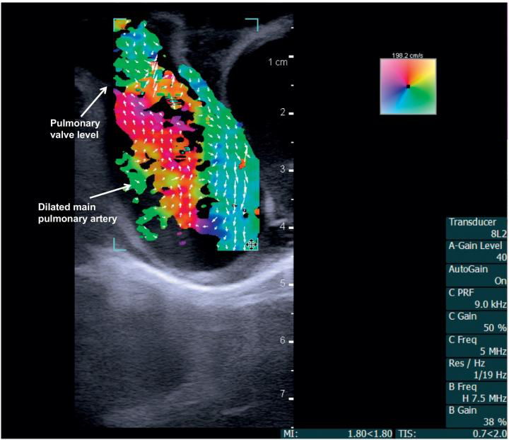
The team used an ultrasound scanner with vector flow imaging to image the hearts of two three-month-old babies, one with a healthy heart and one with congenital heart disease. The technology allowed for complete transthoracic imaging of tissue and blood flow at a depth of 6.5 cm; abnormal flow and cardiac anomalies were clearly visualized in the child with congenital heart disease.
Vector flow imaging demonstrates swirl of blood flow within the dilated main pulmonary artery of a pig.
Vector flow imaging demonstrates swirl of blood flow within the dilated main pulmonary artery of a pig. Image courtesy of Dr. Morten Jensen, PhD.
"Vector flow imaging technology is not yet possible in adults, but we have demonstrated that it is feasible in pediatric patients," Jensen said in a statement released by the university April 3. "Our group demonstrated that this commercially available technology can be used as a bedside imaging method, providing advanced detail of blood flow patterns within cardiac chambers, across valves, and in the great arteries."

Thứ Sáu, 22 tháng 3, 2019

ABUS better than DBT for diagnostic workup.


By Kate Madden Yee, AuntMinnie.com staff writer
March 5, 2019 -- Automated breast ultrasound (ABUS) is more accurate than digital breast tomosynthesis (DBT) as a diagnostic tool for working up positive findings on mammography screening in dense breast tissue, according to a presentation delivered at ECR 2019.
ABUS does have limitations, notably microcalcifications and imaging the retroareolar area of the breast, said presenter Dr. Norran Hussein Said of Cairo University. But its benefits may outweigh them.
"ABUS has the capability of 3D assessment [of dense breasts], and can differentiate solid from cystic lesions," Said told session attendees.
The group used data from the Egypt Breast Cancer National Screening Program to compare the performance of ABUS to tomosynthesis in the workup of 242 women with dense breasts recalled after positive screening mammography. Positive findings included focal asymmetries, masses, distortions, or microcalcifications.
All of the women who participated in the study underwent both tomosynthesis and ABUS. The researchers compared exam results to pathology data.
Tomosynthesis vs. ABUS in women with dense breast tissue
MeasureTomosynthesisABUS
Sensitivity92%92%
Specificity92%98%
Positive predictive value76%92%
Negative predictive value98%98%
Accuracy92%97%
"Our results show that most of our recalled cases consisted of masses, followed by asymmetries," Said told session attendees.
The two modalities agreed on true positives in 43 out of 51 proven cancers, and each modality had four false negatives. ABUS had four false positives, while tomosynthesis had 15. ABUS helped avoid 187 biopsies, while tomosynthesis helped avoid 176.
"In our study, ABUS showed a higher accuracy than DBT," Said concluded.
Works with 2D, too
In a related study, a Romanian team found that, when added to full-field digital mammography (FFDM), ABUS was an effective adjunct tool for finding cancer in women with dense tissue, specifically improving sensitivity and negative predictive value compared with FFDM alone.
Dr. Ioana Boca of Emergency County Hospital in Cluj-Napoca compared the performance of FFDM, 3D mammography, and ABUS for breast cancer detection in women with dense tissue. Two radiologists interpreted 127 ABUS exams procured after FFDM or 3D mammography. Their results were compared to histopathology for biopsied lesions, handheld ultrasound for benign lesions, and follow-up for benign lesions unchanged for at least two years. Nineteen cancers were identified.
ABUS boosted FFDM's sensitivity and negative predictive value, although it did not outperform 3D mammography in this screening context, Boca noted.
FFDM, FFDM + ABUS, and 3D mammography in dense tissue
FFDM2D mammography + ABUS3D mammography
Sensitivity77.8%80%100%
Specificity100%89.2%86.4%
Positive predictive value100%71.4%74.2%
Negative predictive value92%93%100%

"In screening, ABUS added to FFDM compared with FFDM alone improved readers' detection of breast cancers in women with dense breast tissue," Boca concluded.

Thứ Tư, 20 tháng 3, 2019

Ultrasound can regulate inflammatory response.


By AuntMinnie.com staff writers
March 12, 2019 -- Bioelectronic research teams from GE Research in Niskayuna, NY, and the Feinstein Institute for Medical Research in Manhasset, NY, have demonstrated noninvasive methods to regulate dysfunction in the body's metabolic or inflammatory control systems using ultrasound, according to a study published online March 12 in Nature Communications.
Bioelectronic medicine is a combination of neuroscience, molecular biology, and bioengineering that researchers hope will be able to treat disease and injury in the nervous system without pharmaceuticals, the institute said. A team led by study co-author Chris Puleo, PhD, of GE Research, found that using ultrasound in targeted ways reduced inflammatory markers and altered metabolism.
"In our studies, we show that applying ultrasound to a specific target in the spleen altered inflammatory markers that can cause arthritis, inflammatory bowel disease, and other ailments," Puleo said in a statement released by the institute. "And when targeting a specific part of the liver, we were able to modulate blood glucose levels."
The researchers plan to conduct more preclinical studies to understand the use of ultrasound in this way, they said.