Tổng số lượt xem trang

Thứ Tư, 9 tháng 10, 2019

Elastography helps liver transplant patients avoid biopsy.


By Kate Madden Yee, AuntMinnie.com staff writer
October 8, 2019 -- Ultrasound with a shear-wave elastography (SWE) technique can help liver transplant patients avoid biopsy on follow-up, according to a study published online October 7 in the American Journal of Roentgenology.
The findings are good news for a patient population often vulnerable to invasive procedures after transplant -- and can help save healthcare resources, wrote a team led by Dr. Corinne Deurdulian of the University of Southern California in Los Angeles.
"Given the significant resources allotted to perform liver biopsies (e.g., radiologist time, nursing staff, and hospital resources) and patient recovery time, as well as patient discomfort and the possibility of significant postbiopsy complications developing, utilization of a noninvasive tool to determine the degree of hepatic fibrosis would be useful in the initial assessment and follow-up of liver transplant patients," the group wrote.
After liver transplant, patients are monitored for both possible rejection of the new organ and hepatic fibrosis, and they often undergo liver biopsies as part of this follow-up, Deurdulian and colleagues noted.
The researchers sought to determine whether shear-wave elastography could offer a noninvasive alternative to biopsy to assess for liver fibrosis, helping clinicians quantify it in liver transplant recipients.
The study included 111 adult liver transplant patients who underwent 147 SWE exams of the right hepatic lobe followed by biopsies between May 2015 and December 2017. The researchers compared SWE values with fibrosis scores of biopsy samples using the Metavir system: Metavir scores are F0 (no fibrosis), F1 (portal fibrosis without septa), F2 (portal fibrosis with few septa), F3 (numerous septa without cirrhosis), and F4 (cirrhosis). The team tracked SWE's sensitivity, specificity, positive predictive value, negative predictive value, and overall accuracy.
Of the 147 SWE exams and liver biopsies, the researchers found consistent threshold values for patients with Metavir scores of F0 and F1 (no or minimal fibrosis), compared with those with Metavir scores of F2, F3, or F4 (significant fibrosis).
SWE's performance in classifying fibrosis
Performance measureSWE
SWE value
No or minimal fibrosis≤ 1.76 m/sec
Significant fibrosis> 1.76 m/sec
Other performance measures
Sensitivity77%
Positive predictive value33%
Negative predictive value96%
The study results suggest that clinical decisions for liver transplant patients can be based on SWE results rather than biopsy, the researchers concluded.
"If the median SWE value is 1.76 [m/sec] or less, the patient can be classified as having no or minimal fibrosis ... and can avoid biopsy," the group concluded. "According to these results, which have a negative predictive value of 96%  liver biopsies may be obviated in most patients."

Thứ Năm, 3 tháng 10, 2019

Radiofrequency ultrasound , AI predict thyroid cancer.

Radiofrequency ultrasound , AI predict thyroid cancer
By Kate Madden Yee, AuntMinnie.com staff writerRadiofrequency ultrasound and an artificial intelligence (AI) model can be used to effectively predict the malignancy of thyroid nodules, as well as stratify their risk, according to a study set for publication in the November issue of Ultrasonics.

The combination of radiofrequency ultrasound with an artificial neural network (ANN) could also avoid operator dependency issues and help prevent unnecessary thyroid biopsies, according to a group led by Dr. Chunrui Liu of the Affiliated Hospital of Nanjing University Medical School in China.
"The proposed method has no operator dependency; all of the analyses are performed by computer," the team noted (Ultrasonics, November 2019, Vol. 99, pp. 1-9). "Preliminary results indicated that the performance of ANN combined with radiofrequency ultrasound signals is better than that combined with conventional ultrasound images."
Common but not often malignant
Thyroid nodules are common, but only 8% to 16% are actually malignant, according to the researchers. Many ultrasound techniques are used to evaluate nodule malignancy, including strain elastography, acoustic radiation force impulse imaging, and contrast-enhanced ultrasound, but these methods' efficacy remains unclear, the group wrote.
That's where radiofrequency ultrasound comes in. The technique elicits more clinical information than conventional ultrasound by extracting radiofrequency signals from tissues. But how it performs with thyroid nodules has not been studied, Liu and colleagues noted.
"Preliminary studies of radiofrequency ultrasound have been promising, and the method has been shown to have broader prospective applications in identifying prostate and breast cancers and grading fatty liver," they wrote. "To date, few studies on radiofrequency ultrasound's thyroid cancer detection performance have been reported."
The researchers developed their method to predict suspicious thyroid nodules by first gathering radiofrequency data and then creating radiofrequency ultrasound images using Matlab software (MathWorks). After a radiologist outlined regions of interest on the images, textural features were then analyzed using the gray-level co-occurrence matrix (GLCM) algorithm and principal component analysis. The resulting characteristic values from the textural analysis were subsequently used to train the ANN.
The study included 131 pathologically proven thyroid nodules, of which 59 were benign and 72 were malignant. The nodules were randomly divided into training, validation, and testing cohorts. To test their hypothesis that radiofrequency ultrasound could provide more tissue characteristic information than conventional ultrasound, the researchers also performed the same texture and ANN analyses on the B-mode ultrasound images.
The ANN algorithm performed better with radiofrequency ultrasound than it did on conventional ultrasound in all categories except specificity, the group found.
ANN performance for predicting thyroid nodule malignancy
Performance measureANN on conventional ultrasound imagesANN on radiofrequency ultrasound images
Sensitivity94.4%100%
Specificity93.2%91.5%
Accuracy93.9%96.2%
AUC*0.9170.945
*AUC = Area under the curve
The group also used the ANN with radiofrequency ultrasound to characterize new malignancy risk groups for categories 3 (probably benign), 4 (suspicious), and 5 (probably malignant) thyroid nodules as established by the American College of Radiology's Thyroid Imaging Reporting and Data System (TI-RADS). The new categories better distinguished malignant nodules compared with TI-RADS.
ParameterCategory 3Category 4Category 5
No. of samples421673
No. of malignant samples0369
Risk of malignancy
ANN plus radiofrequency ultrasound018.8%94.5%
TI-RADS055.1%88.2%
"The new categories allow for a selection of suspicious nodules to be submitted to fine-needle aspiration, thereby avoiding unnecessary thyroid biopsies," the group wrote.
More research to come
More research needs to be done to establish the benefits of using an ANN and radiofrequency ultrasound, according to Liu and colleagues.
"Of course, although these preliminary results suggested [the use of the ANN and radiofrequency ultrasound] could help sonographers to identify risky thyroid nodules and reduce the number of unnecessary thyroid biopsies, more data will be collected and analyzed in our future study to further confirm the feasibility and accuracy of the proposed method," they concluded.

Thứ Ba, 1 tháng 10, 2019

A I Can Accurately Diagnose Appendicitis.


By Erik L. Ridley, AuntMinnie staff writer
October 1, 2019 -- By analyzing lab values and ultrasound data, an artificial intelligence (AI) algorithm can be highly accurate for diagnosing acute appendicitis and could potentially help avoid unnecessary surgery in two-thirds of patients without appendicitis, according to research published online September 25 in PLOS One.


A team of researchers led by Josephine Reissmann of Charité Universitätsmedizin Berlin trained an AI algorithm to provide an automated diagnosis of appendicitis based on the analysis of full blood counts, C-reactive protein (CRP), and appendiceal diameters on ultrasound examinations. In testing, the algorithm was 90% accurate for diagnosing appendicitis.
"The presented method has the potential to change today's therapeutic approach for appendicitis and demonstrates the capability of algorithms from AI and [machine learning] to significantly improve diagnostics even based on routine diagnostic parameters," the authors wrote.
Acute appendicitis represents one of the major causes for emergency surgery but remains a challenging diagnosis. As a result, the researchers set out to establish a decision-making model for suspected acute appendicitis in children based on reliable nonclinical parameters that are unbiased from interpretation or expert opinion. They also focused on differentiation between uncomplicated (phlegmonous) and complicated (gangrenous/perforated) appendicitis.
"Early diagnosis of complicated inflammation is particularly important, because this severe type of disease primarily requires surgical treatment," they wrote. "In contrast, for uncomplicated appendicitis conservative strategies are under investigation and will most probably be primarily applied in the near future, as shown by a current multicenter randomized controlled trial."
Reissmann and colleagues first gathered data from 590 pediatric patients who had received surgery for suspected acute appendicitis at their institution between December 2006 and September 2016. Of the 590 patients, 473 had histopathologically proven appendicitis and 117 had negative histopathological findings. The classification model was trained on 35% of the patients, with the remaining 65% used for validation. The AI model found two distinct biomarker signatures for diagnosing appendicitis and complicated appendicitis, respectively.
"For the diagnosis of appendicitis, a selective biomarker signature was developed containing basophils, leukocytes, monocytes, neutrophils, CRP, and the appendiceal diameter," they wrote. "For the differential diagnosis of complicated versus uncomplicated appendicitis, a selective biomarker signature was developed including basophils, eosinophils, monocytes, thrombocytes, [and] CRP, supplemented by the appendiceal diameter."
Performance of AI model on pediatric patients with suspected appendicitis
SensitivitySpecificityAccuracy
Diagnosing appendicitis93%67%90%
Identifying complicated inflammation95%33%51%
If used clinically, the model would be capable of avoiding unnecessary surgery in two out of three patients without appendicitis and one out of three patients with uncomplicated appendicitis, according to the researchers.
"Due to the retrospective nature of our study we do not present a ready-to-use clinical algorithm, but our approach demonstrates significant improvements compared to today's diagnosis and enables secure translation into clinical practice," they wrote. "Our approach also demonstrates significant value in ruling out complicated appendicitis with high sensitivity. Investigations on the [omics] level such as genome-wide gene expression profiling of specific cell compartments could be a path to increase the specificity.

Thứ Hai, 30 tháng 9, 2019

Ultrasound apter DBT helpful in women with dense tissue.


By Kate Madden Yee, AuntMinnie.com staff writer
September 30, 2019 -- The cancer detection rate of screening breast ultrasound in women with dense tissue is comparable regardless of whether it's used as an adjunct to conventional digital mammography or digital breast tomosynthesis (DBT), according to a study published online September 25 in the American Journal of Roentgenology.


The study findings suggest that ultrasound is still a good supplemental option for women with dense tissue, whether they've been screened by digital mammography or DBT, wrote a team led by Dr. Elizabeth Dibble of Brown University in Providence, RI.
"Knowing that the cancer yield of screening ultrasound is similar after DBT versus digital mammography may help inform clinical practice, because questions [have abounded] about whether DBT is sufficient screening for women with dense breast tissue," the group noted.
Dense breast tissue is associated with increased risk of breast cancer, and can have a masking effect on mammography, causing some cancers to be missed. Since 2009, more than 30 U.S. states have passed breast density notification laws intended to inform women if they have dense tissue, and if so, to encourage them to discuss supplemental imaging with their doctor.
Ultrasound has been the primary supplemental imaging modality, Dibble's group noted, but its performance has only been assessed after digital mammography, not after DBT -- which due to its 3D nature has been shown to find more cancers than mammography.
"The value ultrasound screening adds in patients who have already undergone mammographic screening with DBT remains unclear," the team wrote. "Given the enhanced cancer detection with DBT, we hypothesize that fewer cancers will be identified by ultrasound after DBT relative to after digital mammography."
To compare the yield of dense breast ultrasound screening after digital mammography versus DBT, Dibble's group reviewed 3,183 breast ultrasound scans performed between October 2014 and September 2016. Of these, 1,434 (45.1%) were done after digital mammography and 1,672 (52.4%) after DBT. Of the 3,183 exams, 2.5% had no prior mammogram available.
Of 122 digital mammography and DBT patients who received recommendations for biopsy, 96.7% had results available, the group noted. Of 36 biopsies after digital mammography, 16.7% were malignant and 83.3% were benign; of 82 biopsies after DBT, 13.4% were malignant and 86.6% were benign.
The researchers found no significant difference in screening ultrasound's additional cancer detection rate following digital mammography versus DBT.
Cancers detected on ultrasound after digital mammography or DBT
 Digital mammographyDBTp-value
No. of cancers detected3.530.99
The results suggest the use of DBT doesn't necessarily eliminate the benefit of additional screening with ultrasound in women with dense tissue, the researchers concluded.
"Our study found no evidence to suggest a difference in additional cancer detection rate with screening ultrasound after digital mammography versus after DBT," the group wrote. "These findings suggest that patients who have undergone screening mammography with DBT maintain a similar benefit of detecting mammographically occult cancers on screening ultrasound compared with patients who have undergone screening mammography with digital mammography."

Thứ Bảy, 28 tháng 9, 2019

Béo phì và Xơ hoá Gan


Abstract

INTRODUCTION: Obesity has been associated with liver fibrosis, yet guidelines do not emphasize it as an independent risk factor in which to have a high index of suspicion of advanced disease. We aimed to elucidate the effect of a raised body mass index on the risk of liver disease using data from a community risk stratification pathway.
METHODS: We prospectively recruited patients from a primary care practice with hazardous alcohol use and/or type 2 diabetes and/or obesity. Subjects were invited for a transient elastography reading. A threshold of ≥8.0 kPa defined an elevated reading consistent with clinically significant liver disease.
RESULTS: Five hundred seventy-six patients participated in the pathway; of which, 533 patients had a reliable reading and 66 (12.4%) had an elevated reading. Thirty-one percent of patients with an elevated reading had obesity as their only risk factor. The proportion of patients with an elevated reading was similar among those with obesity (8.9%) to patients with more recognized solitary risk factors (type 2 diabetes 10.8%; hazardous alcohol use 4.8%). Obesity in combination with other risk factors further increased the proportion of patients with an elevated reading. In multivariate logistic regression, increasing body mass index and type 2 diabetes were significantly associated with an elevated reading.
DISCUSSION: Obesity as a single or additive risk factor for chronic liver disease is significant. Future case-finding strategies using a risk factor approach should incorporate obesity within proposed algorithms.

Thứ Năm, 19 tháng 9, 2019

SWE tracks effects of knee surgery.


September 19, 2019 -- Shear-wave elastography (SWE) can help clinicians track the long-term effects of total knee arthroplasty, according to a study published online September 14 in Academic Radiology.
The findings offer clinicians another option for patient follow-up besides MRI, which has often been used in conjunction with B-mode ultrasound and power Doppler ultrasound, wrote a team led by Dr. Valentin Quack of University Hosptial RWTH Aachen in Germany.
"SWE allows real-time quantification of tissue stiffness, and, by this, the assessment of the mechanical properties of a tendon and its changes during acute disease and tendon healing," Quack and colleagues wrote.
Tracking the effects of total knee arthroplasty has typically been performed with B-mode ultrasound, power Doppler ultrasound, and MRI. But in the past 10 years, shear-wave elastography has become an option for tendon imaging -- offering an alternative to the more expensive and perhaps less effective MRI exam, according to the researchers.
"Ultrasound and [MRI] have a limited correlation with clinical symptoms and do not provide any information about the mechanical properties of the tendons," the group wrote. "Shear-wave elastography is a modern ultrasound technique that allows the real-time quantification of tissue stiffness and, by this, the evaluation of the mechanical properties of a tendon."
Currently, there is no ultrasound-based research that evaluates tendon changes, anatomical and mechanical properties, and tendon healing of the patellar and quadriceps tendon following total knee arthroplasty, according to the group. So Quack and colleagues conducted a study that used SWE to analyze morphologic, vascular, and mechanical properties of patellar and quadriceps tendons in patients following arthroplasty, with a decline in tendon stiffness indicated success of the surgery.
The study included 63 postoperative patients with 76 total knee arthroplasties and 50 nonoperative knees for comparison. Every study participant underwent B-mode ultrasound, power Doppler ultrasound, and shear-wave elastography of the left and right patellar and quadriceps tendons, for a total of 152 postoperative tendons and 100 nonoperative tendons.
The researchers found that SWE showed statistically significant differences in tendon stiffness in the knees of patients who underwent arthroplasty and those who did not, with lower kilopascal (kPa) values indicating less tendon stiffness:
Tissue stiffness of tendons on SWE
 Operated tendonsNonoperated tendons
Quadriceps tendons36.43 kPa52.65 kPa
Patellar tendons45.69 kPa60.08 kPa
"We found a significant reduction of tendon stiffness assessed by SWE after total knee arthroplasty, compared to nonoperated knees," the group wrote.
SWE offers another way to track tendon healing after total knee arthroplasty, the authors concluded.
"SWE was reliably able to measure and display ... tendon changes, which, in line with previous studies, might allow SWE to evaluate and monitor tendon healing," they wrote.

Thứ Bảy, 7 tháng 9, 2019

Chủ Nhật, 1 tháng 9, 2019

DÀY QUANH KHOẢNG CỬA= CHỈ DẤU BỆNH LÝ VIÊM TRONG Ổ BỤNG TRẺ EM

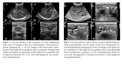
Is the presence of echo-rich periportal cuffing in the liver indicator for abdominal inflammation in pediatric patients? 

Nurdan Fidan1, Esra Ummuhan Mermi Yetis2, Muammer Murat1, Cuneyt Yucesoy3, Ebru Turgal4, Mehmet Metin5


ErPC [echo rich periportal cuffing] appears especially in diseases associated with abdominal inflammation such as gastroenteritis, acute appendicitis, perforated appendicitis and mesenteric lymphadenitis. When evaluated with adequate clinical information, the presence of ErPC is a finding of high sensitivity and specificity in pediatric patients. This could have a significant contribution to correct diagnoses by directing the radiologists or clinicians to further examinations or follow-ups.
DÀY KHOẢNG CỬA: DẤU HIỆU SIÊU ÂM CỦA VIÊM TRONG Ổ BỤNG

MỤC TIÊU HỌC TẬP:
1/ Mô tả được hình ảnh siêu âm dày khoảng cửa và dấu trời sao khi khám siêu âm gan
2/ Trình bày được các nguyên nhân và tình huống lâm sàng của dày khoảng cửa và dấu trời sao trong siêu âm gan
3/ Phân tích được một số tình huống lâm sàng có dày khoảng cửa và dấu trời sao 

NỘI DUNG
1.    DẪN NHẬP:
Đau bụng là lý do để khám siêu âm bụng trong thực hành lâm sàng ngoài các cách khám lâm sàng truyền thống. Tuy không là phương tiện chẩn đoán chính thống, siêu âm bụng thường quy/tại giường/tiếp cận giúp định hướng chẩn đoán và cung cấp hình ảnh bình thường và bệnh lý trong khoang bụng.
Gần đây y văn siêu âm có những bài báo về đau bụng ở trẻ em và người lớn, trong đó ghi nhận vài dấu hiệu siêu âm TRONG GAN như phản ứng quanh cửa [portal reaction], dấu trời sao [starry sign appearance], dày quanh cửa [periportal echo rich] trong một số bệnh lý như viêm gan, viêm dạ dày ruột, viêm ruột dư cấp…
Trong bài này chúng ta sẽ mô tả và tìm hiểu các dấu hiệu siêu âm trên.
2.    CÁC DẤU HIỆU SIÊU ÂM TRONG GAN CỦA ĐAU BỤNG:
A.   PHẢN ỨNG QUANH CỬA [PORTAL REACTION]:
Là các đường echo rich cạnh khoảng cửa gan trên nền nhu mô gan echo poor thường gặp trong viêm gan cấp (t10% đến 13.9% , n=1.367 ca) được cho là các tế bào gan viêm phù nề làm tăng sáng quanh cửa ở trung tâm gan. Dấu hiệu này có thể gặp trong các ổ bụng có hội chứng viêm các cơ quan khác như dạ dày,ruột.


B.   DDẤU TRỜI SAO [STARRY SKY APPEARANCE]:
Là các đường echo rich rải rác trong vùng ngoại biên gan. Dấu hiệu này có thể gặp trong các ổ bụng có hội chứng viêm các cơ quan như gan, dạ dày,ruột. Theo H Tchelepi - ‎2002, dấu hiệu này không có giá trị trong viêm gan


C.   DÀY QUANH CỬA [PERIPORTAL ECHO RICH]:
Dấu hiệu này mới được công bố trong một bài báo siêu âm ở trẻ em (2019). Tác giả không ghi nhận có trong viêm gan, chỉ có ở một số trường hợp viêm trong ổ bụng ở trẻ em cả 2 dấu trời sao và dày khoảng cửa.


3.    BÀN LUẬN:
Cả 3 dấu hiệu này đều ở khoảng quanh cửa trung tâm và ngoại biên của gan. Đây là mô tả của siêu âm dựa vào phù hợp của chẩn đoán lâm sàng sau cùng, chưa được chứng minh bằng các phương tiện chẩn đoán hình ảnh khác.
Khi tìm thấy 3 dấu hiệu trên người làm siêu âm nên tìm thêm các triệu chứng siêu âm bất thường ở gan, dạ dày, ruột, hạch trong ổ bụng để giúp hướng chẩn đoán cho một trường hợp đau bụng trong thực hành lâm sàng.

TÀI LIỆU THAM KHẢO:
SIÊU ÂM CHẨN ĐOÁN TRONG VIÊM GAN, 1997, Nguyễn Thiện Hùng – Phan Thanh Hải – Phạm Thi Thu Thủy Trung tâm Chẩn đoán Y khoa (MEDIC) Thành Phố Hồ Chí Minh
SONOGRAPHY of DIFFUSE LIVER DISEASES, by H Tchelepi - 2002, www.jultrasoundmed.org/content/21/9/1023.full
STARRY SKY APPEARANCE https://radiopaedia.org/articles/starry-sky-appearance-ultrasound-1

IS THE PRESENCE OF ECHO-RICH PERIPORTAL CUFFING IN THE LIVER INDICATOR FOR ABDOMINAL INFLAMMATION IN PEDIATRIC PATIENTS htps://www.medultrason.ro/medultrason/index.php/medultrason/article/view/1940

Thứ Sáu, 23 tháng 8, 2019

FDA adds tranducer check to ultrasound guidance.


By Kate Madden Yee, AuntMinnie.com staff writer
August 23, 2019 -- New ultrasound transducers should include an integrated quality control check that runs every time the probe is turned on, according to final guidance issued by the U.S. Food and Drug Administration (FDA) on August 22

The FDA plans to incorporate the new transducer check recommendation into the agency's 510(k) compliance policy that covers marketing clearance of diagnostic ultrasound systems and transducers. The new policy -- as well as clarifications to guidelines on when the FDA will require modified probes to receive a new 510(k) application -- was explained in a webinar by Shahram Vaezy, PhD, of the FDA's Center for Devices and Radiological Health.
"This guidance supersedes the FDA's 2008 guidance ... and describes the types of modifications to a diagnostic ultrasound device for which the FDA does not intend to enforce the requirement for a premarket notification," Vaezy said. "[It also] includes a new transducer element check that applies to all ultrasound devices."
Transducer element check
The FDA is recommending that manufacturers integrate into transducers for which they are filing new 510(k) applications a way to test probe performance each time it is activated.
"Each device should include some level of testing," the agency wrote. "This integrated test feature would also generate a report on the performance of the probe under test for documentation ... [and] should also be available to the operators to initiate anytime when a particular probe is suspected of failure."
Developing transducer check features will take time, Vaezy acknowledged.
"The FDA is open to discussions with manufacturers about how to implement transducer checks into devices," he said.
Compliance policy
The FDA clarified that it will not require a new 510(k) for modified ultrasound and transducer devices that have already received an initial 510(k) clearance when all of the following apply:
  • The intended use of the modified device is not changed.
  • The device is not a reusable device subject to the requirement for the submission of reprocessing labeling and validation data.
  • The modes of operation for the modified device are well-established.
  • The modifications do not lead to acoustic outputs that exceed the recommended maximum acoustic output levels.
  • The modifications do not result in a range of ultrasound interrogation parameters outside a well-known range.
  • The modifications do not use novel mechanical or thermal effects for imaging or measurements.
  • The measurements and analyses are clearly described, and the user can adjust the associated control parameters.
  • Transducer element check is performed.
  • Transducer surface temperature falls within a well-defined range.
  • Appropriate transducer covers are recommended to users.
During the webinar, Vaezy listed some examples of possible compliance policy applications that would require a new 510(k):
  • Adding continuous-wave and pulsed-wave Doppler methods to the device
  • Adding an algorithm that measures the volume of an organ based on established image segmentation and volume calculation methods
  • Adding a new transducer with similar indications for use and similar acoustic output
  • Adding a B-mode noise reduction filter for general imaging use to a system
"The revised final guidance enables manufacturers with an established track record of ultrasound device development, via submission of 510(k)s for their original devices, to add new safety features and make certain modifications to address clinical needs without [having to] submit another 510(k)," he said.

Thứ Sáu, 16 tháng 8, 2019

USPSTF Opens Review of carotid stenosis screening.


By Kate Madden Yee, AuntMinnie.com staff writer
August 16, 2019 -- The U.S. Preventive Services Task Force (USPSTF) has posted a draft research plan on screening for asymptomatic carotid artery stenosis, an exam that typically involves the use of ultrasound to detect signs of stenosis before a stroke can occur.

The new plan is part of the USPSTF's five-year review of its previous guidance on carotid artery screening, issued in 2014. At that time, the USPSTF gave the procedure a letter grade of D, recommending against its use in asymptomatic individuals.
The low prevalence of carotid artery stenosis would result in many false positives on screening ultrasound, the group concluded in 2014. In addition, a large number of surgical interventions such as carotid endarterectomy could be performed that aren't any better than standard medical therapies such as statins in terms of reducing stroke risk, according to the task force.
The new draft introduces the following questions for review:
  • Is there direct evidence that screening asymptomatic adults for carotid artery stenosis with duplex ultrasonography improves health outcomes?
  • What are the harms associated with screening for asymptomatic carotid artery stenosis?
  • For asymptomatic persons with carotid artery stenosis, does treatment with carotid endarterectomy or carotid angioplasty and stenting provide incremental benefit beyond current standard medical therapy?
  • What are the harms associated with carotid endarterectomy or carotid angioplasty and stenting for the treatment of asymptomatic carotid artery stenosis?
The draft research plan is available for public comment from August 15 through September 11, 2019.