Tổng số lượt xem trang

Thứ Năm, 15 tháng 10, 2020

Will handheld ultrasound bring imaging into the home?

By Alan Stoddart, AuntMinnie.com contributing writer


October 15, 2020 -- For all the changes in medicine, there are some things that seem resolutely stable. Chief among these is the idea that sick people go to doctors' offices or hospitals to be diagnosed and treated. This, the simplest of healthcare tenets, could however begin to be challenged, as one area of imaging -- ultrasound -- begins to be freed.

As identified in Signify Research's recently published Ultrasound Equipment -- World Market Report, the handheld ultrasound market is developing at a rapid pace. But does this growth herald an era of ultrasound at home?

Several things are spurring the growth of the handheld ultrasound market, with one of the most significant being cost. Until recently, a handheld scanner from a trusted vendor was in the region of $5,000 to $10,000. Now, however, largely thanks to the impact of Butterfly Network and its stated aim of democratizing medical imaging, scanners can be picked up for as low as $2,000.

The combination of improved imaging performance, clinical utility. and a lower price has expanded ultrasound's market opportunity beyond hospital departments to also include office-based clinical specialists. Moving forward, for a very modest outlay, a primary care physician can equip a surgical suite with an incredibly useful diagnostic tool. This scenario is predicted to be the biggest mid- to long-term growth driver for the handheld ultrasound product category.

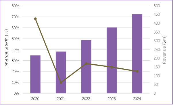
The handheld ultrasound market is set for rapid growth, as shown in the graph below.

The handheld ultrasound market is set for rapid growth

The COVID-19 pandemic has increased demand for handheld ultrasound massively in 2020, with the technology becoming a frontline diagnostic tool for patients with suspected COVID-19. The availability of affordable and fit-for-purpose handheld scanners has helped hospitals to deal with more patients more quickly -- a crucial factor in an exponentially growing pandemic.

Handheld ultrasound scanners, especially wireless versions, are easier to disinfect than traditional cart-based systems and other imaging modalities, and infected patients don't have to move through hospitals to imaging departments, raising the risk of spreading infection.

It is forecast, however, that COVID-19 will diminish as a driver of adoption of handheld ultrasound as the worst effects of the pandemic (hopefully) continue to lessen.

But, COVID-19 might also have a longer-term impact on the handheld ultrasound market in other, less direct ways.

Working from home

The novel coronavirus pandemic and the subsequent lockdown measures enforced by many of the world's governments have forced people to do at home what was thought only possible in specifically designed places. Broadcasters appeared suited and booted in front of the filing cabinets in their spare room and tech support calls were interrupted by the barking of wayward dogs as call center staff worked from their bedrooms. Even politicians weren't exempt, with parliaments having to pass motions from the safety of members' living rooms.

In healthcare, in the exceptional circumstances of the worst hit areas of Italy, the home started to become a work environment in other ways, enabled in part by the growing availability of handheld ultrasound systems. In these areas, people were being scanned in their homes. Suspected COVID-19 patients no longer had to make difficult journeys to overcrowded emergency departments, where they risked either spreading the virus, or, if they weren't already, becoming infected themselves.

This is just the latest example of a trend which -- although in its infancy -- has been steadily gathering pace for several years. Ultrasound is making the jump from a clinical to a domestic setting.

The stage had been set before now, with a number of companies offering the hire of ultrasound machines for the home. Netherlands-based Babywatcher, for example, promises that in conducting a scan, expectant mothers can, "sit back, light the candles and experience the ultimate pregnancy happiness" with the hire of an ultrasound machine being positioned as a perfect pregnancy gift.

Baby-Scan, meanwhile, suggests pregnant women buy its Baby-Scan Baby-Scanner to "take fun scans at home." Medical devices are being offered as consumer items. Even the Butterfly IQ, a professional medical device that includes a warning that it should only be used by qualified practitioners, still features branding and marketing material more reminiscent of a Silicon Valley smartphone than a diagnostic device.

The distinction between serious medical technology and consumer technology was blurred even more this year, when PulseNmore launched in the summer of 2020. The Israeli firm's device is an ultrasound transducer that docks with a smartphone, and it would look right at home alongside the electric toothbrushes, personal wellness devices, and electric shavers that fill people's bathroom cabinets.

What separates the device, though, is that in addition to "enhancing the pregnancy experience," it will help pregnant women "know about the well-being of their baby," according to Lior Wolff, the head of translational innovation at Clalit Health Services, which is offering the device to its members.

Ultrasound has, in just a few years, gone from being a clinical tool to be used in a clinical setting by clinicians to being a technology that can, in theory at least, be used by anyone, anywhere, at any time.

Ultrafreedom

Novice users are being empowered to use a tool in severa; way that, to this day, many primary care physicians can't. First, most handheld scanners come with built-in presets that can be used to quickly garner key measurements. A heart's ejection fraction or the estimated due date of a fetus, for example, can be automatically calculated by the system's tools.

Another enabler is teleultrasound, in which live ultrasound images are transmitted through teleconferencing to experts to guide novice users. Interest in teleultrasound has been heightened during the COVID-19 pandemic and facilitated by the U.S. Food and Drug Administration's emergency use authorization, which has enabled many in the medical field to use ultrasound where they wouldn't have otherwise.

In addition to a typical teleultrasound offering, in which a trained professional can remotely see the scan and talk to an untrained professional through the process, Butterfly Network has also added an augmented reality tool that superimposes arrows on a user's smartphone to visually guide the novice to ensure the scanner itself is positioned correctly.

Artificial intelligence (AI) will also play a key part in assisting novices to use ultrasound, both in terms of AI-enabled image capture assistance alongside automated anatomy navigation. Furthermore, AI can also compare the acquired image to standardized criteria to ensure that it meets clinical standards.

The first example of the latter is GE's Voluson Swift system, which incorporates Intelligent Ultrasound's ScanNav Assist AI technology. While currently only available on more expensive cart-based systems, it is only a matter of time before this capability cascades down to handhelds.

Finally, in what is perhaps the most consumeresque aid of all, vendors have released simple-to-understand YouTube tutorials. Novices can follow along, seeing how to position the Butterfly IQ scanner, what is displayed on the smartphone screen, which settings to press, and what to look for on the scan itself.

These developments, combined with the novel coronavirus pandemic and the need to keep people away from hospitals whenever possible, means that it is increasingly likely that more ultrasound scans are set to be conducted at home. Initially, professionals will be responsible, but for people with certain conditions that require regular scans, it might prove safer, more efficient, and cheaper in the long-term for people at home to be given a handheld unit and basic training and then left to scan themselves at a given frequency, with the images going directly to a doctor.

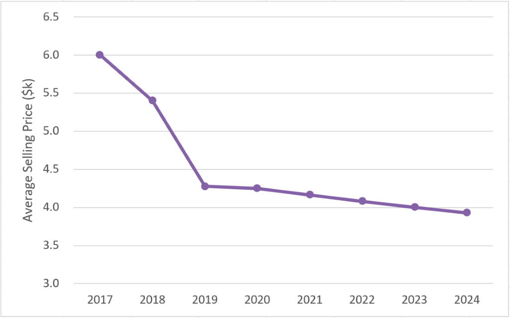
Falling prices make handheld scanners more accessible, as shown in the graph below.

Falling prices make handheld scanners more accessible

Further in the future, it is easy to imagine consumers buying scanners themselves, particularly as costs come down and AI-powered instructional software gets better. This is, in fact, where Butterfly Network's founder, Jonathan Rothberg, PhD, would like to see the market head.

"When the first thermometers were made, when the first blood pressure cuffs were made, they were only in hospitals," he mused when the Butterfly IQ launched.

Hot topic

A little knowledge, so the saying goes, can be a dangerous thing. This is especially true when that small amount of knowledge is on something as complex as a human body, even applying to the far simpler tools that Rothberg hopes Butterfly can follow. Take thermometers, for example. Numerous factors, from genetics to shafts of sunlight and even drafts, can undermine the accuracy of a reading.

When being used by nonexperts, these limitations and inaccuracies can have a huge impact. After all, a medical professional has a wealth of knowledge and experience with which he or she can scrutinize a dubious result. Novice users lack this expertise, and they are therefore susceptible to drawing incorrect conclusions.

This would no doubt also be an issue with ultrasound scanners at home. Women could purchase a scanner to see their unborn babies, and lacking any knowledge other than that shared on the scanner's YouTube channel, they could find the heartbeat and assume everything is fine but miss another indicator of ill health that an expert would pick up. This could be especially prevalent if people depend on the current crop of AI diagnostic tools that are blind to conditions they haven't been specifically developed to detect.

Conversely, the opposite could also be a problem. Stories of people with indigestion or head colds spending an hour on the internet and diagnosing themselves with Ebola or dengue fever are now so common as to be a cliché, but imagine the influx of people into emergency rooms if they also had a live image of their heart, lungs, or bladder to misread. Imagine the panic and emotional upset of people who think they have found a shadow on the lung of their wives or husbands or struggled to find a baby's heartbeat.

It seems inevitable that for every legitimate catch, there are going to be thousands of misdiagnoses, sending thousands of people to hospitals and clinics each year, and placing even greater demands on the time of already overstretched clinicians.

Homeward bound

The prevalence of many of these issues will depend on how ultrasound scanners make their way into people's homes. After all, if a scanner is prescribed by a medical specialist who carefully explains the technology and its limitations, it will be thought of very differently than if it is picked up from a supermarket's health aisle alongside electric toothbrushes and digital scales discounted in the January sales.

Cost will play a big part in deciding which of these paths the technology will take. A price tag of $2,000 might seem cheap to medical professionals used to paying tens of thousands for an ultrasound machine, but to someone at home, that could be more than the cost of their car. As such, the first successful "ultrasound at home" company is likely to be the one that makes it affordable to the mass market.

But, the costs of the devices themselves will, at least in the foreseeable future, only be able to fall to a certain price. Regardless of their ubiquity, they will always be precision medical devices with associated costs like calibration and quality assurance that will never be able to be completely nullified, regardless of production efficiencies. So in all likelihood, this means either a subscription model or as part of a broader health service, perhaps from an insurer, rather than an outright purchase, will be what takes handheld ultrasound mainstream.

This is a good thing. It would bring the cost down to a manageable level for lots of households, getting the devices to people that could benefit from them, but it would also mean that consumers were signing up for a service and not just hardware. It would give them a place to go not only for support during the scan itself but also with their concerns and their homemade diagnoses. A health plan member could conduct their monthly scan, and, if they saw something unusual, flag it for review (perhaps for a small fee), whereupon it would be assessed by a professional, who could then refer the customer to a supported provider.

From a business side, this would also help the vendors. Instead of growth coming from ever greater volume, at ever lower margins as competition in the market increases, an ultrasound at-home subscription service means predictable ongoing revenue and the ability to scale at a more granular pace. Instead of building a new manufacturing hub based on optimistic sales forecasts, investment could be made with the certainty of sticky and repeatable subscription revenues already in the books.

Another avenue for collecting revenue for handheld vendors is an app-store option. If a subscription can get the devices in enough hands, it becomes viable for software developers to offer very specific tools. Consumers at home could have a primary reason for having an ultrasound machine, but if they had a sports injury, for example, they could spend a small amount on downloading an AI app to assess it, with the vendor taking a cut.

Sound outlook

There is still a long way to go before people performing their own ultrasound scans at home is as common as them taking their blood pressure or temperature, but the trend is real. Decentralization is happening, ultrasound has moved from dedicated hospital units to primary care centers and is making its way to people's houses via COVID-19-catalyzed health visits.

How this trend develops is reliant on vendors being able to make their products affordable to customers, yet still profitable for themselves. It will also require careful consideration given the implications and repercussions of allowing a professional tool out in the wild.

In the longer term, these considerations, along with the growing adoption of telehealth services (see Signify Research's Telehealth (Acute, Community, and Home) -- World 2020 report for more information) will mean a shift toward teleultrasound services, in which customers will eventually receive scanners for free as part of a comprehensive ultrasound subscription service, much like today's smartphone business model, with profits made from teleultrasound services and app sales.

Cost is still a barrier to increased adoption. But as that falls, it will just take one vendor, one provider, one insurer, just one company with a novel strategy to strike on the right model, which is both helpful and lucrative, before the floodgates open and ultrasound at home is an everyday occurrence.

Alan Stoddart is an editor at Signify Research.

The comments and observations expressed do not necessarily reflect the opinions of Auntminnie.com or AuntMinnieEurope.com, nor should they be construed as an endorsement or admonishment of any particular vendor, analyst, industry consultant, or consulting group.

Copyright 2020 © Signify Research

Thứ Tư, 14 tháng 10, 2020

Benefits of U S fetal size estimates questioned

By Theresa Pablos, AuntMinnie staff writer


October 14, 2020 -- The benefits of using third-trimester ultrasound scans to predict large infant size may be limited, according to a review published online October 13 in PLOS Medicine. Ultrasound size estimates failed to reliably predict one of the most common complications of delivering large newborns.

Shoulder dystocia is a major perinatal complication associated with macrosomia, a term used to describe infants typically weighing more than 8 lb 13 oz at birth. In an ideal screening program, ultrasound scans would both identify large infants and prevent shoulder dystocia and other size-related perinatal complications.

But the new systematic review of 112,000 pregnancies casts doubt on ultrasound's predictive ability for low- and medium-risk pregnancies. A U.K.-based team found prenatal ultrasound screening correctly identified many cases of macrosomia but only had a weak association with predicting shoulder dystocia.

"There is a debate regarding introducing universal third-trimester screening for macrosomia," wrote the authors, led by Dr. Alexandros Moraitis, an obstetrics and gynecology trainee at the University of Cambridge. "An effective screening program requires two elements: an effective test at predicting a condition and an effective intervention."

The review included 41 studies evaluating the prediction ability of size measurements derived from ultrasound scans performed after 24 weeks of pregnancy. The team specifically excluded studies on high-risk pregnancies, since they are the focus of an ongoing randomized clinical trial in the U.K.

Ultrasound size measurements reliably indicated which infants would have macrosomic birth weights for women with low-risk and mixed-risk pregnancies. The results stayed consistent whether studies calculated birth size by abdominal circumference or estimated fetal weight (EFW) techniques.

Ability of high estimated fetal weight on ultrasound to predict perinatal outcomes
 Birth weight 90+ percentileBirth weight 97+ percentileShoulder dystocia
No. of patients80,04551,68626,264
Sensitivity53.2%67.5%22%
Specificity93.9%89.7%89.6%

The authors also found a significant relationship between large ultrasound size measurements and the occurrence of shoulder dystocia. But they cautioned that the relationship was weak, with large EFW on ultrasound tied to just a 15% greater probability of shoulder dystocia.

"The fact that ultrasonic EFW is relatively poor as a predictor of shoulder dystocia is not unexpected, given that the actual birth weight of the baby is also not a strong predictor of the outcome: The majority of cases of shoulder dystocia involve a normal birth weight infant," the authors wrote.

One of the biggest drawbacks of the review was the quality of the included studies. The studies had largely heterogeneous designs and many were unblinded. In fact, unblinded studies showed a greater association between ultrasound weight predictions and shoulder dystocia than blinded ones, suggesting the presence of bias.

Furthermore, few studies looked at the association between ultrasound-based size predictions and neonatal morbidity. Of the few that did, the researchers found no association between macrosomia prediction and neonatal unit admission.

The authors called on future researchers to study whether other factors can help identify pregnancies most at risk for complications. In the meantime, they can't recommend universal screening for macrosomia -- especially because other studies have found that wrong size predictions can lead to unnecessary caesarean sections.

"We recommend caution prior to introducing universal third-trimester screening for macrosomia, as it would increase the rates of intervention, with potential iatrogenic harm, without clear evidence that it would reduce neonatal morbidity," they wrote.

Thứ Tư, 7 tháng 10, 2020

SIÊU ÂM B-MODE VÀ Viêm Dính Khớp Vai [ACS]


Abstract 

Aims: In this systematic review and meta-analysis, we discuss the value of grey-scale ultrasonography (US) in diagnosing adhesive capsulitis of the shoulder (ACS). 

Material and methods: We retrieved relevant studies from PubMed, Cochrane Library, and Embase before 8 April 2019. We selected 7 studies concerning 446 patients (490 shoulders) that used grey-scale US to diagnose ACS and magnetic resonance imaging (MRI) or arthroscopy as the reference standard. We assessed the diagnostic accuracy of US on the basis of combined sensitivity, specificity, likelihood ratio (LR), and the area under the summary receiver operating characteristic (SROC) curve (AUC). 

Results: The combined sensitivity, specificity, positive LR and negative LR were found to be 88% (95%CI: 74–95), 96% (95%CI: 88–99), 23.89 (95%CI: 6.31–90.51) and 0.12 (95%CI: 0.05–0.29), respectively. The AUC was 0.97 (95%CI: 0.96–0.98). ACS was diagnosed on the basis of four US features: coracohumeral ligament thickening, inferior capsule/axillary recess capsule thickening, rotator interval abnormality, and restriction of the range of motion. The corresponding sensitivities were 64.4 (95%CI: 48.8–78.1), 82.1 (95%CI: 73.8–88.7), 82.6 (95%CI: 74.1–89.2) and 94.3 (95%CI: 84.3–98.8), respectively, and specificities were 88.9 (95%CI: 76.0–96.3), 95.7 (95%CI: 90.3–98.6), 93.9 (95%CI: 89.8–96.7), and 90.9 (95%CI: 75.7–98.1), respectively. 

Conclusions: Our meta-analysis showed that grey-scale US plays a significant role in the diagnosis of ACS. Because of its high sensitivity and specificity, US can be added to the existing clinical diagnosis program. Keywords: ultrasound; adhesive capsulitis; shoulder; systematic review; meta-analysis

Thứ Sáu, 2 tháng 10, 2020

What is POCUS: Leading rads call for new definition


By Theresa Pablos, AuntMinnie staff writer

October 2, 2020 -- A group of radiology thought leaders is calling for a more precise definition of point-of-care ultrasound (POCUS). Their proposal to better distinguish POCUS from other ultrasound technologies was published on September 29 as a white paper in the Journal of the American College of Radiology.


The team behind the paper urged the medical community to see handheld ultrasound as the versatile innovation it is, noting some even liken its use to the stethoscope. But medical terminology and reimbursement codes have not kept up with the rapid and widespread advancement of ultrasound in medicine.

To remedy this, the Society of Radiologists in Ultrasound and the American College of Radiology Commission on Ultrasound came together to better delineate between observational and diagnostic ultrasound use of POCUS. In a new paper, the groups created four unique and distinct ultrasound categories, which include limiting the definition of POCUS to exams performed and reported in conjunction with a patient evaluation and management (E/M) encounter.

"We propose a simple way to broadly classify ultrasound evaluation that is more meaningful and consistent, taking the nomenclature beyond the idea of who performs the evaluation to the more important concept of what evaluation has been performed," wrote the authors, led by Dr. Maitray Patel, a radiologist at the Mayo Clinic Phoenix, AZ, and a member of both ultrasound societies behind the paper.

4 types of ultrasound evaluations: definitions and sample use cases
 Point-of-care ultrasoundNoncomprehensive diagnostic ultrasoundPartial-comprehensive diagnostic ultrasoundComplete-comprehensive diagnostic ultrasound
DefinitionEvaluation performed and reported in conjunction with an E/M encounterNoncompre-
hensive evaluation of any number of elements in anatomic region
Comprehensive evaluation of a partial number of elements in an anatomic regionComprehensive evaluation of a complete set of elements in an anatomic region
Example: pregnant patient in third trimester
  • Determine if fetus is breech or cephalic presentation
  • Document fetal heart motion
  • Evaluate or document lower uterine segment
  • Document fetal heart motion
  • Evaluate nonfetal anatomy components
  • Use of color Doppler when indicated
  • Full OB exam evaluation of fetal and nonfetal components
  • Use of color Doppler when indicated
Example: patient with right upper quadrant pain
  • Determine if gallbladder corresponds to pain area
  • Look for gallstones
  • Evaluate and document gallbladder status
  • Evaluate and document gallbladder and bile ducts
  • Use of color Doppler when indicated
  • Full evaluation of all abdomen elements in region
  • Use of color Doppler when indicated

To create the four classifications, the authors first differentiated between point-of-care ultrasound (POCUS) and diagnostic ultrasound (DXUS) examinations. They asserted that POCUS becomes easier to recognize when defined solely as part of an E/M exam.

This definition of POCUS permits flexibility for what counts as a point-of-care exam. As described, POCUS would include scenarios that typically come to mind when thinking about point-of-care evaluations -- a clinician using a handheld device to scan a patient at bedside. But it also could include exams not obtained with handheld scanners or taken at bedside.

In addition to POCUS, the team created three distinct categories for diagnostic ultrasound use: noncomprehensive diagnostic ultrasound, partial-comprehensive diagnostic ultrasound, and complete-comprehensive diagnostic ultrasound.

The three diagnostic categories differ in relation to the author-defined principles of comprehensiveness and completeness. According to the group's terminology, comprehensiveness refers to the checklists and protocols needed to create, store, and document standardized images, whereas completeness recognizes whether the exam does or does not include all related structures or substructures in an anatomical region.

But this viewpoint is a major departure from how ultrasound is presently viewed in the larger medical infrastructure, particularly when it comes to billing and CPT codes.

"The current CPT framework forces the square POCUS peg exclusively through the round hole of 'diagnostic' imaging rather than considering and reimbursing POCUS as an inherent component or modifier of an E/M service," the authors wrote.

One of the challenges of the current reimbursement structure is that payors typically only pay for one documented and reported ultrasound evaluation of a patient, the authors noted. Instead, the new definition would permit multiple providers to perform POCUS evaluations on the same patient in the same region to answer multiple, relevant clinical questions.

In order to make the framework feasible, payers would have to recognize the benefits of allowing different providers to perform POCUS scans on the same patient. It would also require payors to work with multispeciality groups to define what organs and structures should comprise standardized anatomical subgroups.

"Make no mistake: There is important work to be done to understand the value of POCUS," the authors wrote.

Thứ Năm, 1 tháng 10, 2020

SỎI MẬT CẦN BIẾT

Gallstones top to toe: what the radiologist needs to know


Abstract

Gallstone-related disease can have significant associated morbidity and mortality worldwide. The incidence of gallstone-related disease in the Western world is on the increase. There are multiple different pathological manifestations of gallstone disease: the presentation, diagnosis and associated complications of which vary significantly depending on anatomical location. The role of imaging in gallstone-related disease is broad with radiology playing an essential role in the diagnosis, management and follow-up of gallstone-related pathologies. This paper distills the broad range of gallstone-related pathologies into an anatomical map, discussing the disease processes involved at each point along the biliary tree and reviewing the strengths and weaknesses of different imaging modalities for each distinct disease process.


Key points

  • Gallstone-related pathology is on the increase in the Western world.

  • Gallstones can be located within the gallbladder, migrate into the biliary tree or outside the pancreaticobiliary system altogether with associated pathology.

  • Imaging of gallstones and associated pathology requires a multimodality approach.

Thứ Bảy, 26 tháng 9, 2020

Pocket-sized ultrasound delivers big results for COVID

By Theresa Pablos, AuntMinnie staff writer


September 25, 2020 -- A pocket-sized ultrasound scanner powered by a smartphone performed comparably to a cart-based scanner for lung imaging at the point of care in patients with COVID-19, according to a study published on September 21 in Ultrasound in Medicine and Biology.

In the head-to-head study, a team of Italian authors compared a conventional, portable cart-based ultrasound scanner with a pocket-sized device for point-of-care ultrasound (POCUS) of COVID-19 patients. The smaller -- and cheaper -- system adequately visualized pulmonary regions of patients with COVID-19.

The findings are good news in a pandemic rife with medical equipment shortages.

"Our study confirms the possibilities of portable pocket-sized ultrasound imaging of the lung in COVID-19 patients," wrote the authors, led by Dr. David Bennett from the University Hospital of Siena in Siena, Italy.

Lung ultrasound has repeatedly proved its usefulness as a tool to diagnose and monitor patients with COVID-19. While prior studies on the topic have used portable ultrasound systems, no research has directly compared the performance of pocket-sized ultrasound systems with larger scanners for COVID-19 imaging.

In the latest study, clinicians performed lung ultrasound scans on 18 patients hospitalized with COVID-19 at the University Hospital of Siena. The patients were all symptomatic, tested positive for SARS-CoV-2, and had radiologic evidence of interstitial pneumonia.

Clinicians scanned the patients using two ultrasound systems: (1) Venue Go by GE Healthcare, a cart-based system with built-in artificial intelligence capabilities, and (2) Butterfly iQ by Butterfly Network, a pocket-sized system powered by a smartphone that uses a silicon chip instead of a piezoelectric crystal.

The team used the scans to calculate patients' lung ultrasound scores, a proven method for quantifying pulmonary disease severity in patients with COVID-19. The scoring system works by assigning numerical scores of 0-3 to unique lung regions. Higher scores indicate worse disease severity.

The team found no significant difference between the two systems for patients' overall lung ultrasound scores. Lung scores obtained by both systems also correlated with patients' clinical severity.

In addition, the scores were comparable for most individual pulmonary regions, including the left and right side and vertical location. However, the team did find an 0.082 score difference between the systems for horizontal position, a statistically significant finding they deemed a "practically negligible difference in the posterior side of the thorax."

Further statistical analysis revealed a bias of -0.016 between the systems with very narrow limits of agreement.

"The results of the portable scanner were practically identical to the high-end scanner in the assessment of lung interstitial syndrome," the authors wrote.

While pocket-sized scanners are easy to carry and significantly cheaper than larger models, they also come with a number of drawbacks. Small systems tend to have limited battery life, reduced field of vision, and low penetration, the authors noted.

The authors also cautioned that POCUS should be used with clinical and physiological data for patients with COVID-19. However, they were also confident that pocket-sized systems could accurately assess lung lesions -- and do so more affordably than high-end scanners.

"These ultrasound scanners can play a decisive role when healthcare resources are scarce, during pandemics and in emergency situations, such as the present COVID-19 outbreak," the authors concluded.

Thứ Bảy, 19 tháng 9, 2020

CÓ GÌ MỚI TRONG SIÊU ÂM ỐNG CỔ TAY


https://www.auntminnie.com/index.aspx?Sec=sup&Sub=ult&Pag=dis&ItemId=130216&wf=10305&slid=4B40393935C60377301B37A6321E398B7E93BF974E3458975745C1BD9CB84548

September 16, 2020
 -- Ultrasound imaging shows promise as a painless way to diagnose carpal tunnel syndrome, according to the findings of a small study published on September 11 in Academic Radiology. A combination of ultrasound features produced high
accuracy, sensitivity, and specificity in the study.

Carpal tunnel syndrome is often diagnosed with the aid of electrodiagnostic tests, which can be painful for patients. The new study suggests that ultrasound may be a pain-free and cost-effective alternative for patients.

Building upon prior research, the authors found that ultrasound features like cross-sectional area (CSA) and stiffness and vascularity of the median nerve accurately differentiated between patients with and without carpal tunnel syndrome.

"The [median nerve] stiffness, CSA, and vascularity of [median nerve] showed a high diagnostic performance for diagnosing [carpal tunnel syndrome] independently or in combination; albeit based on a small sample size," wrote the authors, led by Kibo Nam, PhD, an associate professor of radiology at Thomas Jefferson University in Philadelphia.

Researchers have evaluated various ultrasound features for identifying carpal tunnel syndrome in patients. Cross-sectional area has looked particularly promising as a diagnostic tool because symptomatic patients have an enlarged area due to edema and fibrous tissue proliferation.

However, CSA on its own often results in a sensitivity and specificity of under 90%. The authors hypothesized that its effectiveness could be improved by adding other ultrasound features.

Their study included 27 wrists with carpal tunnel syndrome and 20 healthy wrists from 25 total volunteers. All patients underwent strain elastography and shear-wave elastography while sitting with their arm extended and palm facing up.

The authors imaged patients' median nerve at two sites: (1) proximal carpal row and (2) pronator quadratus muscle. They used software built-in to the ultrasound scanners to calculate stiffness and CSA and an offline Matlab script to measure vascularity.

All studied ultrasound features differed significantly between patients with and without carpal tunnel syndrome. Out of the independent features, CSA had the highest diagnostic value, with an accuracy of 94%, specificity of 100%, and sensitivity of 85%.

Accuracy of ultrasound features for diagnosing carpal tunnel
Ultrasound featureSpecificitySensitivityAccuracy
CSA100%85%94%
Color Doppler imaging80%84%84%
Stiffness (site 1)80%85%84%
Stiffness (site 2)65%78%72%

However, the results further improved when the authors combined the CSA measurements with other ultrasound features. A model that combined CSA with stiffness at the pronator quadratus muscle had an accuracy of 95%, specificity of 100%, and sensitivity of 93%.

Similarly, a model that combined CSA, stiffness at either of the measured sites, and color Doppler imaging measurements yielded an accuracy of 96%, specificity of 100%, and sensitivity of 93%.

Accuracy of combined ultrasound features for carpal tunnel
Ultrasound featuresSpecificitySensitivityAccuracy
CSA + stiffness (site 1)100%93%96%
CSA + stiffness (site 2)100%93%95%
CSA + stiffness (either site) + color Doppler imaging100%93%96%

While the results look promising, there are a number of caveats with the research. Notably, the diagnostic accuracy of CSA alone was markedly higher than the values found in other studies. For instance, one meta-analysis cited in the paper found CSA had a pooled specificity of 83% and sensitivity of 87%.

The authors attributed the high diagnostic accuracy of CSA in their study to high-frequency imaging. But they also noted that when CSA alone is such an accurate diagnostic metric, the outcomes aren't as drastically improved by adding other ultrasound features.

The study also had a small sample size, and the ultrasound features could not distinguish between patients with mild, moderate, and severe forms of carpal tunnel syndrome.

Nevertheless, the authors highlighted the promise of using ultrasound features, alone or in combination, as a painless method to diagnose carpal tunnel syndrome. In fact, the results look especially promising when compared with electrodiagnostic tests, which have a sensitivity of 80%-92% and specificity of 80%-99%, according to the authors.

"Although the ultrasound features did not differentiate all the stages of [carpal tunnel] severity, this study suggests the further validation of ultrasound features and standardization of assessment criteria for the use of ultrasonography as a supplement screening tool for [carpal tunnel syndrome]," the authors concluded.