Tổng số lượt xem trang

Chủ Nhật, 22 tháng 2, 2015

ElastPQ and LIVER FIBROSIS, [PQ=point quantification]

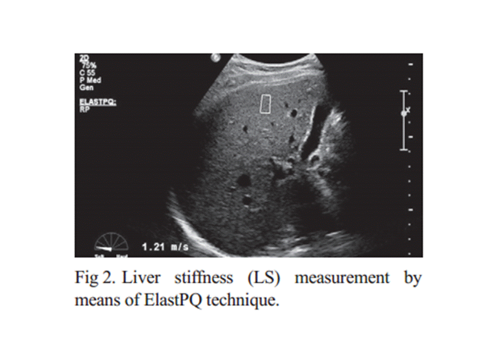
Liver stiffness values obtained by Elast PQ technique are significantly lower than those obtained by ARFI elastography and future studies are needed to establish the best LS cut-offs assessed by ElastPQ for predicting different liver fibrosis stages.


--------------------------------------------------------------------------------------------------

ElastPQ (Philips)

from JSUM Ultrasound Elastography Practice Guideline Liver

 Masatoshi Kudo1*, Tsuyoshi Shiina2, Fuminori Moriyasu3, Hiroko Iijima4, Ryosuke Tateishi5, Norihisa Yada1, Kenji Fujimoto6, 7, Hiroyasu Morikawa8, Masashi Hirooka9, Yasukiyo Sumino10, Takashi Kumada11

 A) Introduction
  Name: ElastPQ (PQ: point quantification)
  Equipment: The iU22 xMATRIX ultrasound system (iU: Intelligent Ultrasound)

 ElastPQ is a non-invasive diagnostic tool to measure tissue stiffness using an ARFI-based technology. Immediately after image acquisition, the screen displays the image and measurement results, including the mean and median values and the deviations in kPa or m/s (Fig. 33).
*If measurement reliability is low, 0.00 kPa will be displayed as the result.
    Elastic value E [kPa] is calculated using the equation E = 3ρVs where Vs [m/s] is defined as the       shear wave propagation velocity and ρ as tissue density (whose approximated value in the human       body is 1). 
    An ROI can be placed anywhere but at a depth of uder  8 cm.

B) Indication

 1. Quantitative assessment of liver fibrosis in diffuse liver diseases
 2. Neoplastic lesions of the liver

C) Procedures (including Tips and Tricks)

 1. Perform right intercostal scanning to visualize the liver
 2. Steadily place the probe with minimum compression
 3. Set the ROI with a depth of under 8 cm.
 4. Ask the patient to breath hold (if not possible, ask the patient to breathe as shallowly as possible).
 5. Push the” Update” button for quantification 
 6. The use of a mean value from more than 10 measurements is recommended.

 Approach the right hepatic lobe from the right intercostal space. Avoid the left hepatic lobe because the measurement is affected by cardiac movement. Breath hold without exerting abdominal pressure. The most appropriate ROI is the center of the image, namely, immediately below the probe, and 3–5 cm from the probe surface. Avoid blood vessels, any necrotic areas, the boundary between organs, and areas influenced by cardiac movement (ex. left hepatic lobe). Three frequencies (R1/RP/P1) are available. The measurement sensitivity of areas deep inside the body can be improved by using a lower frequency. 

 D) Results (What does the value mean?)

 ・Healthy liver: 4 kPa (2.5–4.7 kPa, 1–1.5 m/s) 
 ・Mild fibrosis: 7 kPa (4.7–12.0 kPa, 1.5–2.0 m/s) 
 ・Moderate–severe fibrosis: 12 kPa (12.0–21.0 kPa, 2.0–2.5 m/s) 
 ・Severe fibrosis: over 21 kPa (over 2.5 m/s)

E) Limitations

・There is a limit to measurable depth.
・ElastPQ is affected by respiratory and body movement.
・Cardiac movement also affects the system.
・Accuracy of measurement depends on the skills of the examiner.
・Measurement accuracy is generally low at the sides of an image.
・Ribs may cast lateral acoustic shadows.

F) Recommendations

At present, the number of studies using ElastPQ is not large enough to reach a definitive conclusion. We look forward to having more study results in the near future.

The place of musculoskeletal ultrasonography in gout diagnosis

Daniela Fodor et al


Investigation questions

One of the problems in US investigation of gouty patients is which sites to be investigated (only symptomatic joints or structures or more extensive examination) and what findings to be search (MSU deposits, effusion, synovial hypertrophy, vascularisation) in order to have good sensibility and specificity of the method. In a recent published paper by Naredo et al [17] the authors found that US examination of 12 anatomical sites (bilateral radio-carpal joint, first metatarsal head cartilage, talar cartilage, second metacarpal head, knee femoral condyle cartilage, patellar and triceps tendons) for DC sign and hyperechoic aggregates gives the best results for sensitivity and specificity (84.6% and 83.3%, respectively). This approach had high positive and good negative predictive values (92% and 71%, respectively) for diagnosing gout. Peiteado et al [57] studied the presence of six types of lesions in gouty patients: hyperechoic spots in the synovial fluid, HCA, bright stippled aggregates, DC sign, erosions, and the Doppler signal in 17 joints and 8 tendons. They found that the knees and MTP joints are the most frequent affected sites (in 93% of patients) and a six-minute US examination of four joints (knees and the 1st MTPs) allowed the detection of HCA or DC sign in 97% of cases. Clinical diagnosis of gout is sometimes difficult, even if the manifestations are in MTF-1 (classical podagra). Kienhorst et al [58] recently demonstrated that in patients with MTP-1 arthritis the gout was the correct diagnosed only in 77% of cases but the general practitioner supposed gout in 98% of cases. In these cases US examination of the joint could simplify the diagnosis process. This fact was demonstrated by Lamers-Karnebeek et al [59] in patients with monoarthrithis in which 48% of patients had MSU crystals-proven gout. In this cases sensitivity of DC sign and any US abnormality (DC sign, tophi, or snowstorm appearance of the joint effusion) was 77 and 96 %, respectively. The authors considered that the DC sign is an important informative finding for clinician, with positive predictive value of 74 % and negative predictive value of 78 %.


The new US techniques probably will increase the US capacity to detect the crystals aggregates. MicroPure, a new US image processing function designed to improve the visualization of microcalcifications, was used by Yin et al [60] in patients with gout. Significantly more microcalcifications were seen with MicroPure compared to gray scale US and the level of agreement between investigators was consistently improved. The method seems to be useful especially for the cases where the presence of gray scale US artifacts or unspecific findings make difficult the interpretation. The use of different imaging methods (dual energy CT, MRI, positron emission tomography) may improve the description of lesions [61].


 The main question is if there is a really need in clinical practice for such a complex approach. The need for good education and training for identification and interpretation of the US findings encountered in gout was underline by Filippucci et al [62].The authors highlight the necessity for dedicated training-programme to avoid the false positive and false negative results. After 7 days of training the rheumatologists with limited US experience gain satisfactory skills to identify MSU aggregates in different tissues. Due to the lower diagnostic utility of the clinical gouty features (exception tophi and response to colchicine) [63], there is an incremental need for imaging techniques to confirm the clinicians’ suppositions. The place of the US in patients suspected of gout must be clearly defined [59] due to the high numbers of patients presented with monoarthritis. The research agenda concerning the use of US in gouty patients is still busy despite of the recent achievements in this pathology. In conclusion in the assessment of the gouty patients US is an important and valuable tool. For a complete examination and a correct interpretation of the imagines the examiner need to have solid US knowledge about normal and pathological aspects of the musculoskeletal structures. Knowing the specific US aspects of MSU depositions will permit a rapid diagnosis. Moreover, when combining the clinical examination with US scan in patients with suspicion of gout a proper and rapid decisions for the patient management can be taken.