Tổng số lượt xem trang

Thứ Tư, 28 tháng 3, 2018

AIUM: Can deep learning classify liver fibrosis on US?


By Erik L. Ridley, AuntMinnie staff writer
March 26, 2018 -- NEW YORK CITY - A deep-learning algorithm that analyzes B-mode morphological ultrasound data and patient demographic information can classify the severity of liver fibrosis in cases of chronic liver disease, according to research presented at the American Institute of Ultrasound in Medicine (AIUM) conference.
A team of researchers from Greece trained a deep neural network that could determine if patients were in the early stages of fibrosis or if they had advanced to significant fibrosis. In testing, the algorithm yielded a higher level of performance than existing methods previously reported in the literature, according to presenter Athanasios Angelakis, PhD, from the National Technical University of Athens.
A binary classification problem
Early-stage fibrosis can be reversed with treatment and prevented from reaching cirrhosis. It's also important to be able to diagnose significant fibrosis, classified as Metavir score F0 or F1, from more severe fibrosis (≥ F2).
"Given an individual's biomarkers, we would like to conclude if his or her fibrosis stage is either [Metavir score] ≤ F1 or ≥ F2," he said in a March 24 presentation "It's a significant binary classification problem."
Traditional methods used for this assessment include biopsy; biochemical tests; imaging, such as shear-wave elastography, strain elastography, and FibroScan transient elastography cutoff values; as well as image analysis or radiomic approaches. Recently, machine learning has also been applied in image analysis and radiomics. However, none of the biochemical, imaging, image analysis/radiomics, or machine-learning methods reported in the literature has yielded an area under the curve (AUC) from receiver operating characteristic analysis of more than 0.87, according to Angelakis.
The researchers sought to apply deep learning to this classification task. They used a dataset of 103 patients with chronic liver disease and biopsy-validated fibrosis stages; 62 patients had Metavir scores of F0 or F1, while 41 had scores of at least F2. The deep-learning network was given one demographic parameter (patient gender) and four morphologic parameters -- the longitudinal diameter of the left lobe, caudate lobe, and right lobe of the liver, as well as the spleen. The algorithm then provides its assessment of the patient's fibrosis class (≤ F1 or ≥ F2).
This information could be of particular benefit to inexperienced users, according to the researchers.
"It reduces the experience [needed] for the examiner [to perform these studies]," Angelakis said. "One of the goals of using deep learning in ultrasound in general and shear-wave elastography is to reduce this need for experience with the exam."
After performing tenfold cross-validation, the researchers found that the algorithm yielded a high level of accuracy for liver fibrosis classification:
  • Sensitivity: 90.2%
  • Specificity: 91.9%
  • Positive predictive value: 88.1%
  • Negative predictive value: 93.4%
  • AUC: 0.9126
They acknowledged, though, that more patients are needed to further validate the algorithm.
The future
The ultimate holy grail for a deep-learning algorithm in the liver would be to incorporate patient demographic data such as gender and age, along with morphologic, hemodynamic, biochemical, image analysis, and elastographic data. Such an algorithm, which would have many layers, would not only classify fibrosis but also steatosis, portal hypertension, and inflammation, Angelakis said.
"We have already created algorithms and models that also attack the steatosis problem, because our datasets are bigger now," he said.
Deep learning and data science will likely take on a significant role in ultrasound in the future, according to Angelakis.

"The output from the [deep-learning] model will be one more input for the expert [to consider during interpretation]," he said. "That is how we reduce the expertise [requirement] for the reader. Looking ahead 10 years from now, maybe less, data science will also be used during the manufacturing of ultrasound devices and it will be in every [aspect of the ultrasound acquisition and interpretation process].

Thứ Ba, 27 tháng 3, 2018

NHÂN 03 CA DIFFUSE LARGE B CELL LYMPHOMA CÓ TRIỆU CHỨNG HÀM MẶT

Trong số 20 ca lymphom lan tỏa dòng tế bào B lớn [diffuse large B cell lymphoma] ở Trung tâm Y khoa Medic từ 2008,  có 03 ca có triệu chứng đau răng và hàm mặt điều trị giảm đau, nhổ răng..không bớt, sau đó xuất hiện nhiều tổn thương khác ngoài hạch. Trong khi đó, tìm được một bài báo về đau xương hàm dưới trong lymphom ác. Sau đây là một hồi cứu về đề tài này.

             Diffuse Large B Cell Lymphoma
and Appearences  in Oral Cavity, and Maxillary Area
Nguyen Thien Hung, Jasmine DCB Thanh Xuan, Le van Tai, Le Thong Nhat, Le Thanh Liem, Phan Thanh Hai
Medic Medical Center, HCMC, Vietnam

INTRODUCTION:
May there had been an initial symptom in oral cavity, maxillary and mandibular area for diffuse infiltration of lymphoma?  We will represent some cases of diffuse lymphoma with appearences  in oral cavity,  and maxillary areas.

MATERIALS and METHODS:
We retrospectively reviewed  20 cases of diffuse lymphoma that were histopathologically diagnosed in our center from 2008 in which presented 03 cases  with oral cavity,  maxillary symptoms and  bone involvement [01 case in male 42 yo and 02 cases in female gender, 33 and 40 year-old].

RESULTS:

CASE 1= A 33 year-old female patient with pain in lower maxillary bone for one month and tension in both  2 breats), hyperpigmented edema of areolar area both 2 sides without pregnancy.  ABVS scanning  detected  multiple nodules infiltrating in 2 breasts.
Biopsy of 2 breasts  reported  microscopic with immunohistochemistry scanning, diffuse large B cell lymphoma.
MRI  full body with gado detected  bone marrow changing, 2 breats  hypercaptured contrast,  ascites and kidney infiltration. In pelvis  2 ovarian tumors and big uterine cervix were detected. Blood tests showed   lower platelets,  EGFR  lower  46, beta2 microglobuline raised  3816,  ferritin raised  911, LDH-l  raised  1360.


CASE 2= A 40 year-old female patient with toothache but pain remained  after removing tooth, and in total body examination detected right pleural effusion  and some nodular tumors in thyroid, left breast and subcutaneous area of right neck, left chest all and  lumbar region. Erosions of left clavicle and right 1st rib. Biopsy of left breast tumor reported diffuse large B cell lymphoma.


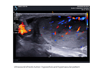
CASE 3= Man 42 year-old, one month ago, pain in oral sinus, difficult eating and 2 days after,  pain appears in left testis. Ultrasound of left big and  hot testis represented  hypoechoic  infiltration,  hypervascular of one part of testis and elastoscan ultrasound value #  10.5 kPa).
 FNAC of this mass of left testis  having abnormal cells. Biopsy of  tumor in oral area represented diffuse large  B cell lymphoma.


DISCUSSION:
There are published reports of pain in mandibular area as initial symptom but symptoms in oral cavity and maxillary bone as onset symptom seems to be not represented in literature. But it exists a case of pain in oral cavity for lymphoma infiltrating in testis and an another case of pain in maxillary area for lymphoma in whole body in this report.Toothache and pain in maxillary region remained with management guided a survey in all body and detected extranodal lesions: bone erosions, perirenal edema, ovarian, uterine cervix, testicular edema, subcutaneous nodules. But in literature, only mandibular lesion with tooth pain were published which not included.

CONCLUSIONS:
Seveval  reports of lymphoma in mandible were published with pain as initial symptom,  but in cases of diffuse infiltration of lymphoma in whole body, the appearences in oral cavity, maxillary and mandibular areas with pain symptom may be added criteria of diagnosis for lymphoma in clinical examination.

REFERENCE

 Mochizuki, Y.,  Harada, H.,  Sakamoto, K.,  Kayamori,  K.,  Nakamura,  S.,  Ikuta, M.,  Kabasawa, Y., Marukawa, E., Shimamoto, H., Tushima, F. and Omura, K. (2015) Malignant Lymphoma with Initial Symptoms in the Mandibular Region. Journal of Cancer Therapy, 6, 554-565


Abstract
Primary intraosseous lymphoma is rare and there are few case reports manifesting with a mass in the mandible. Thus, we retrospectively reviewed and analyzed the clinical characteristics,  treatment, and outcome of extranodal non-Hodgkin’s lymphoma (NHL) with initial mandibular symptoms in our department. At initial treatment of dental clinics, dentists had diagnosed as dental or gingival diseases and had performed dental treatment. Neurological disorder to involvement of the inferior alveolar nerve was present in 80.0% of our cases. On dental or panoramic radiography a specific radiolucent lesion in the mandible was not detected, except for dental lesions. On CT, NHL of the mandible region has no widening and no clear destruction but a slit-like the cortex bone destruction pattern with keeping in shape of the mandibular body  (62.5% of CT-examined cases), and extraosseous soft tissue mass are clearer on MRI  (100.0% of MRI-examined cases).
Histopathologically, 80.0% of our cases were diagnosed as diffuse large B cell lymphoma (DLBCL).
One case as B-cell lymphoblastic lymphoma and one case as B-cell lymphoma unclassifiable with features intermediate between DLBCL and Burkitt lymphoma were Stage IV  (Ann Arbor staging system) and had poor prognosis. The disease-specific survival rate was 77.8% at 5 years. If unexplained non-specific symptoms such as swelling of the jaw, pain, neurological disorder of the inferior alveolar nerve, tooth mobility are observed, oral surgeons and dentists should not perform
dental treatments. CT and MRI show disease specific appearance to be able to give a definitive diasnosis as NHL. PET/CT is useful for scaninng of whole body. A deep bone biopsy is preferred for
suspected malignant lymphoma.