Background: Neonatal sepsis is an important cause of morbidity and mortality among newborns. As there is paucity of literature regarding early alteration of the cerebral blood flow (CBF) in neonatal sepsis our study aims to evaluate the changes in the CBF velocities and Doppler indices in neonates with early‑onset neonatal sepsis (EONS) and to evaluate the predictive accuracy of cerebral blood flow velocities (CBFV) by using ultrasound Doppler as a diagnostic marker of EONS.
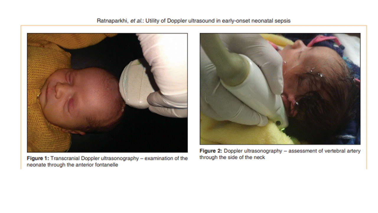
Methods: This cross‑sectional analytical study was conducted over a period of 2 years with 123 neonates enrolled in the study. The neonates were divided into two groups: Group I (with 54 neonates) ‑ neonates with EONS and group II (with 69 neonates) ‑ age‑matched neonates without any signs of sepsis. Ultrasound Doppler examination was performed and the cerebral hemodynamics assessed in neonates during the first seventy two hours of life. Doppler indices and CBFV were measured in the internal carotid artery (ICA), middle cerebral artery (MCA), and vertebral artery (VA) of either side. Data were analyzed using the statistical program SPSS version 23.0 (SPSS Inc., Chicago, IL, USA). Sensitivity, specificity, positive predictive value (PPV), negative predictive value (NPV), and diagnostic accuracy were calculated at different selected cutoff values for CBFV parameters.
Results: Lower resistance and higher peak systolic velocity and end diastolic velocity have been documented in neonates with EONS.
Conclusion: Our study shows that the cerebral hemodynamics in neonates with EONS is altered which can be assessed bedside by noninvasive ultrasound Doppler examination.