Tổng số lượt xem trang

Thứ Năm, 11 tháng 6, 2020

ULTRASOUND and SARCOPENIA


ABSTRACT

Our aim of this study is to compare the thigh muscle thickness measurements obtained using ultrasound and bioelectrical impedance analysis (BIA) methods, and to investigate the validity and cutoff value of the ultrasonography.
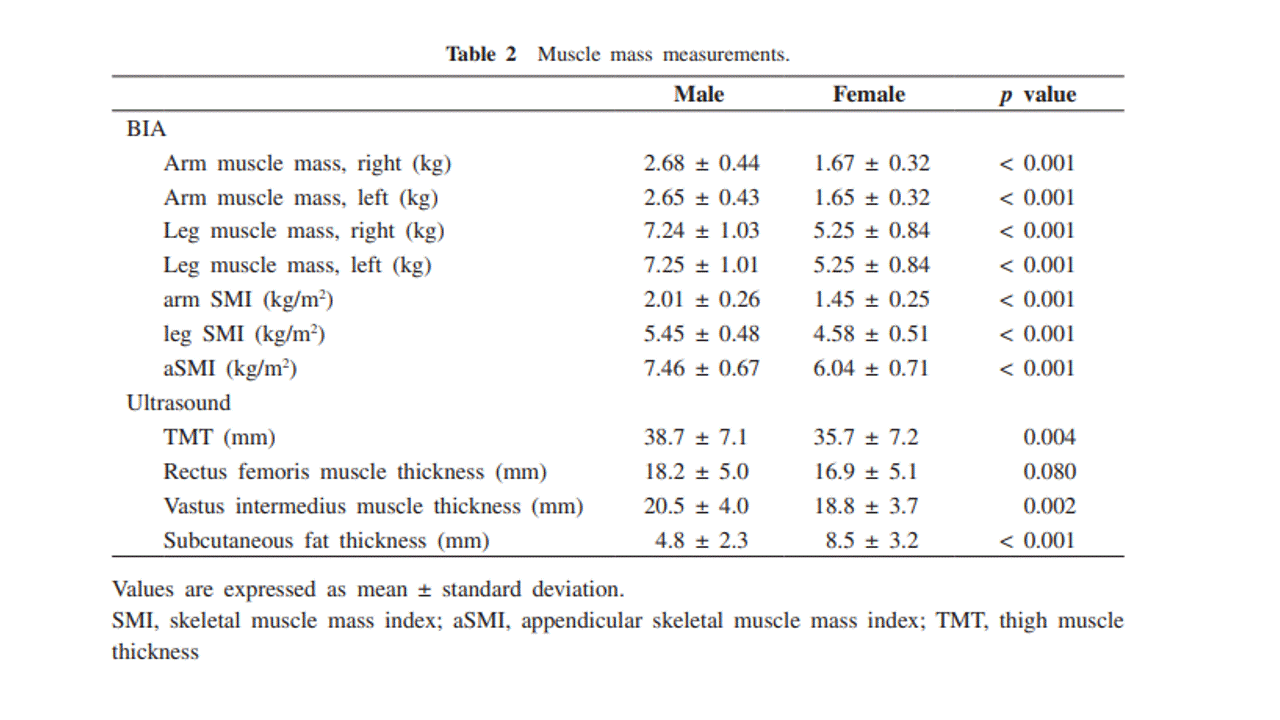
We analyzed a total of 201 participants (99 male and 102 female participants, mean age, 66.2 years) participated in the annual health checkup in the Yakumo Study, 2014. Thigh muscle thickness (TMT, sum of the rectus femoris and vastus intermedius muscle thickness) was measured using ultrasound at mid-thigh in the sitting position. Appendicular skeletal muscle mass (aSMI) was measured using BIA. Cutoff value of TMT was determined through the receiver operating characteristic analysis. We defined sarcopenia with the diagnostic algorithm of Asian Working Group for Sarcopenia.
TMT was significantly reduced in subject with sarcopenia than in those without sarcopenia in both gender. Muscle measurements obtained using the BIA methods (aSMI) and ultrasound methods (TMT) showed a significant correlation, with a correlation coefficient of 0.38 (P < 0.001). Cutoff value, sensitivity, and specificity of TMT in diagnosis of muscle loss were 36 mm, 72.0%, and 73.9%, respectively, for the male participants, and 34 mm, 72.2%, and 72.4%, respectively, for the female participants.
In conclusion, the ultrasonography for thigh muscle might be a simple diagnostic method for sarcopenia.

Keywords: ultrasonography, thigh muscle thickness, sarcopenia, community-dwelling people, cut-off value


Thứ Tư, 10 tháng 6, 2020

C B F in Neonatal Sepsis

 Abstract
Background: Neonatal sepsis is an important cause of morbidity and mortality among newborns. As there is paucity of literature regarding early alteration of the cerebral blood flow (CBF) in neonatal sepsis our study aims to evaluate the changes in the CBF velocities and Doppler indices in neonates with early‑onset neonatal sepsis (EONS) and to evaluate the predictive accuracy of cerebral blood flow velocities (CBFV) by using ultrasound Doppler as a diagnostic marker of EONS.
Methods: This cross‑sectional analytical study was conducted over a period of 2 years with 123 neonates enrolled in the study. The neonates were divided into two groups: Group I (with 54 neonates) ‑ neonates with EONS and group II (with 69 neonates) ‑ age‑matched neonates without any signs of sepsis. Ultrasound Doppler examination was performed and the cerebral hemodynamics assessed in neonates during the first seventy two hours of life. Doppler indices and CBFV were measured in the internal carotid artery (ICA), middle cerebral artery (MCA), and vertebral artery (VA) of either side. Data were analyzed using the statistical program SPSS version 23.0 (SPSS Inc., Chicago, IL, USA). Sensitivity, specificity, positive predictive value (PPV), negative predictive value (NPV), and diagnostic accuracy were calculated at different selected cutoff values for CBFV parameters.
Results: Lower resistance and higher peak systolic velocity and end diastolic velocity have been documented in neonates with EONS.
Conclusion: Our study shows that the cerebral hemodynamics in neonates with EONS is altered which can be assessed bedside by noninvasive ultrasound Doppler examination.