Tổng số lượt xem trang

Thứ Năm, 30 tháng 7, 2020

US correlates with COVID-19 severity, duration


By Theresa Pablos, AuntMinnie staff writer

July 28, 2020 -- Findings on lung ultrasound scans were correlated with COVID-19 severity and duration in a study published on July 23 in the American Journal of Roentgenology. In particular, pulmonary consolidations distinguished between patients with moderate and severe forms of the novel coronavirus disease


The study included dozens of patients who were consecutively treated for COVID-19 at a Chinese hospital in March. The findings may aid physicians in managing patients with moderate-to-severe COVID-19, the authors noted.
"Our results indicate that lung [ultrasound] findings can be used to reflect both the infection duration and disease severity," wrote the authors, led by Dr. Yao Zhang from Ditan Hospital in Beijing.
Zhang and colleagues enrolled 28 patients consecutively hospitalized for COVID-19 at their institution between March 1 and March 30. The patients all tested positive for the novel coronavirus on a nasopharyngeal test and underwent a bedside lung ultrasound scan in a sitting, supine, and decubitus position.
radiology graph
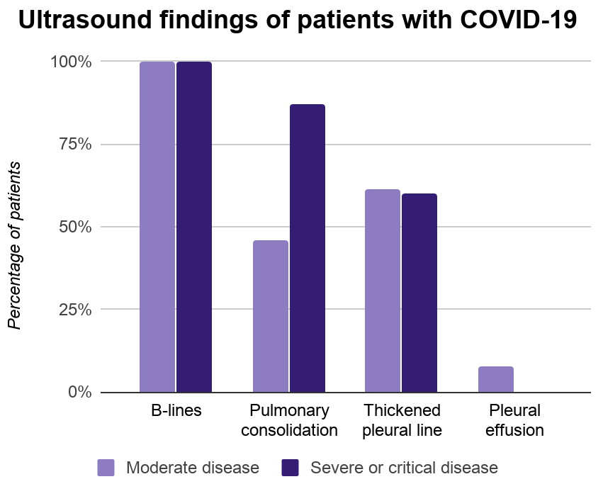
Every patient in the study had B-lines on their ultrasound scans, which indicate areas of increased interstitial fluids and decreased alveolar air. Another two-thirds of patients had pulmonary consolidation, and 61% had a thickened pleural line. Only one patient had a pleural effusion.
Pulmonary consolidation occurred significantly more often in patients with severe or critical COVID-19 than patients with moderate disease, the authors found. Almost 87% of patients with severe or critical disease had pulmonary consolidation on ultrasound, compared with just 46% of patients with moderate COVID-19.
Lung ultrasound image obtained with a convex probe. The outer arrows show confluent B-lines. The middle arrowheads point to a thickened pleural line.
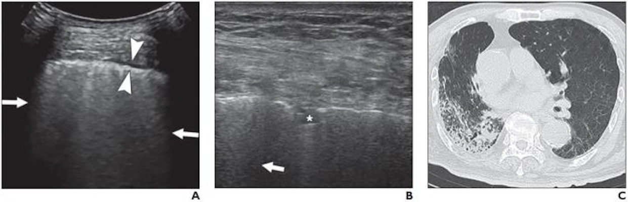
(A) Lung ultrasound image obtained with a convex probe. The outer arrows show confluent B-lines. The middle arrowheads point to a thickened pleural line. (B) Lung ultrasound image obtained with a linear probe. The arrow points to a B-line. The star denotes a patchy pulmonary consolidation. (C) Chest CT image showing reticular and interlobular septal thickening and patchy, focal opacities associated with architectural distortion. Image courtesy of the American Journal of Roentgenology.
Furthermore, patients with a thickened pleural line had experienced a longer infection period than those without a thickened pleural line. Patients with fewer than 20 days between the day they first noticed COVID-19 symptoms and the day of their ultrasound scan were significantly less likely to have a thickened pleural line than those with a difference of 20 days or more.
The study findings add to the growing body of research demonstrating that severity on lung ultrasound scans can predict worse outcomes and even mortality for patients with COVID-19. The authors emphasized that ultrasound also has unique benefits over other imaging modalities, including CT, for treating patients with the novel coronavirus.
"[Ultrasound] is repeatable in critically ill patients, which ensures that monitoring of the severity of the disease and the effects of therapies can be easily carried out," they wrote. "This capability is particularly important in situations in which chest CT is not available, such as in isolation wards and [intensive care units]."
The authors cautioned that their study focused on a small number of patients and did not evaluate how ultrasound findings might have changed over time. They hope future studies will continue to study the use of lung ultrasound for COVID-19, particularly how lung ultrasound scoring systems might improve COVID-19 assessment and treatment.
"Lung [ultrasound] was highly sensitive for detecting abnormalities in patients with COVID-19, and B-lines, a thickened pleural line, and pulmonary consolidation were the most commonly observed features," the authors concluded.

Thứ Sáu, 24 tháng 7, 2020

A New Standardize Doppler waveform reporting


By Theresa Pablos, AuntMinnie staff writerJuly 24, 2020 -- A new set of guidelines aims to standardize the terminology used to report arterial and venous spectral Doppler ultrasound waveforms. The document was jointly published on July 15 in Vascular Medicine and the Journal for Vascular Ultrasound.

The statement creates a designated set of key terms to describe findings on spectral Doppler ultrasound waveforms, the main diagnostic assessment for arterial and venous diseases. It was written by sonographers, vascular specialists, and other experts commissioned by the Society of Vascular Medicine and Society of Vascular Ultrasound.
"The hope of the writing committee is that this document will help us all to 'speak the same language,' and thereby advance the field of vascular ultrasound and improve patient care," stated lead study author Dr. Esther Kim, vascular labs medical director at Vanderbilt University Medical Center, in a press release.
The lack of shared nomenclature has been an ongoing problem for vascular ultrasound professionals. In fact, one out of five ultrasound professionals has had to perform a repeat arterial Doppler ultrasound examination because of terminology differences, according to a survey cited in the consensus statement.
"Over a decade ago, the lack of a standardized nomenclature to describe spectral Doppler waveforms was demonstrated to result in confusion amongst ultrasound professionals," Kim stated. "Not surprisingly, this can lead to negative clinical outcomes."
In the consensus statement, the committee established three major descriptors for ultrasound waveforms: flow direction, phasicity, and resistance for arterial waveforms and flow direction, flow pattern, and spontaneity for venous waveforms.

Major descriptors for arterial ultrasound waveforms
Flow directionAntegrade
  • Blood flows in normal direction
  • Previously known as forward flow
Retrograde
  • Blood flows in opposite direction
  • Previously known as reverse flow
Bidirectional
  • Blood enters and leaves through the same opening
  • Previously known as to-fro
Absent
  • No detected blood flow
PhasicityMultiphasic
  • Waveform crosses zero-flow baseline
  • Previously known as triphasic or biphasic
Monophasic
  • Waveform does not cross zero-flow baseline
  • Blood flows in single direction
ResistanceHigh resistive
  • Sharp upstroke and brisk downstroke
Intermediate resistive
  • Visible end-systolic notch
  • Continuous flow above the zero-flow baseline
Low resistive
  • No end-systolic notch
  • Prolonged downstroke in late systole

Major descriptors for venous ultrasound waveforms
Flow directionAntegrade
  • Blood flows in normal direction
  • Previously known as central or forward flow
Retrograde
  • Blood flows in opposite direction
  • Previously known as peripheral or reverse flow
Absent
  • No detected blood flow
Flow patternRespirophasic
  • Flow velocity related to respiratory cycle
  • Previously known as respiratory phasicity
Decreased
  • Respirophasic flow with less variation than expected
  • Previously known as dampened or blunted
Pulsative
  • Flow velocity is inversely linked to cardiac cycle
  • Previously known as cardiophasic
Continuous
  • Respiratory/cardiac cycles do not affect flow velocity
  • Steady Doppler signal with minimal variation
Regurgitant
  • Flow velocity varies with cardiac cycle
SpontaneitySpontaneous
  • Blood flows without external influence
Nonspontaneous
  • Blood flows only with external maneuvers
The statement also established terms that can be used to modify the main descriptors. For arterial waveforms, the seven modifying terms are as follows:
  1. Rapid upstroke -- Near vertical rise to peak systole
  2. Prolonged upstroke -- Abnormally gradual slope to peak systole; previously known as tardus, delayed, or damped upstroke
  3. Sharp peak -- Single, well-defined peak
  4. Spectral broadening -- Widening of the velocity band or filling in the typically clear area under the systolic peak; previously known as nonlaminar, turbulent, disordered, or chaotic
  5. Staccato -- High-resistance pattern with a short, low-amplitude diastolic signal punctuated by spikes of acceleration and deceleration
  6. Dampened -- Abnormal upstroke and peak, typically with decreased velocity; previously known as parvus et tardus, attenuated, or blunted
  7. Flow reversal -- Flow that changes direction but not as part of normal flow, can be transient or consistent with the cardiac cycle; previously known as pre-steal, competitive flow, or oscillating
For venous waveforms, the three modifying terms as follows:
  1. Augmentation -- Changes in flow velocity related to physical maneuvers, can be described as normal, reduced, or absent augmentation
  2. Reflux -- Persistent retrograde flow beyond normal closure time
  3. Fistula flow -- Flow with an arteriovenous fistula that becomes pulsatile due to communication with artery, sharp peaks often appear as pulsatile; previously known as arterialized or fistulous
In addition to creating the key descriptors and modifiers, the statement defined the reference baseline for spectral Doppler waveforms as the zero-flow baseline. It also advised against using the terms "normal" or "abnormal" to describe a waveform, since what is normal will depend on the part of the body and situation.
The statement also instructed sonographers to use image optimization techniques to acquire quality Doppler waveforms. This includes using an optimal transducer-to-vessel angle, the normal peripheral artery systolic waveform acceleration of 0.2 seconds, and proper transducer support.
Finally, the committee advised sonographers to provide complete descriptions for referring providers, including indication, relevant history, velocity measurements, and waveform characteristics. Sonographers should also include a conclusion with the clinical indication.
"We hope that this new Doppler waveform nomenclature will eliminate confusion and lead to appropriate diagnosis and better patient care," stated Dr. Raghu Kolluri, president of the Society of Vascular Medicine.