Tổng số lượt xem trang

Thứ Tư, 19 tháng 5, 2021

REVERBERATION ARTEFACTS IN LUNG US, WFUMB POSITION PAPER


Abstract
The analysis of vertical reverberation artefacts is an essential component of the differential diagnosis in pulmonary ultrasound. Traditionally, they are often, but not exclusively, called B-line artefacts (BLA) and/or comet tail artefacts (CTA), but this view is misleading.
In this position paper we clarify the terminology and relation of the two lung reverberation artefacts BLA and CTA to specifc clinical scenarios. BLA are defned by a normal pleura line and are a typical hallmark of cardiogenic pulmonary edema after exclusion of certain pathologies including pneumonia or lung contusion, whereas CTAs show an irregular pleura line representing a variety of parenchymal lung diseases. The dual approach using low frequency transducers to determine BLA and high frequency transducer to determine the pleural surface is recommended.

Keywords: lung ultrasound; artefact; B-lines; comet tails; guidelines; misdiagnosis


Suggested approach:

The transducer should be positioned such that the emenating ultrasound beam perpendicularly intersects
the surface of the lung to maximize likelihood of seeing all BLA and CLA as well as A line artifacts (fig 1).
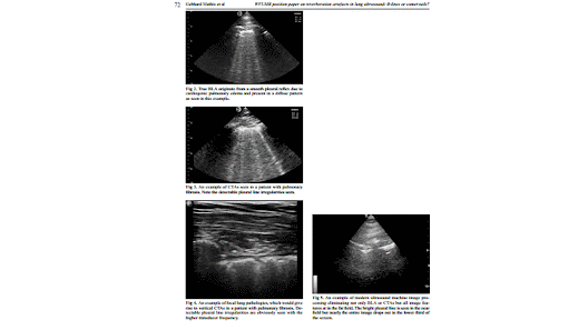
A recent study highlighted the potentially detrimental effects of placing the focal zone below the pleural line,using spatial compounding, higher frequency and tissue harmonics [14]. Once machine settings and transducer orientation have been optimized, we suggest that two most important and distinct vertical lung artefacts should be differentiated: BLA and CTA. While true BLA (fig 2) originate from a smooth pleural reflex due to cardiogenic pulmonary edema and present in a diffuse pattern, CTAs are seen in many lung disorders with irregular and fragmented pleural reflexes and can be focal or diffuse (fig 3).
Hence, the initial step should be to determine if there is evidence for diffuse pulmonary disease or defned focal or localized pathology. Focal lung pathologies by defnition should display vertical artifacts that are consistent with CTAs (fig 4).
Diffusely distributed vertical reverberation artefacts can be divided into two groups: with or without detectable pleural line irregularities and with stable or distally widening width:

1. The reverberation artefact (evaluated by low frequency transducer <5 MHz without interfering presets) is called
BLA if arising from a smooth pleural line (evaluated by high frequency transducer ≥10 MHz). The BLA arises from edema within the interstitium, is well defned with stable width, hyperechoic and extending indefnitely (the entire depth, at least 10 cm), erasing A-lines and moving with lung sliding. It is important to realize that many modern ultrasound machines have post-processing and other features which will eliminate not only BLA but essentially all discernable image detail near the bottom of the screen at greater depths (fig 5).
2. The reverberation artefact is called CTA if arising from an irregular (or fragmented) pleural line (evaluated by high frequency transducer ≥10 MHz), changes in width (such as e a comet with narrow head and wide tail), is well defned, hyperechoic, and extending defnitely (<10 cm in depth) (evaluated by low frequency transducer <5 MHz without interfering presets). It is important to make sure image compounding is turned off to make sure the CTA is not distorted farther field [14].
The differentiation of BLA from CTA is also dependent on the technical adjustments of several external factors, including the type of ultrasound machine, transducers and probe frequencies [6].


In conclusion, the correct diagnosis of pulmonary edema (the etiology of which may be decided upon
through integration of ultrasound data with clinical presentation) in the emergency setting is crucial for the correct management of the patient. The differentiation between ultrasonographic BLA and CTA, using two types (high and low frequency) of transducers allows accurate differentiation between pulmonary edema and other cause of diffuse pulmonary pathology. Both can lead to acute respiratory failure but may require different clinical management. Localized pulmonary diseases representing with CTA are distinguished. Mixed forms of diffuse,but also diffuse and focal lung diseases have to be considered.


 

Thứ Hai, 10 tháng 5, 2021

US accurate in diagnosing hand injuries

By Amerigo Allegretto, AuntMinnie.com staff writer


May 10, 2021 -- Ultrasound can accurately diagnose hand injuries while also being a fast, inexpensive, and potentially indispensable dynamic tool, according to research published April 29 in Ultrasound in Medicine and Biology.

Examining hand tendon injuries with sonography showed 100% accuracy, sensitivity, and specificity for diagnosing full-thickness hand tendon tears, as well as tenosynovitis of hand flexor tendons, according to a study led by Dr. Chris Nabil Hanna Bekhet from Ain Shams University in Cairo, Egypt.

"It also provides data that are important before diagnostic surgical exploration, and the process consumes less time than traditional wound exploration techniques or MRI," the authors wrote.

Hand and wrist injuries make up 28% of all musculoskeletal injuries and account for 14% to 30% of all patients treated in the emergency department. Tendon injuries are the second most common injuries, within injuries to the flexor tendons having debilitating consequences and high rates of reoperation.

Assessing hand injuries through clinical examination can overlook tendon injuries, and surgeons sometimes opt for explorative surgical methods to detect tendon injuries.

While using ultrasound to examine tendon injuries in the emergency department has its advantages, including eliminating the need for surgical approaches, it is not yet readily adopted by surgeons. This could be because clinicians lack education on how to use ultrasound to resolve clinical questions on the state of the tendon.

The study included 35 patients between September 2018 and January 2020 with trauma to the ventral surface of the hand and wrist who were presented to emergency departments or outpatient clinics. The subjects ranged from 18 to 58 years of age, with 24 patients being male and the other 11 being female.

The researchers examined 50 injured tendons in all flexor hand zones.

On ultrasound examination, 21 of the 50 injured tendons were reported to have complete tears, and 10 tendons were partially torn. The most common cause of injury was cut wounds by sharp objects (20 cases), with injury by a knife as the highest incidence.

In all, ultrasound was found to be statistically significant (p < 0.01) in predicting the surgical findings by correctly identifying the 21 fully lacerated tendons. It was also found to be statistically significant (p < 0.01) in predicting the surgical findings by correctly identifying partially torn tendons and determining the degree of the torn fibers.

The study's limitations included tests being performed by a single operating radiologist and the small sample size.

"More studies in this respect can popularize the technique among radiologists and clinicians," the authors wrote. "Our study also helps anchor the notion that musculoskeletal [ultrasound] could be widely employed for soft tissue structures, with their well-recognized advantages compared with other imaging techniques."