Tổng số lượt xem trang

Thứ Tư, 17 tháng 4, 2019

Elastography can help characterize breast tumors.


By Kate Madden Yee, AuntMinnie.com staff writer
April 16, 2019 -- Breast elastography is an overlooked tool for evaluating breast cancer, especially since it shows promise for helping radiologists distinguish between benign and malignant lesions, according to a presentation delivered at the American Institute for Ultrasound in Medicine (AIUM) meeting in Orlando, FL.
Since ultrasound is convenient, easily accessible, and less expensive than some other modalities, it's a helpful tool in the breast cancer detection arsenal. And since it can help characterize breast lesions, breast elastography could have downstream effects on patient care, according to presenter Dr. Stamatia Destounis of Elizabeth Wende Breast Care in Rochester, NY.
"Identifying the relative tissue stiffness can help clinicians distinguish between benign and malignant lesions, which in turn has the potential to reduce unnecessary biopsies," she said.
Although breast elastography has been available for more than 15 years, it's not widely implemented, in part because the various methods lack consistency -- particularly in color scale interpretation, Destounis noted.
"As research continues and elastography is used in clinical practice, there's a need for standardization of the color scale," she said.
Elastography can be performed in static and dynamic modes, and consensus about which mode is better is generally lacking. Static elastography includes strain imaging, while dynamic elastography includes shear wave. Strain elastography is the most widely used technique, estimating the relative stiffness of a particular area compared with other tissue.
In strain elastography, tissue stiffness data are displayed in a color map that is superimposed on a real-time grayscale image. Cancers tend to appear larger on strain elastography than on B-mode ultrasound, and benign lesions tend to appear smaller; this size change between the modes has shown to be highly sensitive and specific for characterizing breast lesions, Destounis said.
Studies have demonstrated that strain elastography is effective for detecting breast cancer, but it does have its drawbacks.
"It can be difficult to measure the amount of force during compression, and absolute elasticity can't be calculated," she said.
As for shear-wave elastography, it offers real-time, quantitative assessment of tissue stiffness; rather than relying on external compression, it uses short acoustic pulses to identify stiffness. To characterize breast lesions, clinicians should focus on the area of highest stiffness in the lesion. But be careful, Destounis cautioned.
"Some breast cancers don't allow for adequate shear-wave generation and may appear black -- that is, no shear-wave speed calculated or with a low shear-wave speed due to noise," she explained.
So which technique should clinicians use? Destounis cited research from Chang et al that found mixed results (American Journal of Roentgenology, August 2013, Vol. 201:2, pp. W347-356). The study compared strain and shear-wave ultrasound elastography for differentiating benign from malignant breast lesions, and it showed that strain elastography is more specific than shear-wave elastography (93.7% compared with 84.8%) but less sensitive (81.7% compared with 95.8%).
It may be a matter of improving elastography technology, Destounis told AuntMinnie.com via email.
"Several vendors have different types of elastography technology, and it needs more consistency in technique and color representation," she stated.

Chủ Nhật, 14 tháng 4, 2019

ML for US


https://www.ncbi.nlm.nih.gov/pmc/articles/PMC5886811/

Abstract

Ultrasound (US) imaging is the most commonly performed cross-sectional diagnostic imaging modality in the practice of medicine. It is low-cost, non-ionizing, portable, and capable of real-time image acquisition and display. US is a rapidly evolving technology with significant challenges and opportunities. Challenges include high inter- and intra-operator variability and limited image quality control. Tremendous opportunities have arisen in the last decade as a result of exponential growth in available computational power coupled with progressive miniaturization of US devices. As US devices become smaller, enhanced computational capability can contribute significantly to decreasing variability through advanced image processing. In this paper, we review leading machine learning (ML) approaches and research directions in US, with an emphasis on recent ML advances.


We also present our outlook on future opportunities for ML
techniques to further improve clinical workflow and US-based disease diagnosis and characterization.

Keywords: Deep Learning, Elastography, Machine Learning, Medical Ultrasound, Sonography

Thứ Ba, 9 tháng 4, 2019

QUANTITATIVE ULTRASOUND [Q U S], EMERGING MODE for CLINICAL ULTRASOUND

By Kate Madden Yee, AuntMinnie.com staff writer
April 8, 2019 -- ORLANDO, FL - Quantitative ultrasound (QUS) shows promise as an emerging mode for clinical ultrasound, offering more specific data compared with conventional ultrasound exams, according to a lecture delivered Monday at the American Institute of Ultrasound in Medicine (AIUM) meeting.

Many conventional ultrasound studies already offer quantitative information, including distance, area, and volume measurements; Doppler-generated velocities and volume flow estimates; cardiac wall motion, strain, and ejection fraction data; and contrast and tissue stiffness analysis. But conventional ultrasound is subject to operator variability, said James Zagzebski, PhD, of the University of Wisconsin in Madison.
"QUS uses bulk acoustic properties and tissue microstructure features to increase ultrasound's sensitivity and specificity," he said during the AIUM's William J. Fry Memorial Lecture. "I believe that the current computational resources on ultrasound systems can be further exploited to provide us even more data than we already have."
QUS technology has continued to advance, replacing conventional ultrasound's beamformer technology with high-capacity computational hardware and software, and synthesized virtual beams, Zagzebski said. Compared with conventional ultrasound, QUS imaging offers more data related to tissue features -- such as attenuation and backscatter coefficients -- that increase the specificity of image findings and can lead to improvements in diagnostic ultrasound. QUS techniques include spectral-based parameterization, elastography, shear-wave imaging, and flow estimation.
"System independent backscatter coefficient and attenuation coefficient estimates can be made accurately using clinical scanners," he told session attendees. "There's evidence that QUS can provide valuable information to assess diffuse liver disease, characterize breast masses, and assess the effects of anesthesia."
QUS may be particularly helpful for breast imaging, according to Zagzebski.
"In vitro studies showed that QUS parameters, attenuation coefficient, and backscatter coefficient can give useful insight into the nature of breast tissue," he said. "And animal studies have shown that the use of QUS parameters such as effective scatter [can] differentiate benign fibroadenomas from malignant breast masses."
Other potential applications for QUS include evaluating changes in the cervix accompanying ripening and breast tumor response to treatment, tracking lymph node involvement in disease, monitoring radiofrequency and microwave ablation, and diagnosing eye and orbital disease, Zagzebski noted.
In any case, as the use of QUS continues, there's a need to set best practices, such as those established through the Quantitative Imaging Biomarkers Alliance (QIBA), he said.

"We need to establish good protocols for QUS, like those developed through the QIBA effort, so that everybody will be operating under the same standards," he concluded.

QUANTITATIVE ULTRASOUND IMAGING

https://www.aapm.org/meetings/amos2/pdf/59-17304-58800-911.pdf


Thứ Sáu, 5 tháng 4, 2019

Noninvasive Evaluation of NAFLD: CURRENT EVIDENCE and PRACTICE


FATTY LIVER - CHRONIC B HEPATITIS and H C C


Researchers test new ultrasound method for heart disease


April 5, 2019 -- Researchers from the University of Arkansas in Fayetteville have published results from a study testing a new ultrasound imaging method for the detection and diagnosis of congenital heart disease in infants and children.
The new technology, called vector flow, creates images of the internal structure and blood flow of children's hearts. It was used for the first time in humans at the Arkansas Children's Hospital in Little Rock, according to a team led by Dr. Morten Jensen, PhD. The study was published March 5 in Progress in Pediatric Cardiology.
About 1% of babies are born with congenital heart defects. Pediatric cardiologists identify congenital heart disease using echocardiography and other processes based on ultrasound, the researchers wrote. Although effective, ultrasound can't accurately obtain details about blood flow within the heart.
The team used an ultrasound scanner with vector flow imaging to image the hearts of two three-month-old babies, one with a healthy heart and one with congenital heart disease. The technology allowed for complete transthoracic imaging of tissue and blood flow at a depth of 6.5 cm; abnormal flow and cardiac anomalies were clearly visualized in the child with congenital heart disease.
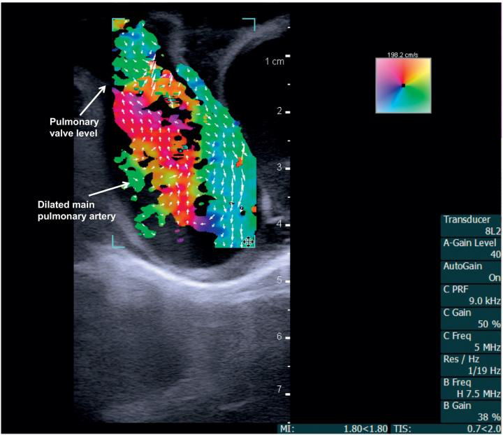
Vector flow imaging demonstrates swirl of blood flow within the dilated main pulmonary artery of a pig.
Vector flow imaging demonstrates swirl of blood flow within the dilated main pulmonary artery of a pig. Image courtesy of Dr. Morten Jensen, PhD.
"Vector flow imaging technology is not yet possible in adults, but we have demonstrated that it is feasible in pediatric patients," Jensen said in a statement released by the university April 3. "Our group demonstrated that this commercially available technology can be used as a bedside imaging method, providing advanced detail of blood flow patterns within cardiac chambers, across valves, and in the great arteries."

Thứ Sáu, 22 tháng 3, 2019

ABUS better than DBT for diagnostic workup.


By Kate Madden Yee, AuntMinnie.com staff writer
March 5, 2019 -- Automated breast ultrasound (ABUS) is more accurate than digital breast tomosynthesis (DBT) as a diagnostic tool for working up positive findings on mammography screening in dense breast tissue, according to a presentation delivered at ECR 2019.
ABUS does have limitations, notably microcalcifications and imaging the retroareolar area of the breast, said presenter Dr. Norran Hussein Said of Cairo University. But its benefits may outweigh them.
"ABUS has the capability of 3D assessment [of dense breasts], and can differentiate solid from cystic lesions," Said told session attendees.
The group used data from the Egypt Breast Cancer National Screening Program to compare the performance of ABUS to tomosynthesis in the workup of 242 women with dense breasts recalled after positive screening mammography. Positive findings included focal asymmetries, masses, distortions, or microcalcifications.
All of the women who participated in the study underwent both tomosynthesis and ABUS. The researchers compared exam results to pathology data.
Tomosynthesis vs. ABUS in women with dense breast tissue
MeasureTomosynthesisABUS
Sensitivity92%92%
Specificity92%98%
Positive predictive value76%92%
Negative predictive value98%98%
Accuracy92%97%
"Our results show that most of our recalled cases consisted of masses, followed by asymmetries," Said told session attendees.
The two modalities agreed on true positives in 43 out of 51 proven cancers, and each modality had four false negatives. ABUS had four false positives, while tomosynthesis had 15. ABUS helped avoid 187 biopsies, while tomosynthesis helped avoid 176.
"In our study, ABUS showed a higher accuracy than DBT," Said concluded.
Works with 2D, too
In a related study, a Romanian team found that, when added to full-field digital mammography (FFDM), ABUS was an effective adjunct tool for finding cancer in women with dense tissue, specifically improving sensitivity and negative predictive value compared with FFDM alone.
Dr. Ioana Boca of Emergency County Hospital in Cluj-Napoca compared the performance of FFDM, 3D mammography, and ABUS for breast cancer detection in women with dense tissue. Two radiologists interpreted 127 ABUS exams procured after FFDM or 3D mammography. Their results were compared to histopathology for biopsied lesions, handheld ultrasound for benign lesions, and follow-up for benign lesions unchanged for at least two years. Nineteen cancers were identified.
ABUS boosted FFDM's sensitivity and negative predictive value, although it did not outperform 3D mammography in this screening context, Boca noted.
FFDM, FFDM + ABUS, and 3D mammography in dense tissue
FFDM2D mammography + ABUS3D mammography
Sensitivity77.8%80%100%
Specificity100%89.2%86.4%
Positive predictive value100%71.4%74.2%
Negative predictive value92%93%100%

"In screening, ABUS added to FFDM compared with FFDM alone improved readers' detection of breast cancers in women with dense breast tissue," Boca concluded.

Thứ Tư, 20 tháng 3, 2019

Ultrasound can regulate inflammatory response.


By AuntMinnie.com staff writers
March 12, 2019 -- Bioelectronic research teams from GE Research in Niskayuna, NY, and the Feinstein Institute for Medical Research in Manhasset, NY, have demonstrated noninvasive methods to regulate dysfunction in the body's metabolic or inflammatory control systems using ultrasound, according to a study published online March 12 in Nature Communications.
Bioelectronic medicine is a combination of neuroscience, molecular biology, and bioengineering that researchers hope will be able to treat disease and injury in the nervous system without pharmaceuticals, the institute said. A team led by study co-author Chris Puleo, PhD, of GE Research, found that using ultrasound in targeted ways reduced inflammatory markers and altered metabolism.
"In our studies, we show that applying ultrasound to a specific target in the spleen altered inflammatory markers that can cause arthritis, inflammatory bowel disease, and other ailments," Puleo said in a statement released by the institute. "And when targeting a specific part of the liver, we were able to modulate blood glucose levels."
The researchers plan to conduct more preclinical studies to understand the use of ultrasound in this way, they said.

Thứ Năm, 14 tháng 3, 2019

SoS for Breast Cancer


Speed of sound ultrasound (SoS)

Abstract

Objectives

To measure speed of sound (SoS) with a novel hand-held ultrasound technique as a quantitative indicator for muscle loss and fatty muscular degeneration.

Methods

Both calf muscles of 11 healthy, young females (mean age 29 years), and 10 elderly females (mean age 82 years) were prospectively examined with a standard ultrasound machine. A flat Plexiglas® reflector, on the opposite side of the probe with the calf in between, was used as timing reference for SoS (m/s) and ΔSoS (variation of SoS, m/s). Handgrip strength (kPA), Tegner activity scores, and 5-point comfort score (1 = comfortable to 5 = never again) were also assessed. Ultrasound parameters (muscle/adipose thickness, echo intensity) were measured for comparison.

Results

Both calves were assessed in less than two minutes. All measurements were successful. The elderly females showed significantly lower SoS (1516 m/s, SD17) compared to the young adults (1545 m/s, SD10; p < 0.01). The ΔSoS of elderly females was significantly higher (12.2 m/s, SD3.6) than for young females (6.4 m/s, SD1.5; p < 0.01). Significant correlations of SoS with hand grip strength (r = 0.644) and Tegner activity score (rs = 0.709) were found, of similar magnitude as the correlation of hand grip strength with Tegner activity score (rs = 0.794). The average comfort score of the elderly was 1.1 and for the young adults 1.4. SoS senior/young classification (AUC = 0.936) was superior to conventional US parameters.

Conclusions

There were significant differences of SoS and ΔSoS between young and elderly females. Measurements were fast and well tolerated. The novel technique shows potential for sarcopenia quantification using a standard ultrasound machine.

Key Points

• Speed of sound ultrasound: a novel technique to identify sarcopenia in seniors.
• Measurements were fast and well tolerated using a standard ultrasound machine.
• The novel technique shows potential for sarcopenia quantification.

Anatomy Lab Trends for 2019


And Now, a Word from Our Instructors: Anatomy Lab Trends for 2019

The results are in! We reached out to anatomy lab instructors across the U.S. and inquired about the tools they use and the challenges they face. We asked about current and future lab setups, trends in educational technology, and how (or if!) the money available and student enrollments were growing or shrinking.
Want to hear what more than 100 instructors of undergraduate and graduate anatomy labs have to say? Keep reading for a summary. You can also download the data to share with colleagues!

1. The State of Anatomy Lab Courses

Student engagement and procuring resources were the two biggest challenges faced by instructors.
Students are not as prepared; lack of critical thinking skills and integrating what they are learning in lab to lecture; distracted by cell phones; would rather take pictures of slides and models and then leave.” — 2-year college anatomy professor
  • 77% of instructors noted student engagement was a problem, with issues ranging from poor study skills to a lack of preparedness for class. 
  • 71% cited the cost of lab materials as a challenge. Respondents reported that over the last three years, they’ve seen a decrease in the availability and quality of biological specimens. Fewer labs are using human cadavers because they are expensive and difficult to maintain, and animal specimens are becoming harder to acquire as well. 
  • Some said that increasing enrollment means they need to find resources that can serve a larger number of students in an affordable way. 
  • 16% of instructors reportedbudget decreases, most of which were due to funding cuts at the university or state level.
The dissection of biological specimens remains a central component of anatomy lab courses.
  • 50% reported that students worked with prosections of human or animal organs.
  • 37% conducted complete animal dissections.
  • 28% of labs dissected human cadavers.
A 2-year college anatomy instructor reports "increased safety concerns when using cadavers for students and faculty. Movement away from dissections due to cost, upkeep and time needed for dissection.”


Synthetic models are also a staple in anatomy labs.
  • Plastic and synthetic models are found in 56% of labs.
Tablets (such as iPads) are the most popular anatomy lab technology.
  • Almost all instructors mentioned traditional classroom technology like monitors, screens, and projectors.
  • By far the most popular newer technological tool in the lab was a tablet, like the iPad. 42% of anatomy instructors mentioned these are used in their labs.

2. The Future of Anatomy Lab Courses

62% of instructors surveyed were at a school with an online anatomy course or a school that will soon offer such a course.

Anatomy lab courses remain in high demand!
  • 56% of instructors reported thatenrollments are steady and 38% reported that they are trending up.
  • 55% noted that limits on lab space posed a challenge.
  • 42% of instructors are already at a school that offers online anatomy courses, and another 20% see such courses on the horizon.
The use of virtual models in anatomy labs is growing.
A medical school lab instructor predicts they will see “more students in the lab and their increased reliance on anatomy sources from the internet as compared to text usage.”

  •  39% of instructors reported that they used digital/virtual models.
  • Many such models are available on tablets. In addition to the 42% who already used tablets, 17% of instructors said they would be adding them to their labs within the next three years.
  • 11% currently utilize technologies like Anatomage tables, Sectra, or Virtual Reality headsets. 7% said they would soon be getting access to Sectra/Anatomage technologies, and 9% would be getting VR headsets

Thứ Ba, 12 tháng 3, 2019

Video from ECR 2019: Erik Ranschaert on AI training


March 5, 2019 -- VIENNA - There's an urgent need for radiologists to become better informed about artificial intelligence (AI), according to Dr. Erik Ranschaert, PhD. He talks about his new book on AI and medical imaging and why it represents excellent value for the money, and he also outlines some future initiatives being organized by the European Society of Medical Imaging Informatics (EuSoMII), of which he is president.

Illumeo with adaptive intelligence, is reshaping imaging
This intelligent solution acts as an assistant, providing technologies and tools to enhance the radiologist’s expertise. Improving a radiologist’s interaction with images and enabling a rich and dynamic output for referring physicians can increase the value of the radiologist to the clinical care team.
Dr. Erik Ranschaert, PhD, from Tilburg, the Netherlands

Thứ Tư, 6 tháng 3, 2019

The Earliest Timing Ultrasound can be Performed in the Screening for Developmental Dysplasia of the Hips (DDH)


Si Heng Sharon Tan, Keng Lin Wong, Andrew Kean Seng Lim and James HoiPo Hui Department of Orthopaedic Surgery, University Orthopaedic, Hand and Reconstructive Microsurgery Cluster, National University Health System (NUHS), Singapore (Full postal address: 1E Kent Ridge Road, NUHS Tower Block, Level 11, Department of Orthopaedic Surgery, Singapore 119074)

 Abstract
  Purpose
 The current study aims to evaluate the results of ultrasound screening done at various weeks of life, to determine the earliest timing ultrasound screening can be performed reliably.
 Methods
 In the 17-year cohort study, all neonates who underwent ultrasound screening prior to the twelfth week of life  with subsequent follow-up radiograph done at one year of life were included. The ultrasound images were  evaluated according to Graf classification, Harcke's dynamic ultrasound screening and Terjesen's femoral head  coverage. The radiographic images were evaluated according to acetabular index and femoral head position. The  accuracy and correlation between the ultrasound findings performed at the various weeks of life with the  radiographic findings performed at one year of life were evaluated.
Results
 A total of 348 neonates were included. When the ultrasound findings were correlated with the radiographic  findings at one year of life, significant differences were identified for ultrasound screening performed prior to the fourth week of life (day 21 and before) (p < 0.05). In contrast, there were no significant differences  identified when the ultrasound was performed between the fourth and twelfth week of life (day 22 and beyond)  (p > 0.05). The accuracy of ultrasound screening was 79.2% and above when performed during or after the  fourth week of life (day 22 and beyond).
Conclusion
The earliest timing that ultrasound screening for DDH can be performed reliably is during the fourth week of life (day 22 and beyond).