Tổng số lượt xem trang

Thứ Hai, 29 tháng 7, 2019

SWE identifies malignant breast microcalcifications.


By Kate Madden Yee, AuntMinnie.com staff writer
July 29, 2019 -- Ultrasound with a shear-wave elastography (SWE) technique can distinguish benign from malignant breast microcalcifications detected on ultrasound, according to a new study published in the August issue of the American Journal of Roentgenology.
The study findings could help physicians better plan a response to microcalcifications, wrote a team led by Dr. Foucauld Chamming's of Institut Bergonié, Bordeaux Cedex, France.
"On SWE, microcalcifications associated with malignant lesions showed significantly higher stiffness values than benign ones," the group wrote. "In the clinical setting, these results are of particular interest for assessment of concordance between radiologic and pathologic findings."
Digital mammography has long been the gold standard for identifying microcalcifications, but its specificity for characterizing them is low -- ranging from 10% to 67% -- leading to a high number of benign biopsies, the authors noted. Suspicious microcalcifications are often worked up with ultrasound to find signs of invasive cancer and any additional lesions, but conventional ultrasound has its limitations, Chamming's and colleagues wrote (AJR, August 2019, Vol. 213:2, pp. W85-W92).
"Although malignant microcalcifications are more likely to be seen on ultrasound than benign ones, B-mode ultrasound does not provide relevant additional information for the characterization of isolated microcalcifications," they wrote. "Shear-wave elastography, which is able to measure tissue stiffness quantitatively, is a relatively recent ultrasound technique and has been shown to improve characterization of breast lesions, especially masses."
Chamming's and colleagues investigated whether SWE could distinguish benign from malignant microcalcifications of the breast by conducting a study that included 74 patients with mammographically detected suspicious microcalcifications who underwent breast ultrasound between February and June 2016. All of the patients also had an SWE exam; those with malignant microcalcifications were biopsied using ultrasound guidance.
The researchers compared qualitative and quantitative elastography results between benign and malignant calcifications, as well as between pure ductal carcinoma in situ (DCIS) and invasive lesions. They used area under the receiver operating characteristic curves (AUC) to evaluate SWE's performance in detecting malignancy and invasive features of breast lesions.
Ultrasound identified 29 groups of microcalcifications in 29 patients. Pathology results showed 16 benign and 13 malignant groups of microcalcifications.
The authors found that the stiffness of malignant calcifications was higher than benign ones (p = 0.0004). SWE had 100% specificity and positive predictive value for both detecting malignant calcifications and identifying invasive features of breast lesions.
SWE's performance for breast lesions
Performance measureDiagnosis of malignancyDetection of invasive features
AUC0.890.93
Sensitivity69%75%
Specificity100%100%
Negative predictive value80%75%
Positive predictive value100%100%
Accuracy86%85%
The study results could help physicians better manage microcalcifications, according to the authors.
"When faced with stiff calcifications, which have a high probability of being malignant, a biopsy yielding benign results should be questioned, and a second biopsy or surgical excision should be considered," the group concluded.

Thứ Ba, 23 tháng 7, 2019

LUS, HOW to AVOID MISTAKES.

How to avoid mistakes 
in Lung Ultrasound

This commentary provides an excellent review of the misconceptions and problems concerning the application of lung ultrasonography in critically ill patients. Lung ultrasound can have a significant clinical impact in acute care, if used in the appropriate way. (Chest)


Chủ Nhật, 21 tháng 7, 2019

ABVS distinguishes malignant from benign breast lesions.


By Kate Madden Yee, AuntMinnie.com staff writer
July 15, 2019 -- The automated breast volume scanner (ABVS) performs just as well as handheld ultrasound for distinguishing between malignant and benign breast lesions, according to a literature review published in the August issue of Ultrasound in Medicine and Biology.
The findings suggest that clinicians can trust ABVS as an alternative to handheld ultrasound -- which can be affected by user skill variability -- and that it shows promise as a breast imaging technique for both screening and diagnosis, wrote co-authors Dr. Liang Wang and Zhen-Hong Qi of Peking Union Medical College Hospital in Beijing.
"Conventional handheld ultrasound has several inherent limitations, including the operator dependence and the time required for whole-breast examination," they wrote (Ultrasound Med Biol, August 2019, Vol. 45:8, pp. 1874-1881). "In contrast, the automated breast volume scanner has several advantages over handheld ultrasound, such as higher reproducibility, less operator dependence, and less time required for image acquisition."
Most radiologists continue to use handheld ultrasound to characterize breast lesions identified on mammography or other screening modalities, and the diagnostic performance of ABVS is controversial, Wang and Qi noted. To better assess this, the two conducted a literature review of PubMed, Embase, and Cochrane Library databases.
Their research included nine studies with 1,985 lesions from 1,774 patients; of the lesions, 628 were malignant and 1,357 were benign. Wang and Qi evaluated the following performance measures for the two techniques: sensitivity, specificity, positive and negative likelihood ratios, and diagnostic odds ratios.
Across all measures, ABVS performed comparably to handheld ultrasound for distinguishing benign from malignant breast lesions, the researchers found.
Comparison of handheld ultrasound and ABVS for distinguishing benign from malignant breast lesions
Performance measureHandheld ultrasoundABVS
Sensitivity90.6%90.8%
Specificity81%82.2%
Positive likelihood ratio5.225.39
Negative likelihood ratio0.110.10
Diagnostic odds ratio52.6061.68
Area under the receiver operating characteristic curve0.940.93
"On the basis of our data, the ABVS and handheld ultrasound exhibited similar sensitivity and specificity in the differentiation of malignant and benign breast lesions," Wang and Qi wrote.
They cautioned that these results were based on grayscale ultrasound interpretation, meaning that only morphological features were displayed, rather than functional information that elastography and Doppler ultrasound offer.
"Both elastography and Doppler ultrasound can provide independent diagnostic information in addition to the grayscale imaging, which could significantly improve the diagnostic accuracy for breast ultrasound," they wrote.
But ABVS does have an advantage over handheld ultrasound that could make it a valuable tool in clinical practice: its ability to gather additional morphological information on the reconstructed coronal plane.
"The retraction phenomenon on the coronal plane of the ABVS is regarded as a highly diagnostic feature in the differentiation of benign and malignant breast lesions," the researchers wrote. "Therefore, it seems reasonable that the ABVS may be superior to handheld ultrasound in differential diagnosis with the help of coronal reconstruction."

Thứ Năm, 4 tháng 7, 2019

US and Astronaut Spinal Anatomy and Disorders.


Abstract


Objectives

Back pain is one of the most common conditions of astronauts during spaceflight and is hypothesized to be attributed to pathologic anatomic changes. Ultrasound (US) represents the only available imaging modality on the International Space Station, but a formal US protocol for imaging the structures of the spinal column does not exist. This investigation developed a method of acquiring diagnostic‐quality images of the anterior lumbar and cervical regions of the spine during long‐duration spaceflight.

Methods

Comprehensive spinal US examinations were conducted on 7 long‐duration spaceflight astronauts before flight, in flight, and after flight and compared to preflight and postflight magnetic resonance imaging data. In‐flight scans were conducted after just‐in‐time training assisted by remote expert tele‐US guidance.


Results

Novice users were able to obtain diagnostic‐quality spinal images with a 92.5% success rate. Thirty‐three anomalous or pathologic findings were identified during the preflight US analysis, and at least 14 new findings or progressions were identified during the postflight US analysis. Common findings included disk desiccation, osteophytes, and qualitative changes in the intervertebral disk height and angle.

Conclusions

Ultrasound has proven efficacy as a portable and versatile diagnostic imaging modality under austere conditions. We demonstrated a potential role for US to evaluate spinal integrity and alterations in the extreme environment of space on the International Space Station. Further investigations should be performed to corroborate this imaging technique and to create a larger database related to in‐flight spinal conditions during long‐duration spaceflight.

Thứ Sáu, 28 tháng 6, 2019

Using LUNG US to diagnose Pediatric Pneumonia.


By Kate Madden Yee, AuntMinnie.com staff writer
June 28, 2019 -- Lung ultrasound is a viable alternative to chest x-ray and CT for diagnosing pneumonia in children -- with a sensitivity of 94% and a specificity of 92% -- but it is dependent on operator experience, according to a study published online June 18 in Academic Emergency Medicine.
Although using ultrasound to diagnose pneumonia in children appears to be effective, sonographer experience must be taken into account, wrote a team led by Dr. Po-Yang Tsou of Driscoll Children's Hospital in Corpus Christi, TX.
"To our knowledge, this is the first article demonstrating significant differences in lung ultrasound's diagnostic accuracy for pneumonia between novice and advanced sonographers," Tsou and colleagues wrote. "[Our] findings suggest that the sonographer's experience level should be considered when using lung ultrasound to diagnose pneumonia in children."
Difficult diagnosis
Pneumonia is a leading cause of death in children around the world, the authors noted. In developed countries, the annual incidence rate is 33 per 10,000 children 0 to 5 years of age, with a mortality rate of less than one per 1,000 children. But in developing countries, the condition is more serious with an annual incidence rate of 2,900 per 10,000 children age 0 to 5 and a mortality rate of 26 per 1,000 children.
To make matters worse, diagnosis of pediatric pneumonia can be challenging, in part due to the variation in signs and symptoms. The illness has typically been identified with chest x-ray and clinical presentation. Chest CT is also used and has excellent diagnostic accuracy, but its drawbacks include radiation exposure, high cost, and the possible need for sedation of pediatric patients.
Past studies have suggested that lung ultrasound may be a viable alternative to these modalities for pediatric pneumonia, but it has been unclear whether operator dependence affects its accuracy.
"Studies measuring the diagnostic accuracy of lung ultrasound for childhood pneumonia generally report excellent sensitivity and specificity," the group wrote. "However, ultrasound's accuracy varies by user skill and training."
The researchers conducted a review study to assess the diagnostic accuracy of the modality and compared performance between novice and experienced sonographers. The team searched PubMed and EMBASE from their inception to February 2018 for studies that evaluated the utility of lung ultrasound in children with suspected pneumonia against the reference standard of either imaging results alone or a combination of clinical, laboratory, and imaging results. The group also assessed diagnostic accuracy between novice (seven days or fewer of training) and advanced sonographers.
The analysis included 25 studies with a total of 3,389 patients presenting with pneumonia symptoms. Of these 25, 18 were prospective cohort studies, five were retrospective cohort studies, one was a randomized controlled trial, and one case was a control study. Subjects were mostly infants, children, and adolescents. Of the studies included in the research, 16 used advanced sonographers, seven used novice sonographers, and two used sonographers of unknown training level in lung ultrasound.
Lung ultrasound showed an overall sensitivity of 94%, specificity of 92%, and an area under the curve (AUC) of 0.97. However, the group did find a significant difference in diagnostic accuracy for pneumonia between less experienced and more experienced sonographers, with novice sonographers showing less sensitivity than their advanced counterparts.
Tsou and colleagues also found that point-of-care lung ultrasound performed better than exams conducted in the radiology department.
Comparison of sonographer experience and location for lung ultrasound
Performance measureNovice sonographersAdvanced sonographersAll sonographersPoint-of-care lung ultrasoundRadiology department lung ultrasound
Sensitivity80%96%94%94%91%
Specificity96%90%92%94%86%
AUC0.970.970.970.980.93
Not only does lung ultrasound show promise as a diagnostic tool for pediatric pneumonia, it also can be used to monitor disease progression without exposing children to radiation, costs less than more invasive tests, and may even decrease the length of stay in the emergency department.
Standardized curriculum
Novice sonographers included in this study had experience ranging from one hour to seven days of training. So what can be done to improve novice sonographers' ability to use lung ultrasound to diagnose pneumonia effectively? It comes down to instituting a standardized curriculum in the department, according to the research team.
"This study also importantly reveals the training of lung ultrasound impacts its diagnostic accuracy for pneumonia," the authors concluded. "[These] results indicate the need for a standardized curriculum, ideally consisting of a certain number of supervised scans and a post-test administered by ultrasound experts. ... Future studies are needed to standardize the curriculum for lung ultrasound training and determine the [number] of scans and duration of training required for a novice to achieve adequate proficiency in lung ultrasound."

Thứ Năm, 27 tháng 6, 2019

ACUTE APPENDICITIS: CT and US


ONSD detects Increased Intracranial Pressure


Purpose: Increased intracranial pressure (ICP) is one of the prevalent symptoms of trauma, especially traumatic headache, which requires quick action for the diagnosis and treatment. The optic nerve sheath diameter (ONSD) is a newly proposed technique for the detection of an increase in ICP. The aim of this study was to assess the efficacy of this new diagnostic method in patients with increased ICP induced by trauma. Methods: This prospective study was conducted between December 2016 and February 2017. The patients with traumatic headache and who had been diagnosed with increased ICP using clinical signs and computed tomography scan were compared to the voluntary healthy group. In each patient, measurements were performed employing ultrasound three times on each eye in an axial region, and the mean of these sizes was obtained as the ONSD. Results: A total of 112 participants were examined. The mean ONSD measurement of the patients and the voluntary healthy group was 6.01 ± 0.76 and 3.41 ± 0.56 mm in the right eye, 6.11 ± 0.75 and 3.39 ± 0.54 mm in the left eye, and 6.06 ± 0.75 and 4.02 ± 1.07 mm in both sides, respectively. The ONSD in the right and left sides had high and significant correlation in the patients (r = 0.929, P < 0.000) and voluntary healthy (r = 0.630, P < 0.000) group. The mean ONSD of one of the patients was 6.24 ± 0.56 mm, and in another patient with no clinical sign of increased ICP, the mean ONSD was 4.61 ± 0.09 mm. Conclusions: Ultrasound performed on the diaphragm of the optic nerve with acceptable sensitivity can detect patients with an increase in ICP and can be efficacious in expediting the action needed to reduce ICP. Due to the sensitivity and specificity of the ultrasound and high accuracy of the diameter of optic nerve sheath in detecting increase in ICP, as well as considering the fact that ultrasound is a noninvasive and available technique; it can be performed at the patient's bedside.

Thứ Bảy, 22 tháng 6, 2019

5 trends shaping the future of global ultrasound


By Simon Harris, AuntMinnie.com contributing writer
June 17, 2019 -- 2018 was a record year for the world ultrasound equipment market, with revenues increasing by 6.8%, tipping the market over the $7 billion mark for the first time. Despite the backdrop of global economic uncertainty, the ultrasound market is forecast to continue to grow relatively strongly in the coming years, with the following five trends driving growth.
1. Ultrasound attracts new users
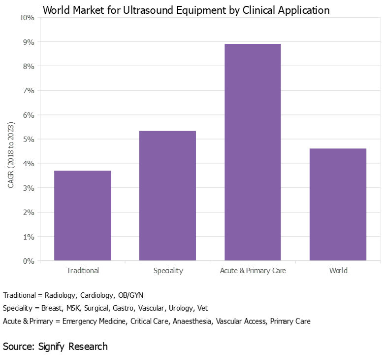
Over the years, the use of ultrasound has gradually expanded beyond radiology, cardiology, and ob/gyn to a wide range of clinical specialties, including surgery, musculoskeletal, and gastroenterology, to name a few, expanding the customer base and driving additional revenue growth.
This trend started out in developed countries, but more recently it has spread to developing markets, particularly India and China, where specialty departments at larger hospitals now often have their own budgets to buy ultrasound scanners.
Additionally, ultrasound is gaining acceptance in acute and primary care settings as both a screening and diagnostic tool, as well as for procedure guidance. With the use of handheld ultrasound gaining pace (see trend No. 4 below), this trend will accelerate in the coming years.
Chart of world market for ultrasound equipment by clinical application
All images courtesy of Signify Research.
2. New clinical applications evolve
Along with the growing customer base, the use of ultrasound with existing customers is expanding, typically as a lower-cost and/or radiation-free alternative to other imaging modalities, such as MRI and CT. The global shift to value-based care as a replacement to the traditional fee-for-service approach will support this trend.
For example, ultrasound is playing an increasingly important role as a screening tool for women with dense breast tissue. In acute care, ultrasound is increasingly being used for lung imaging to diagnose conditions such as pleural effusion, pulmonary edema, and pneumothorax. In another example, the use of shear-wave elastography is expanding beyond hepatology (e.g., liver fibrosis) to other body areas, including the breast, prostate, thyroid, and spleen. Musculoskeletal imaging is another relatively untapped market for ultrasound, including orthopedics, rheumatology, and sports medicine.
3. New markets emerge
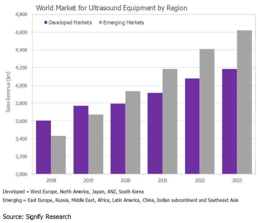
The ultrasound market in developed regions, such as Western Europe, North America, and Japan, is largely saturated, and the outlook is for low-single-digit to midsingle-digit growth. While these markets will continue to account for the lion's share of the world market, developing markets continue to represent a growth opportunity. In 2019, the fastest growth is forecast for Southeast Asia, Brazil, China, and India.
That said, ultrasound market growth is slowing in many of the emerging markets, particularly China, largely due to slowing global economic growth.
Chart of world market for ultrasound equipment by region
4. Handheld ultrasound picks up steam
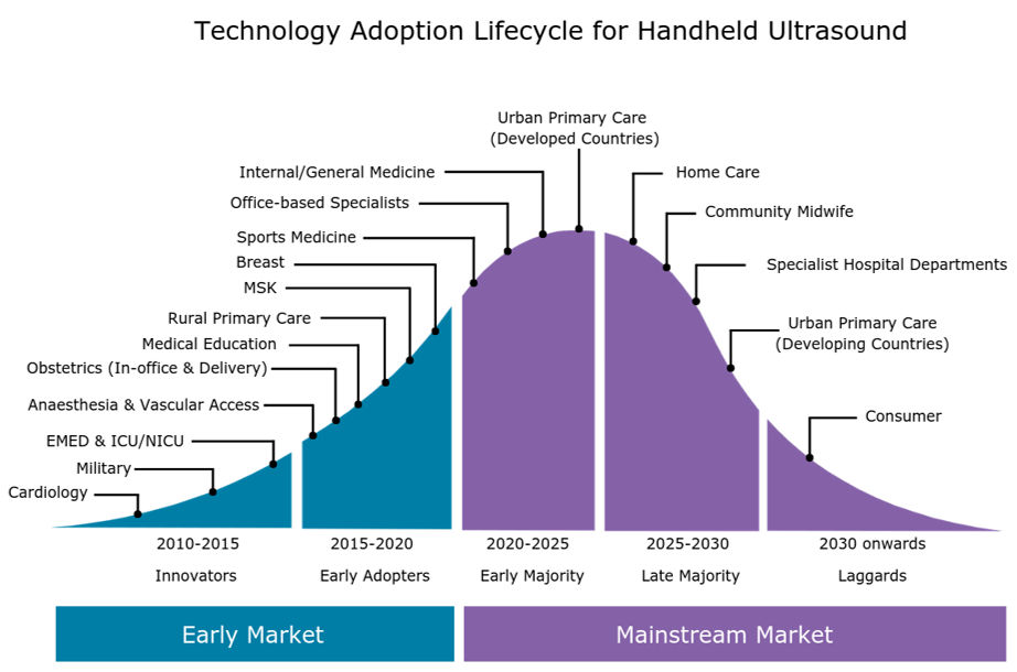
As we discussed in a previous insight ("Handheld Ultrasound Market Poised for Next Wave of Growth"), the handheld ultrasound market is growing rapidly as the latest generation of ultraportable devices gains acceptance among a diverse range of customer groups, from emergency medicine physicians and intensivists to internists, office-based specialists, and -- looking forward -- primary care physicians.
The expanding customer base of handheld systems, coupled with the increased availability of affordable handheld scanners, is forecast to boost global sales of handheld ultrasound by more than 50% in 2019. By 2023, the global market for handheld ultrasound is forecast to exceed $400 million.
Graphic of technology adoption lifecycle for handheld ultrasound
5. Artificial intelligence to transform market
Artificial intelligence (AI) will have a transformative effect on the ultrasound market as AI addresses some of the key limitations associated with ultrasound; namely, the shortage of trained sonographers and the relatively steep learning curve, high operator dependency during image acquisition and interpretation, poor image quality for certain exam types, and relatively lengthy exam time compared with other modalities.
The first wave of ultrasound AI applications are entering the market and are mainly for image optimization (noise reduction) and automation of time-consuming and repetitive tasks, such as anomaly detection, image labeling, and feature quantification. However, the greatest impact of AI will be on guided ultrasound (ultrasound navigation), which will provide real-time support during image acquisition (i.e., probe placement and anatomy detection).
The first AI-enabled guided ultrasound systems are expected to be released in the second half of 2019. These systems are expected to expand the user base by making ultrasound more accessible to novice users, particularly in acute and primary care.

Thứ Ba, 18 tháng 6, 2019

Which biopsy method best detects prostate cancer?


By Wayne Forrest, AuntMinnie.com staff writer
June 18, 2019 -- The combination of ultrasound- and MRI-guided biopsy yields the best chance of detecting prostate cancer, as each method can find malignant lesions missed by the other, researchers from the University of California, Los Angeles (UCLA) reported in a study published online June 12 in JAMA Surgery.
In the three-year Prospective Assessment of Image Registration in the Diagnosis of Prostate Cancer (PAIREDCAP) study, the researchers found that using ultrasound and MRI in tandem can identify up to one-third more cancers than standard techniques.
"Our research suggests that the different biopsy methods identify different tumors," said senior author Dr. Leonard Marks, from UCLA's department of urology, in a statement. "To maximize our ability to identify prostate cancer, we need to take advantage of all the information we can. Our cancer detection rate, while using different methods in tandem, surpasses that from using either method alone."
While the clinical value of MRI-guided prostate biopsy has been proved in previous large prospective studies, some debate remains over the optimum approach with the modality. Clinicians have debated whether biopsy should focus on areas with lesions only visible on MRI or if ultrasound-guided systematic biopsy -- in which 12 biopsy cores are acquired of multiple areas of the prostate -- is sufficient.
"Answers to these questions are vital because of the importance of diagnosing prostate cancer early vis-a-vis cost-effectiveness issues," the authors wrote.
The prospective study included 300 men who were undergoing prostate biopsy for the first time: 248 consecutive subjects (mean age, 65.5 ± 7.7 years) had MRI-visible lesions, while the control population consisted of 52 men (mean age, 63.6 ± 5.9 years) with no visible lesions on MRI. Each biopsy was preceded by a 3-tesla multiparametric MRI scan (Magnetom Trio, Siemens Healthineers) with an abdominal coil within 60 days of the procedure.
The study protocol used three biopsy techniques:
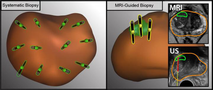
  • Systematic biopsy, performed with a standard handheld transrectal ultrasound-guided technique in which 12 cores were acquired, with the operator blinded to the MRI results
  • Targeted cognitive fusion, in which a radiologist used MR images on a screen next to the patient to guide the urologic biopsy operator to aim an ultrasound-guided biopsy at the area of the prostate corresponding to where the lesion was seen on MRI; three biopsy cores were obtained cognitively from the index region of interest (ROI)
  • Software fusion, in which researchers fused MRI and 3D ultrasound images using commercially available software (Artemis, Eigen), and an operator performed three targeted biopsies on the coregistered images
The difference between ultrasound- and MRI-guided methods to obtain tissue samples from different regions in the prostate
The illustration shows the difference between ultrasound- and MRI-guided methods to obtain tissue samples (black needles) from different regions in the prostate (brown oval object). MRI (right) allows doctors to detect lesions (green oval object) and take tissue samples from specific lesions. Images courtesy of UCLA Health.
The combination of methods produced the highest results, the researchers found.
Cancer detection rate of prostate biopsy methods
Biopsy methodCancer detection rate
Combination of systematic, cognitive, and software fusion biopsy70.2%
Cognitive fusion or software fusion62.1%
Cognitive targeting46.8%
The cancer detection rate of 70% is considerably higher than the cancer detection rate of conventional biopsy in previous studies, likely reflecting the outcomes of the MRI screening, the researchers noted. "These data thus serve to confirm the value of MRI for providing risk assessment when considering biopsy," they wrote.
Indeed, the researchers found that if only a single biopsy method was used, from 11.5% to 33.3% of cancers would have been missed, based on patient population.

Interestingly, among the 52 healthy controls, traditional ultrasound found prostate cancer in eight men (15%). This means that MRI does not identify all tumors, thus heightening the need to also incorporate traditional ultrasound-guided systematic biopsy as well, according to the researchers.

Ultrasound helps assess DVT risk after ablation


By Kate Madden Yee, AuntMinnie.com staff writerJune 18, 2019
The researchers from Stanford University also found that ultrasound helped clarify which ablation technique is safer.
"The rates of DVT decreased over time, suggesting an improvement in overall procedural safety; however, LA demonstrated a considerable decreased risk for DVT when compared to RFA," first author Dr. Nathan Itoga and colleagues wrote.
Studies have shown that both radiofrequency ablation and laser ablation are at least as successful as surgery for treating conditions such as varicose veins, and they can be performed without general anesthesia and tend to have fewer negative effects. But the rates of complications such as DVT after venous ablation procedures have been unclear, the group noted.
"Despite the relative safety of these techniques, LA and RFA may cause endovenous heat-induced thrombosis which ... may extend or propagate, leading to deep vein thrombosis," the researchers wrote. "In rare cases, LA and RFA procedures may also lead to pulmonary embolism. The overall risk for thromboembolic complication after endovenous ablation is controversial, and estimates in the literature vary greatly, from less than 1% to 18%."
The study included data from Truven Health Analytics MarketScan databases from 2007 and 2016 for a cohort of patients who underwent RFA or LA and had a follow-up duplex ultrasound scan within seven days and 30 days of the ablation procedure. Of the patient cohort, 256,999 underwent 433,286 ablations, 44.4% of which were radiofrequency and 55.6% of which were laser.
Of these patients, 1.9% had a newly diagnosed DVT within seven days of the procedure, and 3.1% had a newly diagnosed DVT within 30 days of the procedure. Factors associated with a decreased risk of DVT within 30 days included having had laser ablation, being female, and having sclerotherapy performed on the same day as the ablation; factors associated with an increased risk of DVT included a diagnosis of peripheral artery disease and undergoing stab phlebectomy.
Screening ultrasound revealed laser ablation to be the safer method, showing an 18% decrease in the incidence of DVT at 30 days (2.8%) compared to radiofrequency ablation (3.4%).
The study results demonstrate ultrasound's usefulness in tracking patients who have undergone ablation procedures for complications, according to the researchers.
"Some have suggested that postoperative evaluation of DVT is unnecessary, arguing that ultrasound overdiagnoses DVT, leading to overtreatment," Itoga and colleagues wrote. "However, if postoperative ultrasound were reserved for only those with symptoms, a considerable number of DVT would be missed."

Thứ Bảy, 15 tháng 6, 2019

Better ultrasound protocols slash trauma imaging overuse.


By Kate Madden Yee, AuntMinnie.com staff writer
June 11, 2019 -- When lower extremity duplex ultrasound screening (LEDUS) protocols for the trauma department are refined, overuse of hospital resources is reduced by more than a third -- and without negative effect on patient outcomes, according to a new study published June 5 in the Journal of Surgical Research.
The findings translate to more efficient trauma departments -- and better patient care, wrote a team led by Dr. Jennifer Baker of the University of Cincinnati in Ohio.
"Refinement of LEDUS protocols can decrease overutilization of hospital resources without compromising trauma patient outcomes," the researchers concluded.
A tool called the risk assessment profile (RAP) is typically used in the trauma center to identify patients who would most benefit from lower extremity duplex ultrasound screening. But it can lead to overuse of imaging and needs refinement, Baker's group wrote.
The researchers conducted a study for which they hypothesized that revising their hospital's LEDUS protocol so that screening ultrasound examinations were performed in patients with a RAP score ≥ 8 within 48 hours of admission would reduce the number of screenings performed -- without changing patient outcomes.
The study included 1,014 trauma patients admitted from July 2014 to June 2015 and July 2016 to June 2017. From 2014 to 2015, patients with a RAP score ≥ 5 had weekly LEDUS examinations starting on the fourth hospital day, while from 2016 to 2017, the protocol changed to screening patients with a RAP score ≥ 8 by the second hospital day. Both protocols included screening with weekly ultrasound after the first examination. Baker's team gathered data on demographics, injury characteristics, LEDUS exam findings, and venous thromboembolism incidences.
The group found that from 2014 to 2015, 602 patients underwent LEDUS exams, but from 2016 to 2017, only 412 patients did: a 32% drop. Significantly, the team noted no difference in the number of patients diagnosed with deep vein thrombosis (DVT) or pulmonary embolism between the two protocols; in fact, DVT was often identified in the first LEDUS exam in both year groups.
Regarding those patients who received a DVT diagnosis on first LEDUS exam, the group also found the following:
  • These patients had significantly higher RAP scores than those who did not have a DVT diagnosis (12 versus 10).
  • The time to their first LEDUS exam was shorter (one day versus three days).
  • The DVT diagnosis was made more quickly (two versus four days).
A simple change to protocol can make a big difference without harming patients, according to the researchers.
"[This protocol change caused] no significant difference ... in the number of patients diagnosed with DVT or pulmonary embolism," they concluded.

Thứ Năm, 6 tháng 6, 2019

Ultrasound shows Heart disease Risk from Psoriasis.


By Kate Madden Yee, AuntMinnie.com staff writer
June 6, 2019 -- Ultrasound imaging of the carotid arteries can help identify patients with psoriasis and psoriatic arthritis who are at increased risk for heart disease by revealing the extent of clogging in patients' arteries, according to a new study published online June 5 in Arthritis & Rheumatology.
The results show that combining ultrasound imaging data with traditional cardiovascular risk factors -- as measured by, for example, the Framingham risk score -- could identify which patients with psoriatic disease may benefit from more intensive heart-protective therapies, wrote lead author Dr. Curtis Sobchak, of the University of Toronto, and colleagues.
"Ultrasound is widely used in rheumatology settings as a point of care to detect joint inflammation. Our study suggests that ultrasound can also be used to identify patients that are at high cardiovascular risk who may be missed by the conventional methods such as the Framingham risk score," senior author Dr. Lihi Eder, PhD, said in a statement released by the journal. "This will allow early intervention, such as initiation of lipid lowering therapy, which will ultimately lower the risk of developing cardiovascular events."
Sobchack and colleagues evaluated whether carotid ultrasound could predict incident cardiovascular events in patients with psoriatic disease and whether using ultrasound imaging data would improve the performance of the Framingham risk score for cardiovascular risk prediction.
The study included 559 patients with psoriatic disease. Of these, 23 had experienced cardiovascular events that suggested heart disease. Patients underwent ultrasound of the carotid arteries between December 2009 and December 2015; the researchers then assessed the extent of atherosclerosis by measuring carotid intima media thickness and total plaque area, evaluating the patients' risk of experiencing further cardiovascular events using a hazard ratio (HR) model (with base HR equal to 1).
The team found that the rate at which patients experienced their first cardiovascular event during the study period was 1.11 events per 100 patient years. The group also found the following factors predicted incident cardiovascular events after controlling for the Framingham risk score:
Factors on carotid ultrasound that predict cardiovascular events
 Hazard ratio
Total plaque area3.74
High total plaque area category3.25
Mean carotid intima media thickness1.21
Max carotid intima media thickness1.11
Since carotid atherosclerosis is associated with an increased risk of developing future heart disease -- and since patients with psoriatic disease are known to be at increased risk of heart disease -- using ultrasound-generated vascular imaging data with information on traditional cardiovascular risk factors could improve the accuracy of cardiovascular risk assessment in these patients, according to the group. And ultrasound has many benefits when compared with other imaging modalities.
"The advantage of ultrasound over other modalities for vascular imaging includes lack of radiation, low cost of the examination, and its widespread use in rheumatology for joint evaluation," the team concluded. "Thus, this assessment could potentially be performed 'at the bedside' during consultation to provide immediate valuable information to complement clinical data from history, physical examination, and laboratory data."