Tổng số lượt xem trang
Chủ Nhật, 15 tháng 12, 2019
Thứ Năm, 12 tháng 12, 2019
O-RADS
O-RADS™” is an acronym for an Ovarian-Adnexal Imaging-Reporting-Data System which will function as a quality assurance tool for the standardized description of ovarian/adnexal pathology. The creation of a standardized lexicon permits the development of a practical, uniform vocabulary for describing the imaging characteristics of ovarian masses that can be used to determine malignancy risk, with the ultimate goal of applying it to a risk stratification classification for consistent follow up and management in clinical practice.
The use of internationally agreed upon standardized descriptors should result in consistent interpretations and decrease or eliminate ambiguity in reports resulting in a higher probability of a correct diagnosis. In the case of the adnexal mass, the correct interpretation leading to the correct diagnosis is the key to accuracy in determining risk of malignancy and, finally, optimal patient management.
In the Summer of 2015, under the supervision of the American college of Radiology, the Ovarian-Adnexal Reporting and Data System(O-RADS) Committee was formed with the purpose of creating a standardized lexicon for describing the imaging characteristics of ovarian and adnexal masses and applying it to a risk stratification and management system for evaluation of malignancy. This is an ongoing collaborative effort of an international group of experts in gynecological imaging and management of ovarian/adnexal masses that includes a broad spectrum of experts in radiology, gynecology, pathology, and gynecologic oncology from the US, Canada, Europe, and the United Kingdom.
Since ultrasound is widely considered the primary imaging modality in the evaluation of adnexal masses and MRI the problem-solving tool, parallel working groups (US and MRI) were formed to develop separate but consistent groups of terms specific to each modality. The principal goals of O-RADS are to improve the quality and communication between interpreting and referring physicians, to limit variability in reporting language and ultimately to guide patient management based on actionable information in the imaging report. The committee is sponsored by the American College of Radiology with eventual lexicon trademark by the aforesaid organization.
ABUS deliveres better diagnostic performance
By Wayne Forrest, AuntMinnie.com staff writer
December 9, 2019 -- Both automated breast ultrasound (ABUS) and traditional handheld ultrasound can significantly improve breast cancer detection when used as an adjunct to mammography in women with dense breasts. But ABUS yields better diagnostic performance, according to research presented at RSNA 2019 in Chicago.
In a study involving over 1,200 women, a team of researchers led by Dr. Mengmeng Jia of the Chinese Academy of Medical Sciences in Beijing found that ABUS produced a higher specificity, positive predictive value, and area under the curve (AUC) than handheld ultrasound.
Breast cancer is the most commonly diagnosed cancer in Chinese women, but less than 1% of cases are detected by screening, according to Jia. Compared with Western countries, Chinese women also have a higher proportion of dense breasts, for which mammography is less sensitive, she said. As a result, these patients need adjunctive imaging modalities such as ultrasound, digital breast tomosynthesis, or MRI.
Traditional handheld ultrasound is inexpensive, safe, and suitable for dense breasts. But it's also labor intensive and highly dependent on the operator. ABUS, on the other hand, is specifically designed for finding cancer in dense breast tissue. It's also reproducible due to its operator-independent acquisition method, Jia noted.
"Most importantly, the image acquisition can be separated from interpretation," she said. "That means that images can be taken by the operator and then interpreted by radiologists [at another location]. That is helpful in resource-limited areas [where there are not enough qualified radiologists]."
The researchers sought to evaluate the diagnostic performance of ABUS and handheld ultrasound as an adjunct to mammography in women ages 40 to 69. They also wanted to assess the performance of both methods in mammography-negative dense breasts.
The team enrolled 1,266 women ages 40 to 69 in a multicenter study involving five tertiary hospitals. All women received mammography, as well as handheld ultrasound and ABUS. Of the 1,266 women, 958 were deemed to have dense breasts.
Overall, sensitivity increased from 87.3% with mammography alone to 96.9% for both handheld ultrasound and ABUS. Negative predictive value also increased from 95.5% to 98.8% with handheld ultrasound and 98.8% for ABUS. Mammography alone had an AUC of 0.88, compared with an AUC of 0.92 for mammography and handheld ultrasound and 0.93 for mammography and ABUS.
In women with mammographically negative dense breasts, ABUS and handheld ultrasound detected 31 additional cases of breast cancer. The techniques had comparable sensitivity and negative predictive value, but ABUS had a higher specificity, positive predictive value, and AUC, according to the researchers.
| Performance in women with mammographically negative dense breasts | ||
| Handheld ultrasound | ABUS | |
| Sensitivity | 31/33 (93.9%) | 31/33 (93.9%) |
| Specificity | 619/665 (93.1%) | 635/665 (95.5%) |
| Positive predictive value | 31/77 (40.3%) | 31/61 (50.8%) |
| Negative predictive value | 619/621 (98.8%) | 635/637 (99.7%) |
| Area under the curve | 0.935 | 0.947 |
"More studies are [now] needed to [further] evaluate the performance of adjunctive ultrasonography, including ABUS and handheld ultrasound, in [resource]-limited areas," Jia concluded.
Are clinicians overusing CTA for carotid stenosis?
By Abraham Kim, AuntMinnie.com staff writer
December 9, 2019 -- The use of CT angiography (CTA) as the first-line imaging exam for carotid artery stenosis has increased nearly threefold during the past several years, raising concerns over growing patient costs and radiation exposure, according to a study presented on Friday at RSNA 2019.
The researchers, led by Dr. Jina Pakpoor from Johns Hopkins Hospital, examined imaging requests for the diagnosis of carotid artery stenosis from outpatient centers across the U.S. Their analysis of the data revealed that CTA usage rates increased every year from 2011 to 2016, whereas ultrasound usage steadily trended downward.
"Overall, there is actually high compliance with the current recommendation to use Doppler ultrasound for initial testing," Pakpoor told session attendees. "We did, however, find that there was a shift in the direction, where CTA use is increasing and Doppler ultrasound use is actually decreasing, which is going to have higher costs for patients and higher radiation exposure."
CTA on the rise
Current guidelines from the Society of Vascular Surgery recommend Doppler ultrasound as the first-line imaging exam for carotid stenosis. More advanced imaging exams such as CTA and MR angiography (MRA) are typically reserved as a second-line imaging test for cases requiring more detailed stenosis characterization or urgent therapy.

Dr. Jina Pakpoor.
However, recent studies have shown that CT usage rates have increased dramatically for carotid imaging in general over the past decade, whereas rates for other imaging modalities have been decreasing. These reports motivated Pakpoor and colleagues to determine whether physicians in the U.S. were complying with existing guidelines for the initial imaging workup of suspected carotid artery stenosis in the outpatient setting.
The researchers obtained information from the 2011 to 2016 IBM MarketScan U.S. national commercial claims and insurance database, which includes data submitted by large employers, managed care organizations, hospitals, electronic medical record providers, Medicare, and Medicaid.
The study population included 229,464 patients ages 18 to 65 who underwent neck CT angiography, Doppler ultrasound, or MR angiography for their first carotid stenosis encounter. Approximately half of the patients were male, and their average age was 55.
Over the eight-year period, the vast majority of patients received an ultrasound exam at 95.8%, followed by CTA at 2.4%, and finally MRA at 1.3%.
Though ultrasound remained by far the most used imaging modality overall, a year-by-year analysis showed that the ultrasound usage rate decreased by a statistically significant degree from 2011 to 2016. In contrast, the usage rates roughly tripled for CTA and remained relatively constant for MRA.
| Trends in carotid stenosis detection on ultrasound, MRA, and CTA | ||||||
| 2011 | 2016 | |||||
| Ultrasound | MRA | CTA | Ultrasound | MRA | CTA | |
| Proportion of all imaging exams | 96.9% | 1.2% | 1.6% | 93.8% | 1.5% | 4.7% |
Room for concern
Further analysis revealed that use of CTA and MRA varied depending on the region of the U.S. where the exams were performed. To be specific, combined CTA and MRA use was considerably greater in the western U.S. (5.5%) than in the northeastern U.S. (2%; p < 0.001). In addition, females were more likely than males to receive advanced imaging.
Though most referring providers appear to be complying with current recommendations for the diagnosis of carotid stenosis, the sustained increase in CTA utilization poses a concern, Pakpoor noted. Growing reliance on CTA for first-line imaging may stem from the increasing availability of CT scanners throughout the U.S.
"These days a lot of institutions have CT scanners available in the emergency department, and CT is known as a modality that you can easily and quickly get access to," she said. "And from a provider's perspective, if you're not worried about practice costs and radiation, CTA can provide a lot more information and avoid the need for a second [imaging] study. But it is not the current recommendation, and we don't want people to be moving in that direction."
The findings suggest a possible need to educate outpatient providers on the appropriate protocol in order to prevent this trend from continuing in the same direction, Pakpoor concluded. "It will certainly be something important to look at in the future -- to emphasize the importance of continuing to use Doppler ultrasound as the first modality, despite the fact that we're seeing an increase in use of CT over all aspects of radiology."
Thứ Ba, 10 tháng 12, 2019
FETAL BIOMETRY GUIDELINES
The performance and interpretation of fetal biometry is an important component of obstetric ultrasound practice. In fetuses for which gestational age has been established appropriately, measuring key biometric parameters, together with transformation of these measurements into EFW using one of the many validated formulae, permits detection and monitoring of small fetuses. Serial sonographic assessment of fetal size over time can provide useful information about growth, with the possibility of improving the prediction of SGA infants, particularly those at risk for morbidity. However, errors and approximations that may occur at each step of such a process greatly hamper our ability to detect abnormal growth, and most importantly FGR. Therefore, in clinical practice, fetal biometry should represent only one component of how we screen for abnormal growth. It is reasonable to believe that no single measurement, EFW formula or chart will significantly improve our current practices. Improved FGR screening may be feasible by using a combined approach that includes biometry as well as other clinical, biological and/or imaging markers. This goal will come within reach only when the ‘biometric component’ is better standardized for all those who care for pregnant women.
Thứ Sáu, 6 tháng 12, 2019
Chủ Nhật, 1 tháng 12, 2019
Multiparametric Ultrasound (MPUS) or “the many faces” of ultrasonography
Multiparametric ultrasound (MPUS) or “the many faces” of ultrasonography
Alina Popescu
Department of Gastroenterology and Hepatology, “Victor
Babeș” University of Medicine and Pharmacy Timișoara,Romania
Received Accepted
Med Ultrason
2019, Vol. 21, No 4, 369-370
Ultrasound (US) is still seen as a “Cinderella” of the imaging
techniques, without taking into considerationthe many advantages of the method.
It is a real time, dynamic method, very accessible, rather inexpensive, but (and
maybe more important) a non irradiating technique,repeatable and very well
accepted by patients. Even if for some pathologies the accuracy of conventional
US is not so high (for example the positive diagnosis of focal liver lesions),
in other situations it is the best method for the diagnosis (for example
gallbladder stones, biliary obstruction).
The major drawbacks of US are considered operator dependence
and lack of specificity and accuracy for some diagnoses. On the other hand,
with proper training, it is a very good method for orientation in clinical
practice and a very good screening method. Because it is well accepted by the
patients it is also a good follow up method.
If the drawback of being an operator dependent method can be
overcome by training, the lack of accuracy is a disadvantage that is harder to
overcome.
One of the criticisms brought to US in the past was the
impossibility to characterize the vascularization of different lesions, a
feature necessary for the correct diagnosis of the different pathologies. Even
if the Doppler techniques are established ultrasound techniques as M and
B-mode, the lack of possibility to perform a contrast enhanced study, such as
for contract enhanced computer tomography (CE-CT) or contrast enhanced magnetic
resonance imaging (CE-MRI), limits the possibility of characterization of
different lesions detected by US, and on account of these limits the accuracy
of the technique.
The development of new applications in US in the last period
has improved the position of this imaging technique in the management of
different pathologies. The 3D and 4D US but, more importantly, contrast
enhanced US (CEUS) and US based elastography techniques, provided the missing
data required for a better diagnosis accuracy of US, and created the concept of
multiparametric ultrasound (MPUS) [1], a term borrowed from sectional imaging,
especially MRI.
There are several examples in the literature of the role of
MPUS for the assessment in different pathologies: prostate cancer [2-4],
chronic kidney diseases [5], thyroid nodules [6] or parathyroid lesions [7].
I want to bring attention to another organ – the liver,
where the role of MPUS is already well known and emphasized also by other
authors [8], and more specifically - focal liver lesions. US is an excellent imaging
modality for the detection of liver tumors, but it lacks the necessary
specificity for a correct positive diagnosis.
The evaluation of a focal liver lesions can be very
expensive (CE-CT or CE-MRI), needs time and usually is very stressful for the
patients. CEUS changed this, because is an US technique that can be performed
in the same session as conventional US: it takes 5 more minutes, and has good
accuracy for the characterization of liver tumors [9]. The accuracy increases
if we know if the lesion is developed on a cirrhotic or a non-cirrhotic liver
[9].
US can answer today also this question, more precisely US
based elastography, a technique with a very good accuracy for ruling in or
ruling out liver cirrhosis in a very short time (less than 5 minutes) [10].
Elastography of the focal lesion can also be helpful for the diagnosis in some situations.
Thus, in a three-step algorithm using MPUS, starting with conventional US,
followed by elastography and then CEUS, we can make a complex evaluation of a
focal liver lesion in the same session with a very good accuracy for the
positive diagnosis.
In conclusion, US is here to remain in the big picture of
imaging techniques and should be used, with all its available features, as the
first line diagnostic method in many situations.
Thứ Bảy, 30 tháng 11, 2019
Thứ Năm, 28 tháng 11, 2019
Supersonic to debut quantification tools at RSNA 2019.
By AuntMinnie.com staff writers
November 26, 2019 -- Ultrasound equipment manufacturer SuperSonic Imagine will be debuting three new quantitative liver ultrasound applications at the upcoming RSNA 2019 meeting in Chicago.
The first two tools, Att Plus and SSp Plus, are designed to simultaneously quantify ultrasound attenuation in the liver and intrahepatic speed of sound to reflect fat content for detecting and diagnosing hepatic steatosis, according to the vendor.
The third new tool, Vi Plus, works in combination with real-time elasticity imaging to visualize and quantify tissue viscosity. These viscosity assessments provide clinicians with information for tissue characterization.
Liver Health: Three New Ultrasound Markers Enter the U.S. Market
SuperSonic Imagine, a major actor promoting innovation in ultrasound, will introduce a suite of three ultrasound markers for non-invasive assessment of the severity of chronic liver diseases with quantitative results: Att PLUS, SSp PLUS and Vi PLUS. The first two of these tools allow for the simultaneous quantification of ultrasound attenuation in the liver and intrahepatic sound speed, reflecting fat content, an essential criterion for the detection and diagnosis of hepatic steatosis. Coupled with elasticity imaging in real time, the third one, Vi PLUS, makes it possible to visualise and quantify tissue viscosity, providing clinicians with important information for tissue characterisation.
“Liver diseases including non-alcoholic steatohepatitis (NASH) linked to conditions like diabetes and obesity affect millions of Americans and have become an important public health concern in the space of just a few years. We are very proud to present the fruits of our expertise in liver health — the series of three non-invasive liver markers — for the first time in the United States. All the more so as these markers have just received 510(k) clearance from the FDA. Following the acquisition of SuperSonic Imagine by Hologic, we are also going to present many shared sessions dedicated to women's health,” concludes Michèle Lesieur, the CEO of SuperSonic Imagine.
Thứ Ba, 26 tháng 11, 2019
ULTRASOUND THALAMOTOMY MAY HELP TREAT ESSENTIAL TREMOR.
By AuntMinnie.com staff writers
November 21, 2019 -- A treatment that uses ultrasound may be effective in relieving symptoms of essential tremor by targeting the affected area of the brain, according to a study published online November 20 in Neurology. The treatment, called focused ultrasound thalamotomy, essentially destroys the area of the brain causing the tremor.
More than 7 million people in the U.S. have essential tremor, according to the American Academy of Neurology. Currently, the most common treatment for the disorder when patients don't respond to medication is deep brain stimulation, a procedure that involves incisions and insertion of electrodes or probes into the patient's brain. Ultrasound thalamotomy would offer a less invasive treatment option and remains effective for approximately three years. It does, however, cause an irreversible brain lesion.
For the study, 56 patients received ultrasound thalamotomy and 20 received a fake treatment. The research team measured hand tremors, level of disability, and quality of life at the commencement of the study and after six months, one year, two years, and three years. By the three-year mark, the researchers noted a 50% improvement in hand tremors, a 56% improvement in disability, and a 42% improvement in quality of life.
While senior author Dr. Casey Halpern of Stanford University did note some limitations of the study, he concluded that "this treatment should be considered as a safe and effective option" for those living with essential tremor.
Thứ Hai, 25 tháng 11, 2019
Thứ Bảy, 23 tháng 11, 2019
Thứ Năm, 21 tháng 11, 2019
Ultrasensitive Ultrasound Microvessel Imaging (UMI) boots BI-RADS accuracy
By Kate Madden Yee, AuntMinnie.com staff writerUsing a novel ultrasound technique called ultrasensitive ultrasound microvessel imaging (UMI) with conventional ultrasound significantly improves the diagnostic accuracy of BI-RADS classification of breast lesions, according to a study to be published in the December issue of Ultrasound in Medicine and Biology.
And improved BI-RADS categorization could translate into fewer unnecessary breast biopsies, wrote a team led by Ping Gong, PhD, of the Mayo Clinic in Rochester, MN.
"The microvessel information provided by ultrasound microvessel imaging may add clinical value to supplemental ultrasound screening adjunct to mammography in reducing unnecessary and missed biopsies," the researchers wrote.
Reducing false positives
Combining ultrasound with mammography improves diagnostic sensitivity in women with dense tissue compared with mammography alone, Gong and colleagues noted. But the combination can also increase false positives due to the low specificity of ultrasound -- leading to more unnecessary biopsies. That's why improving ultrasound's performance is important (Ultrasound Med Biol, December 2019, Vol. 45:12, pp. 3128-3136).
"In contrast to normal tissue and benign tumors, malignant tumors typically present a vessel pattern with chaotic distribution, irregular branches, and penetrating peripheral vasculature," the group wrote. "[But] the clinical reliability of conventional Doppler is undermined by limited vessel detection sensitivity. An ultrasensitive ultrasound microvessel imaging technique ... [provides] advanced vessel sensitivity ... without using ultrasound contrast agents."
Instead of the line-by-line scanning of conventional Doppler ultrasound, UMI is based on "ultrafast plane wave imaging ... that leads to at least 10 times more ultrasound frames for blood flow detection than conventional Doppler," the group wrote. This rapid imaging allows for a much higher sensitivity for visualizing microvessels, which helps distinguish between malignant and benign lesions, study co-author Shigao Chen, PhD, also of the Mayo Clinic, told AuntMinnie.com via email.
"Conventional ultrasound has low sensitivity for detecting very small vessels, and thus may miss the microvessels in the tumor which provide important information for diagnosis," he said. "This technology uses a newer ultrasound scanner with very high imaging frame rates so that more data can be acquired to boost sensitivity, and it uses more advanced signal processing to better remove tissue signal -- which is orders of magnitudes higher than blood signal -- to reveal the underlying microvessels," he said.
The researchers sought to explore the feasibility of UMI for assessing breast tumor microvessel distribution and to compare the performance of UMI and conventional ultrasound in differentiating between benign and malignant lesions. The study included 44 women with 51 breast masses, all of whom were imaged with conventional ultrasound and the combination of conventional ultrasound and UMI. Of the 51 masses, 28 were malignant and 23 were benign.
Gong's group found that adding UMI to conventional ultrasound significantly improved the visualization of small vessels compared with Doppler ultrasound alone across a variety of performance measures and that the microvessel structures UMI depicted were associated with benign or malignant tumor characteristics.
| Performance of conventional ultrasound alone compared with ultrasound plus UMI | ||
| Performance measure | Conventional ultrasound | Ultrasound plus UMI |
| Sensitivity | 96.4% | 100% |
| Specificity | 17.4% | 47.8% |
| Positive predictive value | 58.7% | 70% |
| Negative predictive value | 80% | 100% |
| Diagnostic accuracy | 60.8% | 76.5% |
The study also found that the diagnostic accuracy of correct BI-RADS categories improved by 16 percentage points with the combination of conventional ultrasound and UMI compared with ultrasound alone.
"This improvement indicates the potential of UMI in reducing unnecessary benign biopsies and avoiding missed malignant biopsies," Gong and colleagues wrote.
Keep the research coming
More research using this ultrasound technique is needed, Chen told AuntMinnie.com.
"If this preliminary research is confirmed by larger studies, it will imply that information about the microvasculature of the tumor obtained by ultrasensitive Doppler ultrasound can be used to differentiate benign from malignant tumors and thus reduce unnecessary biopsy," he said. "In addition, the technology may be useful for early evaluation of treatment effects to guide adjustment of chemotherapy or medical therapy regimens and avoid unnecessary side effects of ineffective therapies."
PocUS a Septic SHOCK Case
Abstract
A 77-year old male was admitted in
the emergency department for septic shock, yet no clear source of infection was
noted upon physical examination and a portable chest x-ray.
Due to his unstable
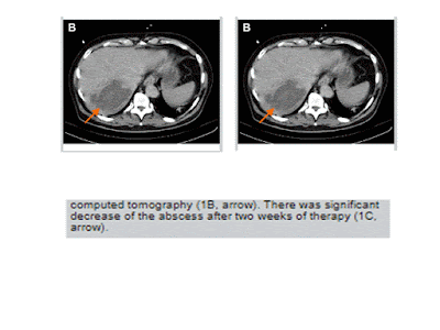
condition, bedside ultrasound was performed. A heterogeneous mass in the liver
was noted, hence a tentative diagnosis of liver abscess was made. This was
latter confirmed by abdominal computed tomography.
This case highlights that
point-of-care ultrasound, when performed by expert physicians, can
significantly decrease time to diagnosis for septic patients.
Thứ Bảy, 16 tháng 11, 2019
Đăng ký:
Bài đăng
(
Atom
)


