Tổng số lượt xem trang

Thứ Bảy, 4 tháng 1, 2020

Noncontact Laser US Imaging.


Abstract

Full noncontact laser ultrasound (LUS) imaging has several distinct advantages over current medical ultrasound (US) technologies: elimination of the coupling mediums (gel/water), operator-independent image quality, improved repeatability, and volumetric imaging. Current light-based ultrasound utilizing tissue-penetrating photoacoustics (PA) generally uses traditional piezoelectric transducers in contact with the imaged tissue or carries an optical fiber detector close to the imaging site. Unlike PA, the LUS design presented here minimizes the optical penetration and specifically restricts optical-to-acoustic energy transduction at the tissue surface, maximizing the generated acoustic source amplitude. With an appropriate optical design and interferometry, any exposed tissue surfaces can become viable acoustic sources and detectors. LUS operates analogously to conventional ultrasound but uses light instead of piezoelectric elements. Here, we present full noncontact LUS results, imaging targets at ~5 cm depths and at a meter-scale standoff from the target surface. Experimental results demonstrating volumetric imaging and the first LUS images on humans are presented, all at eye- and skin-safe optical exposure levels. The progression of LUS imaging from tissue-mimicking phantoms, to excised animal tissue, to humans in vivo is shown, with validation from conventional ultrasound images. The LUS system design insights and results presented here inspire further LUS development and are a significant step toward the clinical implementation of LUS.


Thứ Năm, 2 tháng 1, 2020

Model predicts metastases in endometrial cancer patients


By Rebekah Moan, AuntMinnie.com contributing writer

January 2, 2020 -- Using ultrasound in combination with endometrial biopsy results and clinical characteristics allows clinicians to reliably predict the risk of lymph node metastases in endometrial cancer patients before surgery. Researchers found the model to be superior to risk stratification alone in a study published online December 16 in Ultrasound in Obstetrics and Gynecology.
The prospective multicenter study compared three ways to assess metastases: endometrial biopsy alone, biopsy with ultrasound, and biopsy with ultrasound and risk characteristics. The study team led by Dr. Linda Eriksson from the pelvic cancer department at Karolinska University Hospital in Stockholm found the method using all three showed higher sensitivity and specificity.
"Our risk prediction model can be used to decide which women should or should not be subject to lymphadenectomy or sentinel node biopsy, depending on the individual risk of lymph node metastases," they wrote.
Comparing modalities
Lymphadenectomy is required for surgical staging in endometrial cancer, but it doesn't have any survival advantages in early-stage endometrial cancer and isn't recommended in low-risk cases. Using transvaginal ultrasound or MRI along with preoperative biopsy helps to determine the necessity of lymphadenectomy.
What can further refine the process is a preoperative risk prediction model to assess the risk of lymph node metastases before surgery -- to avoid undertreatment or overtreatment with lymphadenectomy.
Previous research has relied on small, retrospective studies that use MRI to create a prediction model. However, transvaginal ultrasound demonstrates similar accuracy to MRI and has the advantage of being accessible, well-tolerated, less expensive, and less time-consuming.
In the current study, Eriksson and colleagues sought to develop a preoperative risk prediction model with variables from demography, endometrial biopsy, and ultrasound to estimate the individual risk of lymph node metastases in endometrial cancer patients. They then compared its performance with endometrial biopsy results alone or endometrial biopsy results combined with ultrasound findings.
The study included 1,501 women with histologically confirmed endometrial cancer from 16 centers in seven European countries. The women were subjected to transvaginal ultrasound before surgery. The information they included to assess risk was age, duration of abnormal bleeding, result of preoperative endometrial biopsy (histotype and grade), tumor extension according to ultrasound, color content of the endometrial scan, tumor size according to ultrasound, and "undefined tumor with an unmeasurable endometrium."
The individual predicted risk of lymph node metastases was obtained by multiplying each risk factor by its prediction coefficient and adding the intercept.
Clinicians performed lymphadenectomy in 691 women, of whom 127 had lymph node metastases. Endometrial biopsy and endometrial biopsy combined with ultrasound have fixed sensitivities and specificities, but the risk prediction model does not -- it can be shifted upward to obtain a better specificity or downward to obtain a better sensitivity based on different threshold levels. For a comparison, see the table below.
Lymph node metastatic risk model vs. endometrial biopsy and ultrasound
 Endometrial biopsyEndometrial biopsy + ultrasoundRisk model at threshold of 0.196Risk model at threshold of 0.113
Sensitivity35%75%50%80%
Specificity77%52%80%53%
Even when the researchers continued to fiddle with the risk threshold, it outperformed both endometrial biopsy alone and endometrial biopsy combined with ultrasound. They also found the overall area under the curve of the risk prediction model was 0.73.
"The use of a risk prediction model can aid in planning the expected operation room time and the need of a skilled surgeon," Eriksson and colleagues wrote. "If implemented at the point of care, the model can be integrated into ultrasound machines or apps that automatically calculate the risk of lymph node metastases."
Given the large sample size and the multicenter design, the study is likely generalizable. However, further validation on new data is needed, preferably with known sentinel node outcome in cases without lymphadenectomy.
"After successful external validation, an impact analysis (i.e., decision-analytic modeling, a cost-effectiveness analysis, a before-after implementation study, or a randomized trial) would add to the evidence required before introducing the model into clinical practice," the researchers concluded.

Thứ Bảy, 28 tháng 12, 2019

GUIDELINES for OPHTHALMOLOGY COLOR DOPPLER ULTRASOUND


The 5 Coolest Medical Stories of 2019

Visible Body Logo

The 5 Coolest Medical Stories of 2019

By Laura Snider, Thu, Dec 26, 2019 @ 09:00 AM

It’s that time of year again, folks! 2020 is nearly upon us, so it’s time to review some of the coolest medical stories of this year. Get ready to be inspired by these stories of innovation and achievement!

1. Live-streaming brain surgery to educate the public

In October, a 25-year-old occupational therapy Masters student named Jenna Schardt elected to have her awake brain surgery livestreamed on Facebook. The surgery, conducted at Methodist Dallas Medical Center by Dr. Randall Graham and Dr. Bartley Mitchell, was necessary to remove a mass of blood vessels in her brain that was causing her to have seizures and impairing her ability to speak.
This surgery is interesting not only because the patient agreed to have it livestreamed but also because during part of the procedure, she needed to be awake so doctors could map out the parts of her brain related to speech and motor functions. They wanted to make sure their strategy for removing the tangle of blood vessels from Jenna’s brain wouldn’t effect these areas. This type of procedure is called awake brain mapping.
brain-brocas-area-left-ifg
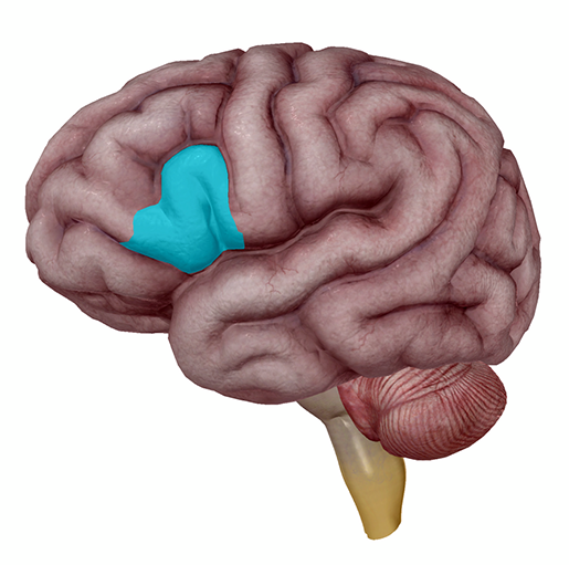
When it comes to speech and the brain, Broca’s area (the pars triangularis and pars opercularis of the left inferior frontal gyrus) is the usual suspect. Image from Human Anatomy Atlas.
If you’ve got about 45 minutes to spare, I’d highly suggest watching the livestream here. Don’t worry—there’s no blood, since the actual surgical site is covered by a sheet. One of the reasons this procedure was livestreamed was to educate the public about awake brain surgeries, and to show that they’re really not as frightening as they seem. 
At the beginning of the livestream, Jenna is under anaesthesia, but she wakes up a couple minutes in. As the commentators (a PR representative and the head of neurosurgery at the hospital) assure viewers, she is in no pain. Local anaesthesia around where her skull was opened ensures this. In addition, the brain itself doesn’t have pain receptors on its surface.  
Once she’s awake, a neurologist shows Jenna words on an iPad, which she reads aloud while the surgeons stimulate different regions of her brain to see whether her speech is impacted. Throughout this time, the operating room staff makes sure she is comfortable. She even has a conversation with the neuro-monitor about her dog before she goes back under anaesthesia so that the surgeons can complete the procedure.
If you’re interested in neuroscience or speech science like I am, you should definitely check this out. This livestreamed surgery is a fantastic piece of patient education, and it’s awesome that Jenna was willing to share her experience with the world. It’s also great that the procedure was successful and Jenna is recovering well. What a technologically amazing world we live in! 


2. Progress on vaccines for Ebola and malaria

You may remember our article on Ebola virus disease from August, in which we mentioned that a vaccine was being used to help control the ongoing outbreak in the Democratic Republic of the Congo (DRC). Ebola is a hemorrhagic fever which spreads via contact with the bodily fluids of infected individuals. 
Earlier this year, a vaccine manufactured by Merck, called Ervebo, was administered in a “ring style,” where people who were at a high risk of contracting Ebola were vaccinated first, followed by people who were in close contact with the group being vaccinated. More than 250,000 people have received the Merck vaccine, which has recently been given marketing authorization by the European Commission, making it the first approved Ebola vaccine. Ervebo has also been prequalified by the WHO, which will make it easier for at-risk countries to get access to it more quickly. 
As of November, a second vaccine—this one manufactured by Johnson & Johnson—will be joining the first in the fight against Ebola. The goal is to conduct a clinical trial of this vaccine in at least 50,000 people. It will be available to adults and children over one year old living in particular regions of the city of Goma. Scientists are optimistic about these two vaccines’ ability to combat Ebola, especially since the J&J one is better for people who haven’t already been exposed to the disease, and the Merck is better used in the middle of an outbreak. 
There has also been progress towards creating a vaccine for another deadly disease, malaria. 
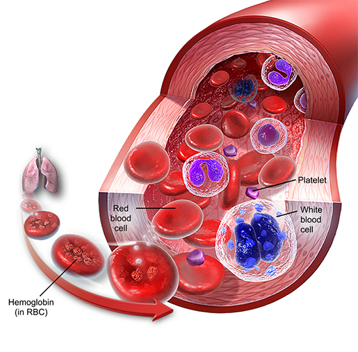
Malaria is caused by a parasite (Plasmodium falciparum) spread by mosquitoes. The parasite infects the red blood cells (erythrocytes) of its human hosts. Malaria claims many lives each year, especially among children in developing countries. 
blood-composition-red-blood-cells
Composition of blood. Image from A&P 6
In April, a pilot program of a vaccine known as RTS,S began in Malawi (and expanded to Ghana and Kenya). In clinical trials, RTS,S was able to prevent 4 in 10 cases of malaria in children, and reduced the level of severe anemia caused by malaria by 60%. The trial is a collaboration between GlaxoSmithKline (the vaccine manufacturers, the UN, the health ministries of Ghana, Kenya, and Malawi, as well as several other national and international organizations. 
The goal of the vaccine pilot program is to vaccinate 360,000 children a year in the three countries involved in the trial—hopefully, the program will not only save lives on its own but will also bring the world closer to finding a reliable way to prevent malaria.

3. A revolutionary drug treatment for cystic fibrosis

Cystic fibrosis (CF) is a genetic disease in which mutations of the CFTR (cystic fibrosis transmembrane conductance regulator) gene cause the CFTR protein to become dysfunctional
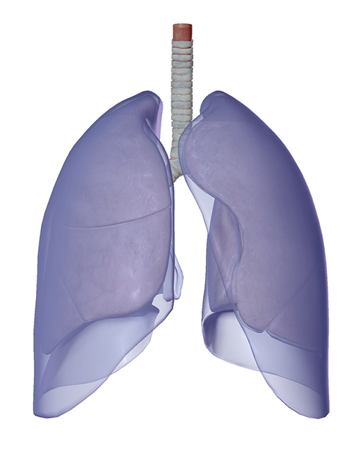
Ultimately, this leads to the body’s mucus becoming thick and sticky. In the respiratory system, the sticky mucus traps germs and clogs up airways, causing frequent lung infections and trouble breathing. 
lungs-and-pleurae
The trachea, lungs, and pleurae. Image from Human Anatomy Atlas.
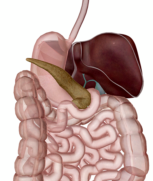
The digestive system can be affected as well: when mucus builds up in the pancreas, the release of digestive enzymes made by the pancreas is impaired. In addition, liver disease can result from mucus blocking the bile duct. 
digestive-system-accessory-organs
The accessory organs of the digestive system: the liver, gall bladder, and pancreas. Image from Human Anatomy Atlas.
Around 70,000 people worldwide have CF. It used to be the case that most people with CF died in childhood, but as treatment options have improved, more and more people with the condition have been able to live to adulthood. Unfortunately, it still often proves fatal. 
There’s good news, though! Just this October, a drug that can help 90% of people with CF was approved. Trikafta, developed by Vertex Pharmaceuticals, is a combination of three medications that drastically improves lung function in people with the most common CFTR mutation (Phe508del or F508del). 
Though people with rarer mutations may not be able to benefit from this new drug, the Cystic Fibrosis Foundation announced an initiative aimed at finding treatments that will help them, too.


4. A new immunotherapy for peanut allergies awaits FDA approval

In September, an advisory panel from the FDA voted to approve a new drug called Palforzia, which is intended to reduce the risk of severe allergic reactions to peanuts in kids aged 4-17. 
The drug contains a small, standardized dose of peanut flour that can be gradually increased, providing children with a way to build up their tolerance to peanuts. Though this treatment is by no means a “cure,” it will allow allergy sufferers and their families some peace of mind because it will decrease the risk of having a severe reaction (such as anaphylaxis) as a result of accidental exposure to peanuts. 
It should be noted that participants in the clinical trials for Palforzia did experience some side effects, including allergic reactions to the peanut powder in the drug as well as “coughing, hives, itching, throat irritation, nausea, and vomiting.” Nine percent of participants dropped out of the trial due to these negative effects. However, two-thirds of the clinical trial participants could handle about 600mg (two peanuts’ worth) of peanut protein following treatment. 
Palforzia is still awaiting final approval (which should happen this coming January), and more research on its safety and efficacy will likely be needed, but it nevertheless represents a step forward in improving quality of life for children and teens suffering from potentially life-threatening peanut allergies. Given that 2.5% of American children have a peanut allergy, this treatment has the potential to help a lot of people.


5. An EEG study of baby hiccups!

That’s right, baby hiccups. A group of researchers from University College London published a paper in the December issue of Clinical Neurophysiology that provides a fascinating explanation for why babies hiccup so much. (Full disclosure: this is easily the most adorable scientific study I’ve read all year.) 
First off, let’s define what a hiccup is. Most simply put, it’s a contraction of the diaphragm, which causes intake of air, and subsequent closing of the vocal folds (that’s where the “hic” sound comes from). 
hiccups-diaphragm-and-vocal-folds
Highlighted in blue: the vocal folds and diaphragm. Posterior view.  Image from Human Anatomy Atlas.
Babies start hiccuping even before they’re born. Moms usually become aware of fetal hiccups during the second or third trimester, but hiccups have been detected via ultrasound as early as nine weeks gestational age.  The authors of the current study state that “hiccups typically occur in bouts, which last for approximately 8 minutes, and happen predominantly during active behavioural states in fetuses, and during wakefulness in neonates.” Previous research suggests that, all in all, preterm infants spend about 1% of their day hiccuping. So yes, babies hiccup A LOT. 
In this study, the researchers used EEG (electroencephalography) to look at the brain response to hiccups in 13 newborns between 30-42 weeks gestational age (both preterm and fullterm infants were included). They examined patterns of electrical activity, measured by electrodes at the scalp, time-locked to hiccup events. 
Their results showed a “clear cortical response” to hiccups—a series of three specific event-related potentials (brain responses time-locked to stimuli). Ultimately, they concluded that the babies’ responses to their hiccups indicated that “contractions of respiratory muscles provide sensory input from the internal body environment to the developing brain and may provide the necessary information for the formation of interoceptive representations.” 
So it seems like babies’ hiccups help them to develop a brain-representation of their own body. Frequent limb muscle contractions serve this function in newborns, so it would make sense that a contraction of respiratory muscles would occur for a similar reason. 
As for why adults hiccup, or how to “cure” hiccups...there’s still not a totally solid answer. 


And there you have it—five of this year’s coolest medical stories. Here’s to a 2020 filled with even more scientific advances! 

SIÊU ÂM PHỔI TÌM BIẾN CHỨNG PHỔI SAU MỔ BỤNG LỚN


Abstract

Background

Postoperative pulmonary complications after major abdominal surgery are associated with adverse outcome. The diagnostic accuracy of chest X-rays (CXR) to detect pulmonary disorders is limited. Alternatively, lung ultrasound (LUS) is an established evidence-based point-of-care diagnostic modality which outperforms CXR in critical care. However, its feasibility and diagnostic ability for postoperative pulmonary complications following abdominal surgery are unknown. In this prospective observational feasibility study, we included consecutive patients undergoing major abdominal surgery with an intermediate or high risk developing postoperative pulmonary complications according to the Assess Respiratory risk In Surgical patients in CATalonia (ARISCAT) score. LUS was routinely performed on postoperative days 0–3 by a researcher blinded for CXR or other clinical findings. Then, reports were drawn up for LUS concerning feasibility and detection rates of postoperative pulmonary complications. CXRs were performed on demand according to daily clinical practice. Subsequently, we compared LUS and CXR findings.

Results

A total of 98 consecutive patients with an ARISCAT score of 41 (34–49) were included in the study. LUS was feasible in all patients. In 94 (95%) patients, LUS detected one or more postoperative pulmonary complications during the first four postoperative days. On day 0, LUS detected 31 out of 43 patients (72.1%) with one or more postoperative pulmonary complications, compared to 13 out of 36 patients (36.1%) with 1 or more postoperative pulmonary complications detected with CXR RR 2.0 (95 CI [1.24–3.20]) (p = 0.004). The number of discordant observations between both modalities was high for atelectasis 23 (43%) and pleural effusion 29 (54%), but not for pneumothorax, respiratory infection and pulmonary edema 8 (15%), 3 (5%), and 5 (9%), respectively.


Thứ Ba, 24 tháng 12, 2019

Does thyroid cancer screening lead to overdiagnosis?

By Wayne Forrest, AuntMinnie.com staff writer


December 23, 2019 -- Two studies from the U.S. and South Korea are raising questions about the role of ultrasound screening and thyroid cancer incidence. Researchers specifically address the issue of overdiagnosis in studies published in the December 24/31 issue of the Journal of the American Medical Association.
Researchers from both countries documented periods of sharply rising incidence of thyroid cancer that followed the implementation of ultrasound-based screening programs. Rising incidence then prompted scrutiny of screening, which was then followed by a decline in incidence (JAMA, December 24/31, 2019, Vol. 322:24, pp. 2440-2443).
"Although a true decline in the occurrence of thyroid cancer is a possible explanation for these changing trends, less intensive workup of thyroid nodules is more likely," wrote the U.S. authors, led by Ann Powers, from the Icahn School of Medicine at Mount Sinai in New York City.
The U.S. study found that the incidence of thyroid cancer tripled from 1974 to 2013, increasing from 4.5 cases per 100,000 people in 1974 to 14.4 cases per 100,000 in 2013. While the gain was attributed primarily to better detection of small subclinical thyroid cancers, Powers and colleagues did not dismiss the possibility of a "concurrent increase in the true incidence of disease."
They cited previous studies in which autopsy results from an estimated 4% to 11% of people with no evidence of thyroid disease uncovered "clinically occult thyroid cancers, suggesting that increasing healthcare utilization and imaging technologies have led to the detection of increasing numbers of cancers, without a change in the actual occurrence of thyroid cancer."
The rate of increase in thyroid cancer cases did slow significantly from 2009 (13.8 per 100,000) to 2014 (14.7 per 100,000) (under 0.001), but it then declined by 2.4% from 2014 to 2016.
Powers and colleagues suggested that the changes over the last 10 years came "during a time of evolving understanding of overdiagnosis and the indolent nature of many small thyroid cancers, reflected in changing clinical practice guidelines, including recommendations against screening for thyroid cancer by the U.S. Preventive Services Task Force in 2017."
On the other side of the world, South Korean researchers, led by Dr. Song Vogue Ahn, PhD, from Ewha Womans University in Seoul, laid the blame for rising thyroid cancer incidence on the launch of a national cancer screening program in 1999 in which ultrasonography was used to detect thyroid cancer. As a result, the incidence rate for the disease rose from 7.2 per 100,000 in 1999 to 68.7 per 100,000 in 2011, with most of those patients treated surgically through thyroidectomies.
Given the meteoric 12-year rise, the country became concerned about overdiagnosis and discouraged ultrasound screening in 2014. This change of heart coincided with lower incidence of thyroid cancer and fewer thyroidectomies, Ahn and colleagues discovered. For example, they calculated a rate of 70.1 thyroidectomies per 100,000 in 2012, compared with 23.6 thyroidectomies per 100,000 population in 2016.
Therefore, the researchers concluded that the initial increase in thyroid cancer cases in the last decade "might be linked to the initiation of a national cancer screening program [in 1999], while the subsequent decrease may be related to concerns about overdiagnosis."

Tìm hiểu ứng dụng “Trí tuệ nhân tạo” trong y tế


Tóm lược những lợi ích của công nghệ AI trong y tế “The Application of Medical Artificial Intelligence Technology in Rural Areas of Developing Countries”, Jonathan Guo và Bin Li  , Health Equity, vol 2.1, 2018:

AI trong ra quyết định lâm sàng:

Với sự phát triển nhanh chóng của công nghệ, lượng thông tin mới từ các công trình nghiên cứu ngày càng nhiều và nhanh hơn. Hiện nay, lượng thông tin y khoa đã tăng gấp đôi cứ sau mỗi 3 năm. Người ta ước tính rằng nếu một bác sĩ muốn cập nhật toàn bộ thông tin y khoa thì phải đọc 29 giờ mỗi ngày. Ngoài ra, nguồn dữ liệu lớn (big data), bao gồm các dữ liệu từ hồ sơ sức khoẻ điện tử (EHR), các dữ liệu “omic” - dữ liệu về di truyền học (genomics), dữ liệu về chuyển hóa (metabolomics) và dữ liệu về protein (proteomics), và dữ liệu về xã hội học và lối sống là những nguồn dữ liệu sẽ không có ích nếu không được phân tích toàn diện. Giải pháp duy nhất để có thể tiếp cận và sử dụng khối lượng thông tin khổng lồ trong lĩnh vực y tế đó là sử dụng công nghệ trí tuệ nhân tạo (Artificial Intelligence – AI). IBM Watson là một trong những hệ thống hàng đầu hỗ trợ chăm sóc sức khỏe bằng AI, giúp các bác sĩ đưa ra quyết định hiệu quả. Với khả năng máy học (machine learning) và khả năng xử lý ngôn ngữ tự nhiên, hệ thống này giúp các bác sĩ xem xét hồ sơ sức khoẻ điện tử của bệnh nhân và cập nhật thông tin hướng dẫn điều trị từ các công trình nghiên cứu y khoa. Một nghiên cứu mù đôi đã so sánh các quyết định của hội đồng các chuyên gia ung thư với quyết định của trí tuệ nhân tạo từ hệ thống ung thư Watson. Kết quả cho thấy 90% các khuyến nghị mà hệ thống đưa ra là phù hợp với các khuyến nghị của hội đồng các chuyên gia, nhưng hệ thống chỉ mất 40 giây để hoàn tất quy trình.

AI trong hồ sơ sức khoẻ điện tử:

Năm 2009, Bộ Y tế Hoa Kỳ bắt đầu khuyến khích áp dụng EHRs. Tuy nhiên, quá trình thực hiện đã gặp nhiều thách thức, các rào cản chính bao gồm sự hài lòng thấp của nhân viên y tế với hệ thống EHR, nhất là các vấn đề về khả năng tương tác và độ trễ trong thực hành hồ sơ sức khoẻ điện tử, nhất là tại các cơ sở chăm sóc ban đầu. Hiện nay, EHR đã trở thành một trong những công việc tiêu tốn nhiều thời gian nhất của nhân viên y tế tại  các cơ sở chăm sóc sức khỏe. Công nghệ AI hứa hẹn có thể giúp các nhà cung cấp dịch vụ chăm sóc sức khỏe thu thập, lưu trữ, định dạng lại và theo dõi dữ liệu lâm sàng, cũng như các kế hoạch và đánh giá cá nhân hóa.

AI trong chẩn đoán:

Lỗi chẩn đoán là một mối đe dọa nghiêm trọng đối với chất lượng và an toàn trong chăm sóc sức khỏe. Tại Mỹ, ước tính tỷ lệ lỗi chẩn đoán ngoại trú là 5,08% tương đương 12 triệu người mỗi năm. Khoảng một nửa trong số các lỗi này là có khả năng gây hại. Công nghệ AI đã được sử dụng để cải thiện chất lượng chẩn đoán, đặc biệt là trong X quang. AI dựa trên nguồn dữ liệu 129.450 hình ảnh lâm sàng để chẩn đoán bệnh ngoài da, kết quả đã chứng minh rằng hệ thống này có thể phân loại ung thư da ở mức tương đương với các bác sĩ da liễu. Một thuật toán dựa trên hình ảnh cộng hưởng từ của chuyển động tim cho phép dự đoán chính xác kết quả bệnh nhân bị tăng áp phổi; một phương pháp phân loại nhịp tự động trong phân tích điện tâm đồ liên tục (ECGs) ở những bệnh nhân mắc bệnh nghiêm trọng. Một nghiên cứu khác đã xem xét các kết quả đầy hứa hẹn sử dụng AI trong hình ảnh đột quỵ và cho rằng công nghệ AI có thể đóng một vai trò quan trọng trong việc quản lý bệnh nhân đột quỵ.

AI trong robots y học:

Các ứng dụng của công nghệ AI y tế cũng bao gồm các robot và thiết bị y tế hỗ trợ. Ví dụ, robot điện thoại có thể tạo điều kiện giao tiếp giữa bệnh nhân với các chuyên gia y tế; thiết bị đi bộ hỗ trợ có thể giúp điều phối đi, đứng hoặc ngồi; và robot giống như động vật có thể giao tiếp và xác định bệnh nhân. Robot cũng có thể được sử dụng trong phẫu thuật với tư cách là trợ lý bác sĩ phẫu thuật, hệ thống phẫu thuật da Vinci là một trong những hệ thống phẫu thuật robot được sử dụng phổ biến nhất.

AI trong y học cá thể:

Y học cá thể (Personized medicine) là một mô hình chăm sóc sức khỏe mới, trong đó việc điều trị và phòng ngừa bệnh dựa trên các điều kiện cá nhân, bao gồm thông tin di truyền, đặc điểm tâm lý xã hội, môi trường và lối sống. Tất cả thông tin này sẽ tạo ra một lượng dữ liệu khổng lồ, chỉ có thể được phân tích và tích hợp bởi công nghệ AI.

AI trong quản lý khám, chữa bệnh:

Việc đưa công nghệ AI vào các hệ thống quản lý chăm sóc sức khỏe có thể giúp xác định các chẩn đoán và điều trị không cần thiết. Do đó, công nghệ AI y tế không chỉ tập trung vào các tương tác cổ điển giữa bệnh nhân với nhà cung cấp dịch vụ chăm sóc sức khỏe mà còn có thể được sử dụng trong quản lý hệ thống y tế cho các tổ chức quy mô lớn. Các hệ thống có thể theo dõi chi tiêu y tế, phục hồi chi phí và đáp ứng với điều trị, do đó làm tăng sức khỏe dân số và chất lượng chăm sóc trong khi giảm chi phí.

Công nghệ AI trong y tế có tiềm năng làm cải thiện khả năng tiếp cận chăm sóc sức khỏe và chất lượng chăm sóc sức khỏe, theo các tác giả hoàn toàn có thể triển khai tại các nước đang phát triển. Để thực hiện mục tiêu này, các tác giả đề xuất phát triển một mạng lưới dịch vụ AI y tế đa cấp tại các nước đang phát triển, bao gồm: hệ thống ứng dụng AI trong y tế tuyến đầu (cấp độ đầu tiên), hệ thống ứng dụng AI trong y tế tuyến khu vực (cấp độ trung bình) và trung tâm phát triển AI trong y tế cấp quốc gia (cấp độ cao nhất)
Nguồn = SỞ Y TẾ TP.HCM

Chủ Nhật, 15 tháng 12, 2019

CHỈ SỐ TRỞ KHÁNG THẬN, R R I


SIÊU ÂM ACUTE ABDOMEN


FAST EXAM

The FAST exam is a specific ultrasound examination used by medical professionals to look for hemorrhage or abnormal fluid in the pericardium, pleural cavity, and peritoneal cavity after traumatic injury. The eFAST (extended FAST) exam is basically the regular FAST with additional views to examine the lungs. It’s a bedside ultrasound, which means the imaging equipment can be brought to where the patient is. 


Peritoneum landmarks in Human Anatomy Atlas 2020

When a FAST/eFAST exam is being conducted, there are typically a few key views that focus on a number of important structures, such as particular organs and regions of the peritoneum. 
The Right Upper Quadrant (RUQ) view “uses the liver as an ultrasound window” to look for fluid in the hepatorenal space (Morison’s pouch). The right subphrenic space, which lies between the liver and diaphragm, is another place fluid can accumulate. In addition, moving the ultrasound probe towards the head (cephalad) provides a view of the right pleural space and caudal movement provides a view of the right paracolic gutter. 
peritoneum-landmarks-right-upper-quadrant-fRight Upper Quadrant peritoneum landmarks (female model). Image from Human Anatomy Atlas.
The Left Upper Quadrant (LUQ) view uses the spleen to look at the left subphrenic space (the space between the spleen and the diaphragm) and the splenorenal recess (the space between the spleen and left kidney). The left pleural space and left paracolic gutter can also be examined via movement of the probe.
peritoneum-landmarks-left-upper-quadrant-f
Left Upper Quadrant peritoneum landmarks (female model). Image from Human Anatomy Atlas.
The Pelvic view is used to examine the rectovesical recess or rectouterine recess. The rectovesical recess is the space between the rectum and the bladder in the male pelvis, while the rectouterine recess is the space between the rectum and the posterior uterine wall in the female pelvis.
peritoneum-landmarks-pelvis-rectouterine-fperitoneum-landmarks-pelvis-rectovesical-m
Rectouterine recess (f) and rectovesical recess (m). Images from Human Anatomy Atlas.