Tổng số lượt xem trang

Thứ Sáu, 7 tháng 8, 2020

Abdominal POCUS in Critical Care


The secrets of the abdomen

Overview of abdominal point-of-care ultrasound use in the ICU, potential diagnoses and findings common to the critical care patient population.

The use of point-of-care ultrasound (POCUS) in critical care as a diagnostic and monitoring tool is rapidly expanding. While its role in cardiovascular and respiratory assessment is well established (within critical care), abdominal ultrasonography is less so; perhaps because of the myriad of potential diagnoses that can be made and the fact that the abdomen is often less accessible due to gaseous interposition. Regardless of modality, the key difference between radiology department scans and scans performed by intensivists is that the latter are more focused and aim to answer a specific clinical question in the context of a specific clinical situation.
It is without the scope of this article to describe every single (potential) use of abdominal POCUS; the aim is to provide an overview of the potential diagnosis and findings common to the critical care patient population.

Basic B-mode ultrasound

1. Trauma


One of the earliest uses of ultrasound (US) outside of radiology was in the detection of intra-abdominal free fluid (or blood) in the context of trauma. The Focused Assessment with Sonography in Trauma (FAST) scan has been consistently included as part of the Advanced Trauma Life Support (ATLS) course over the latest editions (Royal College of Surgeons 2017). The original FAST scan included assessment of the hepato-renal recess (right upper quadrant a.k.a Morison’s pouch), the spleno-renal recess (left upper quadrant) and the pelvis for the presence of free fluid/blood (Carroll et al. n.d.).This has been expanded to include the subcostal views (pericardial fluid/tamponade), anterior thoracic views (to rule out pneumothorax) and the detection of pleural fluid in the so-called extended FAST or eFAST (123sonography.com n.d.) (Figure 1).

The sensitivity and specificity of FAST for the detection of free intraperitoneal fluid were 64–98% and 86–100%, respectively (Bloom and Gibbons 2018). This range may be explained by differences in the levels of clinical experience and in the reference standards.

2. Abdominal free fluid


Ultrasound allows for the identification of free fluid, quantification of the volume and potentially, the underlying aetiology.

The differential diagnosis of the presence of abdominal free fluid is summarised in Table 1. In the critically ill the main cause for abdominal fluid is in the setting of sepsis, capillary leak and massive fluid resuscitation, as seen in severely burned patients. The aetiology of spontaneous haemoperitoneum can vary, and the causes may be classified as gynaecologic, hepatic, splenic, vascular, or coagulopathic conditions.

US is not sensitive at identifying a focus of extravasation from a vessel or organ (Schmidt et al. 205). Therefore, FAST may be an option for the initial evaluation of a patient to detect haemoperitoneum in non-trauma patients, but it does not replace computed tomography (CT) scanning.

3. Assessment of gastric content


Dysfunctional gastric emptying in critically ill patients can contribute to complications during procedures related to airway management and can result in unsuccessful enteral feeding as well as an increased risk of aspiration (Marik 2001). A 6-hour fasting period (2 hours for clear fluid) has been recommended for patients undergoing elective surgery to reduce the risk of aspiration during anaesthesia (s.n. 2017). In the ICU, gastric emptying is frequently altered and influenced by several factors, including age, diagnosis on admission (Hsu et al. 2011) underlying disease processes (e.g. diabetes, porphyria, shock)(Nguyen et al. 2007), therapeutic interventions (e.g. mechanical ventilation), medications (e.g. opioids, sedatives, neuromuscular blockers, vasopressors) (Nimmo et al. 1975; Steyn et al. 1997), electrolyte and metabolic disturbances and mechanical ventilation (Mutlu et al. 2001).

Epidural anaesthesia, on the contrary, improves gastric emptying and peristalsis. The measure of the antral cross-sectional area (CSA) by US is feasible in most critically ill patients and would allow for direct visualisation of stomach content. On average, a CSA > 15-25 cm2 corresponds to a gastric residual volume (GRV) > 300 mL. The same principle has been studied with regard to assessment of preoperative fasting status amongst surgical patients (Van der Putte and Perlas 2014; Perlas et al. 2009).

Gastric US can also identify other pathologies such as gastric tumours (carcinomas and rarely teratomas), hypertrophic pyloric stenosis and even bezoar related to enteral nutrition.

Normal stomach wall anatomy consists of five layers, referred to as the gut signature:
  1. Serosa (hyperechogenic)
  2. Muscularis propria (hypoechogenic)
  3. Submucosa (hyperechogenic)
  4. Muscularis mucosa (hypoechogenic)
  5. Mucosa (hyperechogenic)

4. Bowel obstruction


Features of the bowel which can be assessed using US include:
  • Wall thickness
  • Diameter and intraluminal contents
  • Peristalsis
  • Vascularity

The diameter of the bowel and its contents may vary according to site, fasting/feeding state and bowel function. In adults, the normal small bowel measures under 30mm in diameter and the normal large bowel under 60 mm in diameter (Reintam Blaser et al. 2012). Dilated loops may show thickened walls (normally up to 3 mm in the small bowel), or thickened valvulae conniventes (normally up to 2mm in the large bowel). The exceptions to this are the duodenal bulb and rectum, which are less than 3 and 4mm in thickness respectively (Lichtenstein et al. 2014).Ultrasound patterns can aid in the differentiation of small from large bowel (Table 2).


Assessment of bowel peristalsis is difficult and subjective, but may provide useful information in several intestinal diseases. Increased small bowel peristalsis has been described in coeliac disease and acute mechanical bowel obstruction (increased to-and-fro motion of the bowel contents) (Hefny et al. 2012). In later phases, one may detect a fluid-filled lumen, thinning and spasm of the bowel wall, evidence of extraluminal fluid and decreased or absent peristalsis.

Types of peristalsis:
  • Absent peristalsis
  • Present ineffective peristalsis
  • Present effective peristalsis
  • Augmented peristalsis

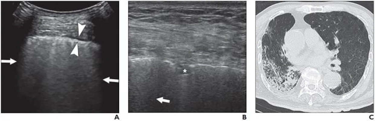
5. Viscus perforation


Physiologic air can be seen in the lumen of the bowel as small stars. Larger air bubbles can appear as hyperechoic stripes generating comet tail artefacts (these are rare in the small bowel but frequent in the large bowel), much like a linear view of the lung would look. Air artefacts can emanate from the thoracic cavity and the lung over the liver. Pathological air, however, may produce an enhanced peritoneal stripe sign (EPSS), reverberation artefacts and ring-down artefacts (Figures 2a and 2b) (Hoffmann et al. 2012).

6. Renal dysfunction


International guidelines recommend that all patients who present in acute renal failure undergo an ultrasound examination to ascertain its cause (Kidney Disease: Improving Global Outcomes 2012). Hydronephrosis due to obstructive uropathy is a reasonably straightforward diagnosis to make.
More advanced techniques include the assessment of blood flow within the renal artery and vein using Doppler analysis.

Analysis of the urinary bladder should be performed as well. The bladder can be empty, filled or distended (globus). The position of the bladder catheter balloon can be checked, as well as the presence of hyperechogenic structures (debris, tumour, blood clot etc).

7. Liver and spleen 


Ultrasound of the liver is divided into general US views including anatomic views of the liver, gallbladder and biliary tree. Pathology within these organs e.g. acute liver failure, can result in intensive care admission, but is beyond the scope of this paper.

Advanced modalities

1. Doppler and colour Doppler techniques


Doppler US is used to assess the signal from visceral vessels that supply the gastrointestinal (GI) tract and smaller vessels within the intestinal wall. Although the technique cannot be used to assess capillary flow, it can be used to analyse all the major visceral vessels e.g. renal, hepatic, mesenteric. It has to be noted that normal bowel wall perfusion cannot be demonstrated by colour or power Doppler. The presence of flow in the bowel wall points towards pathologic perfusion (e.g. hyperaemia in actively inflamed segments, as seen in appendicitis).
Commonly used measurements which can be performed include:
  • Systolic, diastolic and mean velocities
  • Pulsatility index
  • Resistance index (peak systolic velocity – end diastolic velocity)/peak systolic velocity
  • Blood flow volume

GI tract blood flow


Colour Doppler allows for the assessment of mural flow, the absence of which is a sign of ischaemia. Unfortunately, this finding is only reported in 20–50% of patients with a proven diagnosis of ischaemic colitis (Danse et al. 2000a; Danse et al. 2000b). Doppler US can show stenosis, emboli, and thrombosis in the near visible parts of the coeliac trunk, the superior mesenteric artery (SMA) and the inferior mesenteric artery (IMA). In the early phase of bowel ischaemia, US examinations may show SMA occlusion, hyperaemic segments and bowel spasm. Collateral vessels cannot be reliably displayed using ultrasound. Systolic velocities of more than 250–300 cm/s are sensitive indicators of severe mesenteric arterial stenosis (Hamada et al. 2014; Koenig et al. 2011). The spectral analysis of Doppler signals of arteries supplying the GI tract (truncus coeliacus, superior and inferior mesenteric arteries) and the vessels draining the intestine, can be used to estimate bowel perfusion (see below). Assessment in transverse and longitudinal plane should be performed. Low flow states can also be identified by the presence of spontaneous contrast and turbulent flow in the large vessels.

Hepatic blood flow


Portal vein: the normal main portal vein (MPV) is gently undulating with peak systolic velocities ranging between 20 cm/s and 40 cm/s. A low flow velocity of <16 cm/s in addition to a calibre increase in the MPV are diagnostic features of portal hypertension (PH). Further worsening of PH leads to a to-and-fro flow pattern, whereby the nearly stagnant blood column in the portal veins is seen to shift into and out of the liver with the respiratory cycle. In the end stages, stagnation of the blood column can lead to thrombosis or progress to a frank flow reversal or non-forward portal flow (NFPF). This is considered to have grave prognostic significance, indicating severe and irreversible liver failure (Wachsberg et al. 2002).

Hepatic vein: the normal flow is triphasic with two hepatofugal phases related to atrial and ventricular diastole. Fibrotic or inflammatory changes may create a monophasic flow pattern. Early waveform changes in cirrhosis patients include spectral broadening and dampening of the normal, retrograde, pre-systolic wave of the hepatic vein waveform. Later, the normal triphasic waveform pattern may be diminished or replaced with a monophasic pattern. Therefore the monophasic hepatic vein waveform indicates relatively high portal pressures (Ralls 1990).
Hepatic artery: hepatic arterial resistance changes with increasing portal pressure values, but hepatic arterial resistive indices correlate poorly with the severity of cirrhosis and will not be further discussed here.

Renal blood flow


Doppler US can be used to assess renal perfusion. Normal resistive index (RI) is approximately 0.58 ± 0.10 and values >0.70 are considered to be abnormal. A renal Doppler RI may also help in detecting early renal dysfunction or predicting short-term reversibility of acute kidney injury (AKI) in critically ill patients. A recent meta-analysis suggested that RI may be a predictor of persistent AKI in critically ill patients with a pooled sensitivity and specificity of 0.83 (95% CI, 0.77-0.88) and 0.84 (95% CI, 0.79-0.88) (Ninet et al. 2015).

Increased renal resistive index (RRI) has been proven to be an independent predictor of worse cardiovascular and renal outcomes, especially when combined with reduced glomerular filtration rate (GFR), thus providing a useful diagnostic complement to the assessment of renal function in these patients. High RRI has also been correlated with the presence of hypertensive and atherosclerotic organ damage. Values >0.80 have been reported to be predictive of all-cause mortality in chronic kidney disease patients and may indicate impending renal transplant failure in this patient subset (Barozzi et al. 2007; Guinot et al. 2013; Ninet et al. 2015; Schnell et al. 2012).

Gastrointestinal and urinary tract sonography (GUTS) protocol


Gastrointestinal function can be assessed with US, using a combination of anatomical, functional and blood flow evaluation (Schmidt et al. 2005).
  1. Function: peristalsis, bowel motility, gastroparesis, small bowel ileus, large bowel paralysis
  2. Dimensions: bowel dilatation, bowel obstruction, Ogilvie syndrome, bacterial overgrowth, toxic megacolon, bowel wall oedema, abdominal wall oedema
  3. Collections: bowel content (blood, liquid, air, solid), haematoma, gastrointestinal bleeding, ascites
  4. Perfusion: bowel ischaemia, hepatosplanchnic perfusion, shock state (spontaneous contrast), renal resistive index, abdominal perfusion pressure (APP) = mean arterial pressure (MAP)-intraabdominal pressure (IAP)
This approach is summarised by the GUTS (Gastrointestinal and Urinary Tract Sonography) protocol (Figure 3). The structured and stepwise approach may lead to improved practical management of adult ICU patients with acute gastrointestinal injury (AGI), as graded by the European Consensus Definitions (Reintam Blaser et al. 2012). Such a management strategy has not been shown to improve patient outcome, however.


The European Consensus Definition of AGI suggests a graded severity score:
  1. AGI grade I represents a self-limiting condition with increased risk of developing GI dysfunction or failure
  2. AGI grade II (GI dysfunction) represents a condition requiring interventions to restore GI function
  3. AGI grade III (GI failure) represents a condition when GI function cannot be restored with interventions
  4. AGI grade IV represents a dramatically manifesting GI failure, which is immediately life-threatening (e.g. abdominal compartment syndrome with organ dysfunction) (Reintam Blaser et al. 2012).


Future works


Contrast-enhanced ultrasound


Contrast-enhanced ultrasound (CEUS) involves the use of contrast agents containing gas-filled microbubbles administered intravenously, producing an image with greater contrast and/or highlighting more vascular areas. Although reasonably well established in radiology, and more recently cardiology departments, its use in intensive care is in its infancy. Unlike CT contrast agents, CEUS appears safe for patients with renal dysfunction and the modality itself remains free of radiation exposure to patients. Possible use within critical care includes enhanced echocardiography and in blunt abdominal trauma to assess solid-organ injuries (Dietrich 2017).

 
Doppler analysis as a marker of fluid status and venous congestion


As mentioned, Doppler analysis of the vasculature of specific abdominal organs allows for assessment of its perfusion. Some early work showed that this modality could also be used as a marker of systemic vascular congestion (Lewis et al. 1989).

Conclusion

 This paper summarises the multiple uses of abdominal US on the ICU and highlights future work and development. It must be remembered however that despite the myriad of potential diagnosis, utilisation and interpretation of such techniques requires training and experience.

Acknowledgements and conflicts of interest 


Jonny Wilkinson is a member of the International Fluid Academy (IFA) faculty. Manu Malbrain is founding President of WSACS (The Abdominal Compartment Society) and current Treasurer, he is also member of the medical advisory Board of Getinge (former Pulsion Medical Systems) and Serenno medical, and consults for ConvaTec, Acelity, Spiegelberg and Holtech Medical. He is co-founder and member of the executive committee of the International Fluid Academy (IFA). 

Adrian Wong is a member of the executive committee of the IFA.

Abbreviations 

FAST Focused Assessment with Sonography in Trauma 
GI gastrointestinal 
POCUS point-of-care ultrasound 
US ultrasound

Thứ Năm, 6 tháng 8, 2020

Pediatric EDs using more ultrasound, MRI instead of CT

By Erik L. Ridley, AuntMinnie staff writer

August 3, 2020 -- Although pediatric emergency departments (EDs) performed fewer CT exams between 2009 and 2018, their utilization of advanced imaging increased overall due to expanded use of MRI and ultrasound exams, according to research published online August 3 in JAMA Pediatrics

A research team led by Dr. Jennifer Marin of UPMC Children's Hospital of Pittsburgh assessed the change in CT, ultrasound, and MRI rates in over 26 million ED visits archived in the Pediatric Health Information System administrative database from January 1, 2009, to December 31, 2018.

They found that the proportion of pediatric ED encounters that resulted in a CT exam decreased amid expanded use of ultrasound and MRI. But there was also considerable variation in utilization between practices.

"The magnitude of the decrease in CT use varied by site and was associated, in part, with the use of alternative advanced imaging modalities for certain diagnoses, most notably ultrasonography for abdominal conditions," they wrote. "Future efforts appear to be needed to standardize imaging approaches and evaluate the effect of the changing

landscape of advanced imaging on patient-level outcomes."

Change in use of advanced imaging in pediatric ED
 20092018
At least one advanced imaging study6.4%8.7%
CT3.9%2.9%
Ultrasound2.5%5.8%
MRI0.3%0.6%
*All differences were statistically significant (p < 0.001, p = 0.001, p < 0.001, and p < 0.001, respectively).

Delving further into the data, the researchers found that most of the decrease in CT occurred in eight all patient-refined diagnosis-related groups (APR-DRGs), led by concussion (-23%), appendectomy (-14.9%), ventricular shunt procedures (-13.3%), and migraine and other headaches (-12.4%). Declines in CT use also occurred in other disorders of the nervous system (-10.1%); abdominal pain (-6.1%); other ear, nose, mouth, throat, and cranial or facial diagnoses (-5.9%); and seizure (-5.3%).

The researchers also found that strategies to reduce abdominal CT use by performing ultrasound first for evaluating abdominal pain may be associated with the higher use of ultrasound for that application.

"Interestingly, the increase in the use of ultrasonography for these conditions was greater than the decrease in the use of CT," they wrote. "Therefore, a proportion of these ultrasonographic examinations likely represent overuse of a typically widely available and nonradiating imaging tool."

In other findings, the researchers observed wide variation among EDs in the use of ultrasound for appendectomy (median 57.5%), as well as MRI (median 15.8%) and CT (median 69.5%) for ventricular shunt procedures.

In terms of outcomes, the researchers found that hospitalization and three-day ED revisits decreased during the study period. However, the ED length of stay did not change.

While it's encouraging that lower utilization of CT avoided radiation exposure in nearly 28,000 fewer kids, the increase in the number of children receiving at least one form of advanced imaging was less encouraging from a healthcare value perspective, according to an accompanying editorial by Dr. Alan Schroeder and Dr. Alan Imler of Stanford University in Palo Alto, CA.

Although outcomes appeared to have improved somewhat during the study period, other secular trends -- such as heightened public awareness of concussions -- might also explain those outcomes, wrote Schroeder and Imler. Increased imaging among patients who were ultimately hospitalized also further suggests that the more imaging is not associated with reduced hospitalization. 

Thứ Tư, 5 tháng 8, 2020

USPSTF advises against carotid artery stenosis screening


By Theresa Pablos, AuntMinnie staff writer

August 5, 2020 -- The U.S. Preventive Services Task Force (USPSTF) is poised to once again recommend against screening for asymptomatic carotid artery stenosis. The task force reaffirmed its D rating in a draft recommendation statement published on August 4.

The USPSTF last weighed in on the topic in 2014, concluding with moderate certainty that the harms of screening for carotid artery stenosis in the general population outweighed the benefits. In its new draft recommendation statement, the agency reaffirmed that position, stating there was not enough new evidence to change its previous recommendation against screening with either carotid duplex ultrasound, CT angiography, or MR angiography.

"The USPSTF found no new substantial evidence that could change its recommendation and therefore reaffirms its recommendation," the task force wrote.

In theory, screening the general population for stenosis could lead to early detection of narrowed blood vessels, thus enabling medical professionals to conduct potentially life-saving interventions, such as carotid endarterectomy (CEA) and carotid artery stenting (CAS). But the USPSTF concluded that the evidence it reviewed didn't readily support that hypothesis.

The task force has consistently found limited evidence in favor of asymptomatic carotid artery stenosis screening, especially when compared with other medical therapies, such as statins and antihypertensive agents. And the evidence has been particularly lacking since the USPSTF's last review in 2014.

USPSTF draft recommendation rationale for asymptomatic carotid artery stenosis
DetectionUltrasonography has reasonable sensitivity and specificity for detecting clinically relevant carotid artery stenosis, but it also yields many false-positive results in the general population.
Scanning the neck for carotid bruits has poor accuracy for clinically relevant carotid artery stenosis.
BenefitsDirect evidence does not indicate that screening for asymptomatic carotid artery stenosis can improve stroke, mortality, or other adverse health outcomes.
Carotid endarterectomy (CEA) or carotid artery angioplasty and stenting (CAS) provides little or no benefit for improving stroke, myocardial infarction, mortality, or other adverse outcomes compared with current medical therapy.
HarmsWhile direct evidence does not show that screening for asymptomatic carotid artery stenosis can cause harm, there are known harms with confirmatory testing and interventions.
Direct evidence supports that treating asymptomatic patients with CEA or CAS could cause harms, including stroke or death.
Harms related to screening and treating asymptomatic carotid artery stenosis have small-to-moderate magnitude.

After searching the scientific literature, USPSTF investigators found no recent eligible studies that directly investigated the benefits or harms of asymptomatic carotid artery stenosis screening. The two studies that were conducted on the topic in the past six years were both prematurely terminated and produced mixed results.

When looking at the benefits and harms of CEA or CAS, the authors found an additional two national datasets and three surgical registries that met their inclusion criteria. Rates of 30-day postoperative stroke or death after CEA ranged from 1.4% to 3.5% depending on the registry or database. Similarly, 30-day stroke or death after CAS ranged from 2.6% to 5.1%.

Based on the evidence -- or lack thereof -- the investigators concluded there wasn't enough new information to change the D rating for asymptomatic carotid artery stenosis screening. However, they pointed out that two clinical trials are currently underway, which may shed light on the topic in the future.

"There were few new trials, all with methodologic concerns, examining the important question of the comparative effectiveness and harms of revascularization plus best medical treatment compared with best medical treatment alone," they wrote. "The ongoing CREST-2 and ECST-2 trials will be the largest trials to address this issue."

The draft recommendation is available for public comment through August 31. After the comment period has ended, the task force will publish its final recommendation.

Thứ Hai, 3 tháng 8, 2020

COVID-19 pandemic driving increase in ultrasound use


By Theresa Pablos, AuntMinnie staff writer

August 3, 2020 -- The COVID-19 pandemic is driving an increase in the use of lung ultrasonography among physicians in Italy, according to the results of a small survey published in the Journal of Ultrasound in Medicine.
More physicians said they're using ultrasound equipment now than before the pandemic, and experienced physicians are performing more lung ultrasound scans than ever before. The findings support anecdotal evidence that COVID-19 has increased interest in ultrasound, including among clinicians with no prior ultrasound experience.
"Thanks to online resources, many operators could e-learn and apply the technique," wrote the authors, Dr. Allessandro Zanforlin and Dr. Francesco Tursi, who are both members of the Italian thoracic ultrasound academy behind the survey (July 16, 2020, Journal of Ultrasound in Medicine).
The informal, one-week-long survey was conducted by Italian ultrasound society Academia di Ecografia Thoracia after the group noticed its membership spiked from 1,700 members in February to 4,000 members in May. The academy promoted its survey on social media and received 123 responses.
Survey respondents worked in a variety of settings, including on COVID-19 floors (34%), in intensive care units (31%), and in the emergency department (20%). The majority said they used lung ultrasound to monitor pneumonia (63%) and screen for COVID-19 (60%).
A total of 14% of respondents started using lung ultrasound exclusively because of the COVID-19 pandemic. Among these respondents, 81% said they learned how to perform lung ultrasound scans by following video tutorials or participating in webinars. The remaining 19% of respondents gained experience through expert mentoring or local courses.
As the number of patients with COVID-19 surged at hospitals in Italy, so too did the number of lung ultrasound exams. Respondents said the number of daily chest scans increased from an average of three per day before the pandemic to seven per day during the outbreak. For so-called "expert operators" with at least five years of lung ultrasound experience, the number of exams rose from five per day to nine per day.
The majority of respondents also said their lung ultrasound exams had increased in quality (58%) and accuracy (66%) during the pandemic. These percentages were even higher for participants who took an online course or webinar, with 83% of these respondents saying their exams increased in both quality and accuracy.
One factor driving the increase in lung ultrasound exams could be the availability of additional equipment thanks to donations and emergency purchase approvals. More than half of respondents said they acquired new ultrasound equipment during the pandemic, namely portable wheeled systems (37%) or handheld/wireless systems (19%).
The survey adds to the evidence that ultrasound is becoming an invaluable tool for care teams, especially those in Italy, who are treating patients with the novel coronavirus. However, the increase in skilled operators and new equipment may mean the modality will remain prominent for lung imaging even after the pandemic is over.
"What we are learning from this pandemic is the importance of [lung ultrasound] in the diagnosis, evaluations, and monitoring of pneumonia, which, in the hands of many physicians ... could improve the quality of the treatment of respiratory patients," the authors concluded.

Thứ Năm, 30 tháng 7, 2020

US correlates with COVID-19 severity, duration


By Theresa Pablos, AuntMinnie staff writer

July 28, 2020 -- Findings on lung ultrasound scans were correlated with COVID-19 severity and duration in a study published on July 23 in the American Journal of Roentgenology. In particular, pulmonary consolidations distinguished between patients with moderate and severe forms of the novel coronavirus disease


The study included dozens of patients who were consecutively treated for COVID-19 at a Chinese hospital in March. The findings may aid physicians in managing patients with moderate-to-severe COVID-19, the authors noted.
"Our results indicate that lung [ultrasound] findings can be used to reflect both the infection duration and disease severity," wrote the authors, led by Dr. Yao Zhang from Ditan Hospital in Beijing.
Zhang and colleagues enrolled 28 patients consecutively hospitalized for COVID-19 at their institution between March 1 and March 30. The patients all tested positive for the novel coronavirus on a nasopharyngeal test and underwent a bedside lung ultrasound scan in a sitting, supine, and decubitus position.
radiology graph
Every patient in the study had B-lines on their ultrasound scans, which indicate areas of increased interstitial fluids and decreased alveolar air. Another two-thirds of patients had pulmonary consolidation, and 61% had a thickened pleural line. Only one patient had a pleural effusion.
Pulmonary consolidation occurred significantly more often in patients with severe or critical COVID-19 than patients with moderate disease, the authors found. Almost 87% of patients with severe or critical disease had pulmonary consolidation on ultrasound, compared with just 46% of patients with moderate COVID-19.
Lung ultrasound image obtained with a convex probe. The outer arrows show confluent B-lines. The middle arrowheads point to a thickened pleural line.
(A) Lung ultrasound image obtained with a convex probe. The outer arrows show confluent B-lines. The middle arrowheads point to a thickened pleural line. (B) Lung ultrasound image obtained with a linear probe. The arrow points to a B-line. The star denotes a patchy pulmonary consolidation. (C) Chest CT image showing reticular and interlobular septal thickening and patchy, focal opacities associated with architectural distortion. Image courtesy of the American Journal of Roentgenology.
Furthermore, patients with a thickened pleural line had experienced a longer infection period than those without a thickened pleural line. Patients with fewer than 20 days between the day they first noticed COVID-19 symptoms and the day of their ultrasound scan were significantly less likely to have a thickened pleural line than those with a difference of 20 days or more.
The study findings add to the growing body of research demonstrating that severity on lung ultrasound scans can predict worse outcomes and even mortality for patients with COVID-19. The authors emphasized that ultrasound also has unique benefits over other imaging modalities, including CT, for treating patients with the novel coronavirus.
"[Ultrasound] is repeatable in critically ill patients, which ensures that monitoring of the severity of the disease and the effects of therapies can be easily carried out," they wrote. "This capability is particularly important in situations in which chest CT is not available, such as in isolation wards and [intensive care units]."
The authors cautioned that their study focused on a small number of patients and did not evaluate how ultrasound findings might have changed over time. They hope future studies will continue to study the use of lung ultrasound for COVID-19, particularly how lung ultrasound scoring systems might improve COVID-19 assessment and treatment.
"Lung [ultrasound] was highly sensitive for detecting abnormalities in patients with COVID-19, and B-lines, a thickened pleural line, and pulmonary consolidation were the most commonly observed features," the authors concluded.

Thứ Sáu, 24 tháng 7, 2020

A New Standardize Doppler waveform reporting


By Theresa Pablos, AuntMinnie staff writerJuly 24, 2020 -- A new set of guidelines aims to standardize the terminology used to report arterial and venous spectral Doppler ultrasound waveforms. The document was jointly published on July 15 in Vascular Medicine and the Journal for Vascular Ultrasound.

The statement creates a designated set of key terms to describe findings on spectral Doppler ultrasound waveforms, the main diagnostic assessment for arterial and venous diseases. It was written by sonographers, vascular specialists, and other experts commissioned by the Society of Vascular Medicine and Society of Vascular Ultrasound.
"The hope of the writing committee is that this document will help us all to 'speak the same language,' and thereby advance the field of vascular ultrasound and improve patient care," stated lead study author Dr. Esther Kim, vascular labs medical director at Vanderbilt University Medical Center, in a press release.
The lack of shared nomenclature has been an ongoing problem for vascular ultrasound professionals. In fact, one out of five ultrasound professionals has had to perform a repeat arterial Doppler ultrasound examination because of terminology differences, according to a survey cited in the consensus statement.
"Over a decade ago, the lack of a standardized nomenclature to describe spectral Doppler waveforms was demonstrated to result in confusion amongst ultrasound professionals," Kim stated. "Not surprisingly, this can lead to negative clinical outcomes."
In the consensus statement, the committee established three major descriptors for ultrasound waveforms: flow direction, phasicity, and resistance for arterial waveforms and flow direction, flow pattern, and spontaneity for venous waveforms.

Major descriptors for arterial ultrasound waveforms
Flow directionAntegrade
  • Blood flows in normal direction
  • Previously known as forward flow
Retrograde
  • Blood flows in opposite direction
  • Previously known as reverse flow
Bidirectional
  • Blood enters and leaves through the same opening
  • Previously known as to-fro
Absent
  • No detected blood flow
PhasicityMultiphasic
  • Waveform crosses zero-flow baseline
  • Previously known as triphasic or biphasic
Monophasic
  • Waveform does not cross zero-flow baseline
  • Blood flows in single direction
ResistanceHigh resistive
  • Sharp upstroke and brisk downstroke
Intermediate resistive
  • Visible end-systolic notch
  • Continuous flow above the zero-flow baseline
Low resistive
  • No end-systolic notch
  • Prolonged downstroke in late systole

Major descriptors for venous ultrasound waveforms
Flow directionAntegrade
  • Blood flows in normal direction
  • Previously known as central or forward flow
Retrograde
  • Blood flows in opposite direction
  • Previously known as peripheral or reverse flow
Absent
  • No detected blood flow
Flow patternRespirophasic
  • Flow velocity related to respiratory cycle
  • Previously known as respiratory phasicity
Decreased
  • Respirophasic flow with less variation than expected
  • Previously known as dampened or blunted
Pulsative
  • Flow velocity is inversely linked to cardiac cycle
  • Previously known as cardiophasic
Continuous
  • Respiratory/cardiac cycles do not affect flow velocity
  • Steady Doppler signal with minimal variation
Regurgitant
  • Flow velocity varies with cardiac cycle
SpontaneitySpontaneous
  • Blood flows without external influence
Nonspontaneous
  • Blood flows only with external maneuvers
The statement also established terms that can be used to modify the main descriptors. For arterial waveforms, the seven modifying terms are as follows:
  1. Rapid upstroke -- Near vertical rise to peak systole
  2. Prolonged upstroke -- Abnormally gradual slope to peak systole; previously known as tardus, delayed, or damped upstroke
  3. Sharp peak -- Single, well-defined peak
  4. Spectral broadening -- Widening of the velocity band or filling in the typically clear area under the systolic peak; previously known as nonlaminar, turbulent, disordered, or chaotic
  5. Staccato -- High-resistance pattern with a short, low-amplitude diastolic signal punctuated by spikes of acceleration and deceleration
  6. Dampened -- Abnormal upstroke and peak, typically with decreased velocity; previously known as parvus et tardus, attenuated, or blunted
  7. Flow reversal -- Flow that changes direction but not as part of normal flow, can be transient or consistent with the cardiac cycle; previously known as pre-steal, competitive flow, or oscillating
For venous waveforms, the three modifying terms as follows:
  1. Augmentation -- Changes in flow velocity related to physical maneuvers, can be described as normal, reduced, or absent augmentation
  2. Reflux -- Persistent retrograde flow beyond normal closure time
  3. Fistula flow -- Flow with an arteriovenous fistula that becomes pulsatile due to communication with artery, sharp peaks often appear as pulsatile; previously known as arterialized or fistulous
In addition to creating the key descriptors and modifiers, the statement defined the reference baseline for spectral Doppler waveforms as the zero-flow baseline. It also advised against using the terms "normal" or "abnormal" to describe a waveform, since what is normal will depend on the part of the body and situation.
The statement also instructed sonographers to use image optimization techniques to acquire quality Doppler waveforms. This includes using an optimal transducer-to-vessel angle, the normal peripheral artery systolic waveform acceleration of 0.2 seconds, and proper transducer support.
Finally, the committee advised sonographers to provide complete descriptions for referring providers, including indication, relevant history, velocity measurements, and waveform characteristics. Sonographers should also include a conclusion with the clinical indication.
"We hope that this new Doppler waveform nomenclature will eliminate confusion and lead to appropriate diagnosis and better patient care," stated Dr. Raghu Kolluri, president of the Society of Vascular Medicine.

Thứ Sáu, 17 tháng 7, 2020

NEGATIVE Y SIGN=NONRECURRENT LARYNGEAL NERVE


POCUS Findings can Predict COVID-19 Death Risk


By Theresa Pablos, AuntMinnie staff writer
July 17, 2020 -- The findings on initial lung scans with point-of-care ultrasound (POCUS) can predict which patients with COVID-19 are at a greater risk of death, according to a prospective study from Italy published on July 15 in Ultrasound in Medicine & Biology.
Physicians in Rome performed lung ultrasound scans on 41 adult patients who visited a tertiary emergency department with symptoms of the novel coronavirus disease. Patients who later died or were later admitted to the intensive care unit (ICU) had significantly worse pathological findings on their initial scan.
"Our study shows that [lung ultrasound scan] is able to detect COVID-19 pneumonia and to predict, during the first evaluation in the emergency department, patients at risk of intensive care unit admission and death," wrote the authors, led by Nicola Bonadia, from the department of emergency medicine at Agostino Gemelli University Policlinic.
Physicians performed point-of-care ultrasound (POCUS) scans on all patients with suspected cases of COVID-19 who visited the emergency department in March. The emergency department staff used a pocket device with a wireless 6-MHz convex probe and followed a previously described lung ultrasound protocol that includes 14 chest areas.
Each scanned area received a numeric score of 0 to 3 based on the severity of the findings in that section. A higher lung ultrasound score (LUS) signified worse disease severity, with a score of 3 indicating dense or large areas of white lung with or without subpleural consolidations.
The authors analyzed the lung ultrasound findings from 41 patients with a positive SARS-CoV-2 test result and known outcomes. They specifically excluded children and patients with less than six months life expectancy due to preexisting chronic conditions, such as advanced cancer or dementia.
More than 90% of adult patients with COVID-19 had at least one area with an abnormal lung ultrasound finding. Pathological findings occurred in all 14 scanned areas but were most prominent in the lateral lung regions, the authors noted.
radiology graph
Patients with fatal cases of COVID-19 had pathological findings in 100% of scanned areas, compared with just 50% of scanned areas in discharged patients. These patients also had a mean lung ultrasound score of 1.43, compared with 1.0 in discharged cases.
Similarly, patients admitted to the ICU had pathological findings in 93% of scanned areas, compared with just 20% of areas in patients not admitted to the ICU. ICU patients also had a mean LUS of 1.36, compared with 1.0 in non-ICU patients.
Based on their findings, the authors determined the cutoff for a strict definition of COVID-19 pneumonia should be an LUS of 0.4 and a pathological findings rate of at least 20%. Furthermore, no study participants died if they had a mean LUS less than 1.1 and an average pathological rate under 70%.
The authors cautioned their study took place at one institution and had a small sample size. Nevertheless, the findings may help health professionals better triage patients with COVID-19 and spur future studies to evaluate whether lung ultrasound scores can guide patient treatment and admission decisions.
"We found a significant correlation between ultrasound findings and severity of the disease, assessed as mortality and need for ICU admission," the authors wrote. "To our knowledge, this is the first study describing the predictive role of LUS in patients with COVID-19."

Thứ Tư, 8 tháng 7, 2020

US SWE ở trẻ em


In fact, for pSWE and 2D-SWE experience in B-mode US is mandatory. Data acquisition should be undertaken by specialists. Operators experienced both in ultrasonography and elastography are needed to obtain reliable liver stiffness measurements in children, considering the different anatomy, especially in babies (liver situated lower in the abdomen), and the fact that cooperation from a small child is sometimes difficult. The location for measurements can be more difficult to establish in children and here the operator’s experience can play a role.


Neonatal brainSome early reports on the use of transcranial SWE of the periventricular brain parenchyma, in preterm infants and infants with hydrocephalus, suggest that SWE is possible and technically feasible [101,102] (fg 5, fg 6).
Albayrak et al showed that differences between brain stiffness values in preterm and term neonates can be demonstrated by using 2D-SWE. Brain stiffness measured from both the thalamus and periventricular white matter were found to be signifcantly lower in preterm neonates compared with term neonates (cut-off values for determining prematurity less than 8.28 kPa for mean
thalamus stiffness and less than 6.59 kPa for periventricular white matter stiffness). The authors suggested that the results might be reference points for evaluating neonatal brain stiffness in research on patients with various illnesses. 2D-SWE also seems to have the ability to depict increased intracranial pressure (ICP) in infants, with a positive linear correlation between SWE values and ICP
[102]. Infants with ICP seem to have increased 2D-SWE values (mean 24.2±5.1 kPa) compared to healthy infants (mean 14.1±6.6 kPa). However, larger prospective studies are still not available. If these preliminary observations of the benefts of transcranial SWE of the neonatal brain will be confrmed by further studies, SWE might be a useful method for additional diagnostic imaging and
monitoring in premature infants and children with proven or suspected increased ICP. When performing SWE of the neonatal brain, potential risks and harms of applying high energy levels by US to the neonatal brain should be considered. Recently, an experimental study on mice dealing with the potential biological effects associated with 2D-SWE on the neonatal brain was published [103].
The results indicated that 2D-SWE does not cause detectable histologic changes in the brain of neonatal mice, nor does it have long-term effects on the learning and memory abilities. However, some temporary effects were observed when the scanning lasted for more than 30 min. Thus, it is recommended to pay attention to the scanning duration when assessing neonatal brains with 2D-     SWE elastography.
  
The examiner should acquire appropriate knowledge and training in US elastography [104,105]. The operator Fig 5. SWE of the neonatal brain in a healthy newborn (14 days old). Sagittal view of the periventricular region in a healthy newborn. B-mode shows no abnormalities 
(a). 2D-SWE shows a mean periventricular tissue stiffness of 13.5 kPa and a maximum value of 14.8 kPa 
(b). must distinguish a good B mode US image from suboptimal images. 

Thứ Tư, 1 tháng 7, 2020

3D ultrasound is effective for measuring blood flow


AuntMinnie.com  

July 1, 2020 -- Researchers from across the U.S. confirmed the efficacy of 3D color-flow ultrasound for cheaply and reliably measuring blood flow. The findings, which were published on June 30 in Radiology, could help physicians precisely measure blood flow for people with chronic conditions.
The new approach resulted in as little as a 3%-5% difference in measurements between laboratories and operators. Based on the findings, lead study author Oliver Kripfgans, PhD, said the question of the technology's clinical adoption isn't an "if" but a "when."
"These are fantastic results that show that, from a technology point of view, some systems could be ready to go to the clinic," stated Kripfgans, an associate professor of radiology at Michigan Medicine in Ann Arbor, MI, in a press release.
2D ultrasound technology is rarely used to gather precise, quantitative blood flow measurements because results can greatly vary between facilities and operators. In addition, other noninvasive techniques to measure blood flow, such as blood pressure monitoring, can only provide qualitative data.
Kripfgans and his colleagues at Michigan Medicine had previously developed a 3D color-flow ultrasound approach to overcome the limitations of alternative measurement solutions. The researchers tested the method in the new, prospective study, which received funding from the U.S. National Institutes of Health, RSNA, and the American Institute of Ultrasound in Medicine.
For the study, Canon Medical Systems, Philips Healthcare, and GE Healthcare donated 3D ultrasound systems. The vendors modified the systems with data access software and provided the researchers with color-flow velocity, color-flow power, and scan geometry.
Trained medical or engineering staff at three different locations performed the same experimental steps using the three systems:
1.They measured volume flow from 1 - 12 mL/sec in steps of 1 mL/sec at a depth of 4 cm.
2.They measured depth-dependence from 2.5 - 7.5 cm in steps of 0.5 cm.
3.They stepped color flow gain from no color to full blooming.
4.They measured flow volume distal to a lumen stenosis. They also measured three flows at each poststenosis position.

Example screenshot for poststenotic flow. Image courtesy of the RSNA.
The experiments were first performed with a steady, constant flow, then repeated again with a flow simulating a pulse of 60 beats per minute. In the end, the researchers had 730 datasets with 18,450 images.


The use of 3D color-flow ultrasound produced measurements that were accurate and reproducible. Two out of the three ultrasound systems tracked within 10% for measuring flow response, the authors noted.
The approach is promising because it requires no hardware changes and can lead to practical clinical uses for measuring peripheral vascular flow and cerebral blood flow. While it will need to be further evaluated and may not work for all types of blood flow measurements, Kripfgans is optimistic about its clinical potential.
"Once the technique becomes available commercially on scanners, clinical adoption will be much faster because then it's not a research project anymore -- it's something that's readily available, and after that, it's just a matter of time before it reaches the clinic," Kripfgans stated.