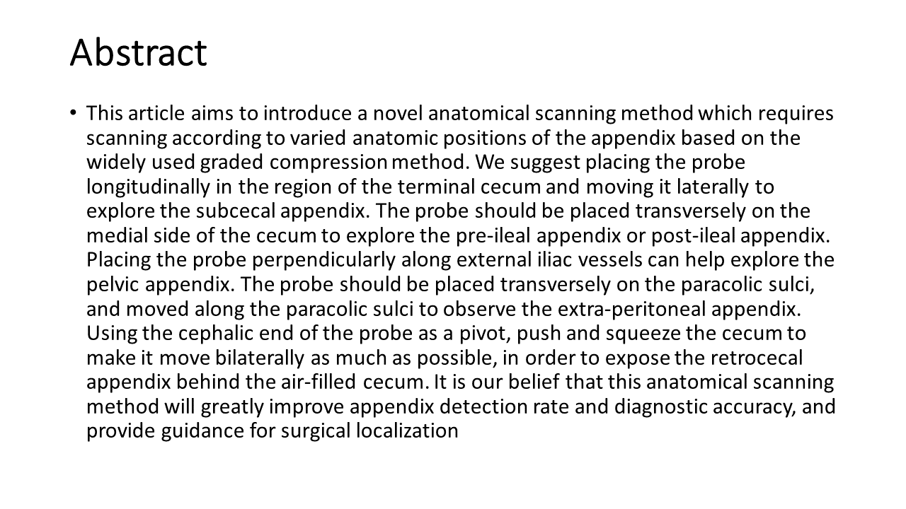
By Theresa Pablos, AuntMinnie staff writer
August 21, 2020 -- Could ultrasound measurements of the carotid lumen diameter be a better measure than carotid intima-media thickness (CIMT) for predicting mortality from cardiovascular disease? Lumen diameter indeed could add new information, according to a study in the
Journal of the American Heart Association.
Researchers from Germany found that lumen diameter measurements of the carotid artery derived from ultrasound scans predicted mortality from both cardiovascular disease and from all causes. And they believe that it provides more information than CIMT.
CIMT has been used as a noninvasive biomarker for
cardiovascular disease risk stratification and the risk of future cardiovascular events, the authors report. Both the coronary and carotid arteries distend during the early stages of atherosclerosis, a phenomenon that can be detected and measured on ultrasound scans.
But recent research has raised doubts about the reliability of CIMT for predicting individual outcomes. The new evidence, published on August 4, demonstrates that lumen size might be a better predictor of death from both cardiovascular and noncardiovascular events, with larger lumens indicating higher risk.
"Our results suggest that [lumen diameter] may be superior to CIMT," wrote the authors, led by Dr. Felix Fritze from the University of Greifswald's medical school in Greifswald, Germany.
The team of German researchers compared the effectiveness of CIMT and lumen diameter using data from a prior study that conducted baseline screenings and 10- and 15-year follow-up exams on the population of a German village.
As part of the baseline assessment, individuals underwent carotid ultrasonography. The original research team also recorded relevant health information, including cholesterol levels, diabetes status, and mortality outcomes.
For the new analysis, Fritze and colleagues created various models to analyze data from 2,751 participants, including 506 who died during the original study. Their further cardiovascular mortality analysis used outcomes from all but 214 of the participants with unknown causes of death.
The researchers found that individuals with the largest CIMT measurements had the highest hazard ratio (HR) for all-cause mortality, at 1.73, compared with a hazard ratio for lumen diameter of 1.29 and the combination of CIMT and lumen diameter at 1.26.
The model using lumen diameter alone was significantly associated with death from all causes, deaths attributed to cardiovascular events, and deaths attributed to noncardiovascular events. On the other hand, the model using CIMT alone was significantly associated with all-cause mortality and noncardiovascular mortality -- but not cardiovascular mortality.
To help determine the likelihood of their models predicting future values, the authors conducted an Akaike information criterion (AIC) analysis. In this analysis, the lumen diameter model came out on top for both all-cause mortality and cardiovascular mortality. It also came in second for cardiovascular mortality, just behind a model that combined lumen size and CIMT values.
Furthermore, the lumen diameter model remained significant for all-cause mortality even after the researchers excluded people with chronic kidney disease, prior myocardial infarction, and type 2 diabetes. The same wasn't true for the CIMT model.
| Lumen diameter vs. CIMT for predicting mortality |
| Model | Rank (AIC) |
| All-cause mortality | Cardiovascular mortality | Noncardiovascular mortality |
| Lumen diameter | No. 1 | No. 1 | No. 2 |
| Lumen diameter + CIMT | No. 2 | No. 2 | No. 1 |
| CIMT | No. 3 | No. 5 | No. 3 |
| None | No. 4 | No. 4 | No. 4 |
| Lumen diameter ÷ CIMT | No. 5 | No. 3 | No. 5 |
The authors do not know why the model using lumen diameter performed much better than CIMT in their analysis. They hypothesized it could be because lumen size is related to CIMT but is also much easier to measure.
"The larger caliber of [lumen diameter] compared with CIMT may improve manual measurement accuracy and thus may be more applicable for an outpatient setting," they wrote.
It's important to note the study only included white participants from one part of Germany, so the results may not be applicable to a more diverse population. As a result, the authors called for follow-up research with more robust patient populations to verify their findings.
"To the best of our knowledge, this is the first study to compare the informative value of CIMT and [lumen diameter] with regard to all-cause, cardiovascular, and noncardiovascular mortality associations," they concluded. "We report that [lumen diameter] provides more information than CIMT."