Tổng số lượt xem trang

Thứ Tư, 26 tháng 5, 2021

US AI model can help evaluate chronic kidney disease


By Erik L. Ridley, AuntMinnie.com staff writer

May 24, 2021 -- Artificial intelligence (AI)-based analysis of kidney ultrasound studies could serve as a first-line method for evaluating patients with chronic kidney disease, according to research published online May 24 in JAMA Network Open.


team of researchers led by Dr. Ambarish Athavale of Cook County Health in Chicago developed a deep-learning algorithm that yielded approximately 90% accuracy on a test set for quantifying interstitial fibrosis and tubular atrophy (IFTA).

"This article provides proof-of-principle that a [deep-learning] system can be used to noninvasively, accurately, and independently predict IFTA grade in patients with kidney disease," the authors wrote. "Although the system in its current form may not be an alternative to kidney biopsy, after robust external validation, a [deep learning]-based, noninvasive assessment of IFTA has the potential to significantly enhance clinical decision-making and prognostication in patients with CKD."

A strong indicator for decline in kidney function, interstitial fibrosis and tubular atrophy is currently measured using histopathological assessment of a kidney biopsy core. There currently isn't a noninvasive test for IFTA, according to the researchers.

The authors utilized AI to test their hypothesis that subtle signs of IFTA are ingrained within kidney ultrasound images and could be quantitatively extracted and analyzed. A deep-learning algorithm was trained and tested to segment the kidney and classify IFTA using 6,135 consecutive Crimmins-filtered kidney ultrasound images acquired at their institution between January 1, 2014, and December 31, 2018. The longitudinal images were obtained from both kidneys and were acquired between six months before and two weeks after kidney biopsy.

Of the total image dataset, 5,122 were used for training and 401 were used for validation. The researchers then tested the model on 612 images. The algorithm was 91% accurate for segmenting the kidney ultrasound images.

Performance of AI algorithm for quantifying IFTA on kidney ultrasound
 Image levelPatient level
Precision0.8930.900
Recall0.8040.842
Accuracy0.8680.896
F1 score0.8390.864

In other results, the researchers noted that the algorithm's accuracy remained high irrespective of the timing of the ultrasound studies and the biopsy diagnosis. Also, adding baseline clinical characteristics into the model's analysis didn't significantly improve its performance.

"From a clinical standpoint, it is foreseeable that a [deep-learning] system such as the one developed in this study has the potential to act as a gatekeeper for rationalizing the decision to conduct a kidney biopsy in patients with CKD," the authors wrote. "We anticipate that because of the ability of this system to provide [a] probabilistic estimate of IFTA in real-time, the system is likely to be acceptable (because it is unlikely to put any time burden on the technicians) and can also reduce the costs associated with kidney biopsy."

The researchers acknowledged that more work is needed to improve the accuracy of the model before it's ready for clinical use. Furthermore, the algorithm needs to be validated on external datasets to assess its performance across varying clinical settings.

Thứ Tư, 19 tháng 5, 2021

REVERBERATION ARTEFACTS IN LUNG US, WFUMB POSITION PAPER


Abstract
The analysis of vertical reverberation artefacts is an essential component of the differential diagnosis in pulmonary ultrasound. Traditionally, they are often, but not exclusively, called B-line artefacts (BLA) and/or comet tail artefacts (CTA), but this view is misleading.
In this position paper we clarify the terminology and relation of the two lung reverberation artefacts BLA and CTA to specifc clinical scenarios. BLA are defned by a normal pleura line and are a typical hallmark of cardiogenic pulmonary edema after exclusion of certain pathologies including pneumonia or lung contusion, whereas CTAs show an irregular pleura line representing a variety of parenchymal lung diseases. The dual approach using low frequency transducers to determine BLA and high frequency transducer to determine the pleural surface is recommended.

Keywords: lung ultrasound; artefact; B-lines; comet tails; guidelines; misdiagnosis


Suggested approach:

The transducer should be positioned such that the emenating ultrasound beam perpendicularly intersects
the surface of the lung to maximize likelihood of seeing all BLA and CLA as well as A line artifacts (fig 1).
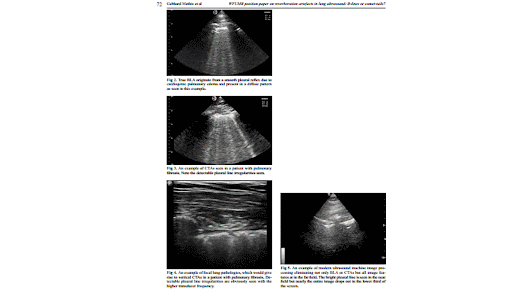
A recent study highlighted the potentially detrimental effects of placing the focal zone below the pleural line,using spatial compounding, higher frequency and tissue harmonics [14]. Once machine settings and transducer orientation have been optimized, we suggest that two most important and distinct vertical lung artefacts should be differentiated: BLA and CTA. While true BLA (fig 2) originate from a smooth pleural reflex due to cardiogenic pulmonary edema and present in a diffuse pattern, CTAs are seen in many lung disorders with irregular and fragmented pleural reflexes and can be focal or diffuse (fig 3).
Hence, the initial step should be to determine if there is evidence for diffuse pulmonary disease or defned focal or localized pathology. Focal lung pathologies by defnition should display vertical artifacts that are consistent with CTAs (fig 4).
Diffusely distributed vertical reverberation artefacts can be divided into two groups: with or without detectable pleural line irregularities and with stable or distally widening width:

1. The reverberation artefact (evaluated by low frequency transducer <5 MHz without interfering presets) is called
BLA if arising from a smooth pleural line (evaluated by high frequency transducer ≥10 MHz). The BLA arises from edema within the interstitium, is well defned with stable width, hyperechoic and extending indefnitely (the entire depth, at least 10 cm), erasing A-lines and moving with lung sliding. It is important to realize that many modern ultrasound machines have post-processing and other features which will eliminate not only BLA but essentially all discernable image detail near the bottom of the screen at greater depths (fig 5).
2. The reverberation artefact is called CTA if arising from an irregular (or fragmented) pleural line (evaluated by high frequency transducer ≥10 MHz), changes in width (such as e a comet with narrow head and wide tail), is well defned, hyperechoic, and extending defnitely (<10 cm in depth) (evaluated by low frequency transducer <5 MHz without interfering presets). It is important to make sure image compounding is turned off to make sure the CTA is not distorted farther field [14].
The differentiation of BLA from CTA is also dependent on the technical adjustments of several external factors, including the type of ultrasound machine, transducers and probe frequencies [6].


In conclusion, the correct diagnosis of pulmonary edema (the etiology of which may be decided upon
through integration of ultrasound data with clinical presentation) in the emergency setting is crucial for the correct management of the patient. The differentiation between ultrasonographic BLA and CTA, using two types (high and low frequency) of transducers allows accurate differentiation between pulmonary edema and other cause of diffuse pulmonary pathology. Both can lead to acute respiratory failure but may require different clinical management. Localized pulmonary diseases representing with CTA are distinguished. Mixed forms of diffuse,but also diffuse and focal lung diseases have to be considered.


Thứ Hai, 10 tháng 5, 2021

US accurate in diagnosing hand injuries

By Amerigo Allegretto, AuntMinnie.com staff writer


May 10, 2021 -- Ultrasound can accurately diagnose hand injuries while also being a fast, inexpensive, and potentially indispensable dynamic tool, according to research published April 29 in Ultrasound in Medicine and Biology.

Examining hand tendon injuries with sonography showed 100% accuracy, sensitivity, and specificity for diagnosing full-thickness hand tendon tears, as well as tenosynovitis of hand flexor tendons, according to a study led by Dr. Chris Nabil Hanna Bekhet from Ain Shams University in Cairo, Egypt.

"It also provides data that are important before diagnostic surgical exploration, and the process consumes less time than traditional wound exploration techniques or MRI," the authors wrote.

Hand and wrist injuries make up 28% of all musculoskeletal injuries and account for 14% to 30% of all patients treated in the emergency department. Tendon injuries are the second most common injuries, within injuries to the flexor tendons having debilitating consequences and high rates of reoperation.

Assessing hand injuries through clinical examination can overlook tendon injuries, and surgeons sometimes opt for explorative surgical methods to detect tendon injuries.

While using ultrasound to examine tendon injuries in the emergency department has its advantages, including eliminating the need for surgical approaches, it is not yet readily adopted by surgeons. This could be because clinicians lack education on how to use ultrasound to resolve clinical questions on the state of the tendon.

The study included 35 patients between September 2018 and January 2020 with trauma to the ventral surface of the hand and wrist who were presented to emergency departments or outpatient clinics. The subjects ranged from 18 to 58 years of age, with 24 patients being male and the other 11 being female.

The researchers examined 50 injured tendons in all flexor hand zones.

On ultrasound examination, 21 of the 50 injured tendons were reported to have complete tears, and 10 tendons were partially torn. The most common cause of injury was cut wounds by sharp objects (20 cases), with injury by a knife as the highest incidence.

In all, ultrasound was found to be statistically significant (p < 0.01) in predicting the surgical findings by correctly identifying the 21 fully lacerated tendons. It was also found to be statistically significant (p < 0.01) in predicting the surgical findings by correctly identifying partially torn tendons and determining the degree of the torn fibers.

The study's limitations included tests being performed by a single operating radiologist and the small sample size.

"More studies in this respect can popularize the technique among radiologists and clinicians," the authors wrote. "Our study also helps anchor the notion that musculoskeletal [ultrasound] could be widely employed for soft tissue structures, with their well-recognized advantages compared with other imaging techniques."

Thứ Tư, 5 tháng 5, 2021

Sonographer vs. radiologist: What does it make?


May 4, 2021 -- The clinical outcomes of children presenting with suspected acute appendicitis were similar regardless of whether they were scanned by sonographers or radiologists, according to research published April 29 in the Journal of the American College of Radiology.

In a study led by Dr. Leah Gilligan from Northwestern University in Illinois, researchers found that differences between radiologists and sonographers did not lead to clinically important outcomes in children undergoing ultrasound for suspected acute appendicitis. This also includes hospital readmission, surgical consultation, and appendectomy performance.

The team noted that this could also be because clinical care pathways could be sufficiently robust and that deviations in the performance of sonographers or radiologists are corrected by redundant safety nets.

"In other words, although sonographers and radiologists are known to vary in performance and interpretation skill, those differences do not seem to translate into meaningful differences in major clinical care outcomes for this indication," Gilligan colleagues wrote.

The team analyzed 9,283 appendix ultrasound scans with a mean patient age of 9.9 years; 58.2% of the patients were boys. For the study, ultrasound scans were performed by 31 sonographers and interpreted by another 31 radiologists.

The group found no statistically significant difference in outcomes between sonographers and radiologists. For example, children had the same appendectomy rate (20.3%) for both sonographers and radiologists, while the hospital admission frequency was also similar: 34% for sonographers and 33.5% for radiologists.

Despite the differences between sonographers and radiologists not being statistically significant, numerous other clinical and system factors do seem to be associated with these outcomes, the researchers found. Some of these were anticipated, such as ultrasound report impression, degree of abdominal tenderness, and white blood cell count.

One unanticipated factor that researchers noted was whether or not imaging was performed at the main hospital versus a satellite hospital.

They found that presentation to the satellite emergency department was associated with a decreased odds of hospital admission and surgical consultation, as well as an increased odds of hospital readmission within 30 days after adjusting for numerous clinical variables and system factors. The satellite hospital is staffed by the same sonographers and radiologists that work at the main hospital.

"As our study was not primarily designed to specifically investigate the impact of the location of imaging, the exact cause of these differences is unknown," they said.

The team also showed that assessing differences "probably" should not be used as a meaningful quality indicator in radiology department members performing and interpreting appendix ultrasound.

These results are potentially important because appendix ultrasound is widely performed and is a first-line test for suspected appendicitis at most dedicated pediatric hospitals, Gilligan and et al wrote.

AI can help to classify masses found on breast ultrasound


By Erik L. Ridley, AuntMinnie.com staff writer

May 3, 2021 -- Artificial intelligence (AI) software can aid radiologists in characterizing masses on screening breast ultrasound exams by improving cancer detection and reducing false positives, according to research from Yale University.

A research team led by Dr. Liane Philpotts retrospectively compared the performance of a commercial AI software algorithm with the original interpreting radiologist on over 200 lesions found on breast ultrasound. The group found that the software would have correctly classified all malignant cases and downgraded many lesions deemed initially to be suspicious.

"AI software appears to be a complementary tool for radiologists," Philpotts said during a presentation at the recent annual meeting of the American Roentgen Ray Society (ARRS). "Utilization of an AI decision support tool for whole-breast ultrasound findings could result in shifts away from the BI-RADS 3 category with the potential to increase the percentage of lesions characterized as benign, therefore increasing the sensitivity for malignant lesions."

Whole-breast screening ultrasound is becoming more commonplace across the U.S. and around the world, Philpotts said. Many states have passed laws regarding the notification of women with dense breasts, and in 2019, the U.S. Food and Drug Administration proposed national changes to the Mammography Quality Standards Act (MQSA) to require that women be notified of their breast density status.

"While these changes have increased the utilization of whole-breast screening ultrasound, the management of incidental solid masses found during these examinations is not well established," Philpotts added.

In their study, the researchers sought to establish a baseline performance for radiologists managing these masses and to determine whether an AI system -- Koios DS for Breast from Koios Medical -- could be used to improve diagnostic accuracy, Philpotts said. Lev Barinov, PhD, of Koios was also a co-author on the study.

Although the software is intended for use as an adjunct during radiologist interpretation, the researchers wanted to evaluate its theoretical benefit by retrospectively and independently assessing its potential impact, if any, on lesion management recommendations, Philpotts said.

"This type of analysis allows us to begin to set the bounds on the impact such systems will have on the interpretation of ultrasound studies," she said.

The researchers gathered cases from October 1, 2017, to September 30, 2018, of women with dense breasts that were interpreted as negative on digital breast tomosynthesis and who subsequently received whole-breast screening ultrasound. A total of 206 lesions of BI-RADS 3 or higher from 206 patients were included in the analysis. For the purposes of the study, ground truth was established via pathological results or an average of 15 months follow-up.

Of the 206 lesions, 162 were diagnosed as BI-RADS 3 (probably benign) by the radiologist and 44 were deemed to be BI-RADS 4 (suspicious). There were seven malignant lesions, two of which were classified by the original interpreting radiologist as BI-RADS 3 and five of which were categorized as BI-RADS 4. The remaining 109 lesions were benign. 

All identified lesions were anonymized and annotated with regions of interest by dedicated breast imagers in two orthogonal planes. The AI software then processed the two orthogonal B-mode views of each lesion to generate a likelihood of malignancy -- benign, probably benign, suspicious, and probably malignant -- that aligned to BI-RADS categories 2-5.

Each software assessment category can then be further subdivided by a confidence level indicator, which displays where within each risk category the lesion falls and provides a continuous probability of malignancy that can be used for subsequent data analysis, Philpotts noted.

Of the BI-RADS 3 lesions in the study that were actually benign, the AI software would have downgraded 41% to BI-RADS 2 and upgraded 32% to BI-RADS 4. The remaining 27% remained as BI-RADS 3. The software identified all malignant lesions, including the two lesions originally categorized as BI-RADS 3 by the initial interpreting radiologist.

Performance of AI software on assessing masses on screening breast ultrasound
 RadiologistsAI software
Sensitivity71.4%100%
Area under the curve0.790.89

Larger and prospective studies will be needed, however, to assess how the software integrates into clinical workflow and influences patient management, according to Philpotts. 

She acknowledged the limitations of their study, including its use of only B-mode lesions. In addition, the group only examined the software's standalone output and didn't evaluate joint physician/AI decision-making.

"Additional clinical information, mammographic findings, or Doppler diagnostic evaluation would [also] be incorporated by radiologists when using the AI software in actual clinical practice," she said.

Thứ Năm, 18 tháng 3, 2021

USPSTF advises against carotid artery stenosis screening


By Kate Madden Yee, AuntMinnie.com staff writer


February 2, 2021 -- The U.S. Preventive Services Task Force (USPSTF) is advising against screening for asymptomatic carotid artery stenosis in the general adult population in a final recommendation statement published February 2 in JAMA.


The recommendation is consistent with the task force's 2014 statement, which graded carotid artery stenosis screening a D. The reason for the low grade is that the harms of carotid artery screening outweigh the benefits, according to USPSTF member Dr. Michael Barry of Massachusetts General Hospital in Boston.

"The Task Force wants to help prevent people from having a stroke, but screening for coronary artery stenosis is not an effective way to do so," he said in a USPSTF statement. "Unfortunately, screening for coronary artery stenosis in adults without symptoms does more harm than good, and we continue to recommend against it."

Carotid artery stenosis affects extracranial carotid arteries, and asymptomatic carotid artery stenosis refers to stenosis in persons without a history of ischemic stroke, transient ischemic attack, or other neurologic symptoms referable to the carotid arteries, the task force wrote in the JAMA article. Screening for the disease is accomplished through a variety of imaging modalities, including carotid duplex ultrasonography, MRI angiography, and CT angiography.

But these screening exams just aren't effective, according to the task force.

"The USPSTF found no externally validated risk stratification tools that could reliably distinguish between asymptomatic persons who have clinically important carotid artery stenosis and persons who do not, or the risk of stroke related to carotid artery stenosis," it wrote.

In an editorial also published in JAMA on February 2, Dr. Larry Goldstein, a neurologist at the University of Kentucky in Lexington, lauded the USPSTF's position.

"The estimated population-attributable risk for stroke related to asymptomatic carotid artery stenosis is approximately 0.7%, a risk considerably lower than for other stroke risk factors such as hypertension, atrial fibrillation, cigarette smoking, and hyperlipidemia," Goldstein wrote. "There remains no validated risk stratification tool for identifying a subpopulation of adults with a prevalence of asymptomatic carotid artery stenosis with a higher population-attributable risk that would lead to a benefit of intervention beyond risk factor management.'

Others pushed back, suggesting that the task force did not include evidence of the benefits of screening in its evaluation process -- or consideration of risk factors that occur in almost a third of U.S. adults, such as high blood pressure, hyperlipidemia, diabetes, smoking, limited physical activity, and poor diet.

"Without a true randomized clinical trial to directly answer the question, there is a lack of consensus and clarity on the role of carotid ultrasonographic screening for prevention of stroke in individuals who are asymptomatic," wrote Dr. Rebecca Smith-Bindman of the University of California, San Francisco in a JAMA Network Open editorial. "Nearly all of the specialty societies actually agree with the USPSTF and recommend against screening with carotid ultrasonography. However, these strong statements against screenings are all followed by the list of exceptions of individuals who, based on risk factors or characteristics, should in fact be screened."

Thứ Năm, 11 tháng 3, 2021

UGAP và CAP định lượng gan thấm mỡ


UGAP measurements UGAP measurements were performed using a LOGIQ E10 ultrasound machine (GE Healthcare, Wauwatosa, WI, USA), using a C1-6-D convex array probe. All measurements were performed in fasting conditions for more than 4 hours, on patients in a supine position, with the right arm in maximum abduction, by intercostal approach, in the right liver lobe. A large colored-coded attenuation map, automatically adjusted by the system, was positioned in the right liver lobe, in a homogenous area of the liver, free of large vessels (fig1). Using the quality map option, the best image was selected in order to acquire the attenuation coefficient measurement. Ten measurements were performed using one or two selected images of the liver and the values were automatically stored in the system. Reliable UGAP measurements were defined as the median value of 10measurements performed in a homogeneous area of liver parenchyma, with an IQR/M <0.30. UGAP values are expressed in dB/cm/ MHz or in dB/m. LOGIQ E10 ultrasound machine can also perform liver stiffness measurements for fibrosis evaluation using an accurate 2D-Shear Wave Elastography (2D-SWE) technique [20-23], but this type of evaluation was not included in the present study.


Thứ Sáu, 19 tháng 2, 2021

AI-guided echo helps novice nurses perform ultrasound

 By Emily Hayes, AuntMinnie.com contributing writer


February 19, 2021

Nurses with no training in ultrasound were able to acquire diagnostic-quality echocardiography images thanks to the guidance of an artificial intelligence (AI)-based software application, according to a study published February 18 in JAMA Cardiology.

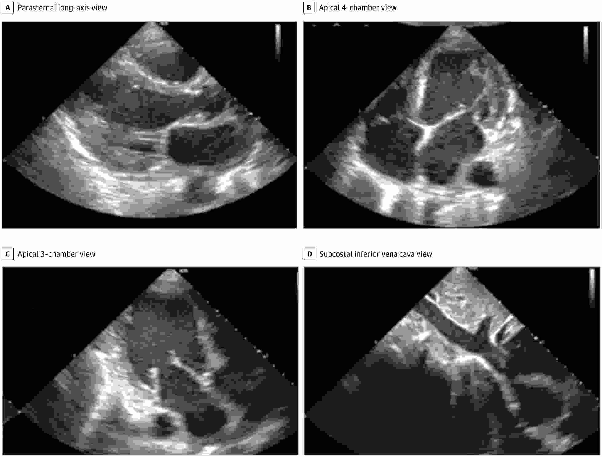
In the prospective study of 240 patients at academic medical centers, nurses were able to acquire images of high quality using a commercially available software application (Caption Guidance, Caption Health) installed on a portable ultrasound system. Images were judged to be of diagnostic quality on four key endpoints: quality for ventricular size, assessment of left ventricular size and function, right ventricular size and function, and presence of pericardial effusion.

"The ability to provide echocardiography outside the traditional laboratory setting is largely limited by a lack of trained sonographers and cardiologists to acquire and interpret images," Northwestern University cardiologist Dr. James D. Thomas and colleagues noted in their report about the data. "Using this AI-based technology, individuals with no previous training may be able to obtain diagnostic echocardiographic clips of several key cardiac parameters."

The study was conducted for regulatory purposes with the U.S. Food and Drug Administration (FDA). It exceeded the requirements for accuracy and the software was cleared in February 2020 through the agency's de novo pathway for novel products.

Each nurse performed 30 scans with using the guidance software installed on a portable ultrasound scanner (uSmart 3200t Plus, Terason). The mean acquisition time was 30 minutes. Ten standard transthoracic echocardiography views were obtained for each patient.

Separately, scans were conducted by sonographers on the same ultrasound unit but without the AI guidance software. Then the diagnostic quality of the scans was independently evaluated by a panel of five expert echocardiographers.

Still images of standard transthoracic echocardiographic views acquired by a nurse using the deep-learning algorithm were judged to be of diagnostic quality
Still images of standard transthoracic echocardiographic views acquired by a nurse using the deep-learning algorithm were judged to be of diagnostic quality. Image courtesy of Caption Health.

Per the FDA, the software needed to demonstrate diagnostic quality enabling diagnosis in at least 80% of patients on four key outcome measures. The nurses' scans were of diagnostic quality in from 92.5% to 98.8% of patients (see table), with no significant difference compared with scans by sonographers.

"Our study met all FDA-prespecified primary end points, with consistent results across [body mass index] categories and cardiac pathology, including potential distractors, such as pacemakers and prosthetic valves, with little difference between the nurse and sonographer scans," Smith and colleagues wrote.

AI-guided echocardiography, performance by nurses on four key outcome measures
EndpointPercent, diagnostic quality
Left ventricular size98.8%
Global left ventricular function98.8%
Right ventricular size92.5%
Nontrivial pericardial effusion98.8%

Performance of nurses was also on par with sonographers on a range of secondary endpoints. However, performance of sonographers was better than nurses when it came to determining the size of the inferior vena cava (91.5% diagnostic quality compared with 57.4%) and this is a "clear target for further algorithm development," the authors wrote.

The authors also acknowledged that limitations of the study included a relatively small number of patients and nurses and the lack of recruitment from intensive care units and emergency departments.

Smith and colleagues believe the results complement prior research, which has largely focused on the application of AI in medical imaging following the acquisition of images, as opposed to use in guiding image acquisition.

"Improvements in ultrasonography and computer hardware have led to the downsizing and cost reduction of ultrasonography machines, with handheld devices commercially available including standalone transducers interfacing with smart phones," they noted. "The [deep learning] algorithm developed in this study is relatively compact (approximately 1.5 GB) and trained on images from multiple vendors, and it therefore could be ported to work on multiple platforms."

Thứ Năm, 4 tháng 2, 2021

USPSTF advises against carotid artery stenosis screening

 By Kate Madden Yee, AuntMinnie.com staff writer


February 2, 2021 -- The U.S. Preventive Services Task Force (USPSTF) is advising against screening for asymptomatic carotid artery stenosis in the general adult population in a final recommendation statementpublished February 2 in JAMA.


The recommendation is consistent with the task force's 2014 statement, which graded carotid artery stenosis screening a D. The reason for the low grade is that the harms of carotid artery screening outweigh the benefits, according to USPSTF member Dr. Michael Barry of Massachusetts General Hospital in Boston.

"The Task Force wants to help prevent people from having a stroke, but screening for coronary artery stenosis is not an effective way to do so," he said in a USPSTF statement. "Unfortunately, screening for coronary artery stenosis in adults without symptoms does more harm than good, and we continue to recommend against it."

Carotid artery stenosis affects extracranial carotid arteries, and asymptomatic carotid artery stenosis refers to stenosis in persons without a history of ischemic stroke, transient ischemic attack, or other neurologic symptoms referable to the carotid arteries, the task force wrote in the JAMA article. Screening for the disease is accomplished through a variety of imaging modalities, including carotid duplex ultrasonography, MRI angiography, and CT angiography.

But these screening exams just aren't effective, according to the task force.

"The USPSTF found no externally validated risk stratification tools that could reliably distinguish between asymptomatic persons who have clinically important carotid artery stenosis and persons who do not, or the risk of stroke related to carotid artery stenosis," it wrote.

In an editorial also published in JAMA on February 2, Dr. Larry Goldstein, a neurologist at the University of Kentucky in Lexington, lauded the USPSTF's position.

"The estimated population-attributable risk for stroke related to asymptomatic carotid artery stenosis is approximately 0.7%, a risk considerably lower than for other stroke risk factors such as hypertension, atrial fibrillation, cigarette smoking, and hyperlipidemia," Goldstein wrote. "There remains no validated risk stratification tool for identifying a subpopulation of adults with a prevalence of asymptomatic carotid artery stenosis with a higher population-attributable risk that would lead to a benefit of intervention beyond risk factor management.'

Others pushed back, suggesting that the task force did not include evidence of the benefits of screening in its evaluation process -- or consideration of risk factors that occur in almost a third of U.S. adults, such as high blood pressure, hyperlipidemia, diabetes, smoking, limited physical activity, and poor diet.

"Without a true randomized clinical trial to directly answer the question, there is a lack of consensus and clarity on the role of carotid ultrasonographic screening for prevention of stroke in individuals who are asymptomatic," wrote Dr. Rebecca Smith-Bindman of the University of California, San Francisco in a JAMA Network Open editorial. "Nearly all of the specialty societies actually agree with the USPSTF and recommend against screening with carotid ultrasonography. However, these strong statements against screenings are all followed by the list of exceptions of individuals who, based on risk factors or characteristics, should in fact be screened."

Thứ Sáu, 29 tháng 1, 2021

Philips debuts ultrasound tools for AAA

By AuntMinnie.com staff writers


January 27, 2021 -- Philips Healthcare parent Royal Philips has introduced 3D ultrasound-based technology to monitor abdominal aortic aneurysms (AAAs)

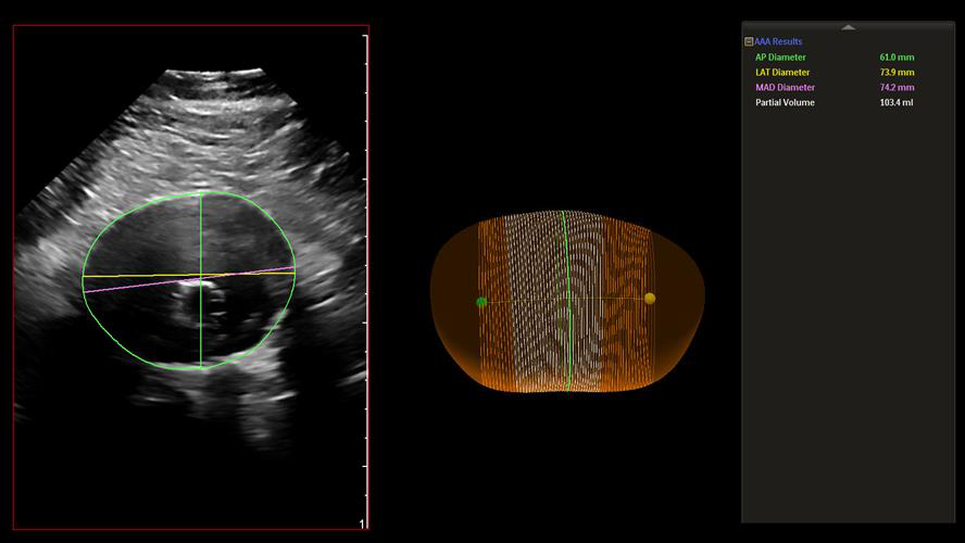
The company's AAA model is designed to assist clinicians by providing key measurements, including the partial volume and centerline of aneurysms.

The AAA model has received the CE Mark in Europe, and the U.S. Food and Drug Administration has cleared it for sale in the U.S. The model is making its debut at the virtual Leipzig Interventional Course (LINC), which is currently underway.

The AAA model provides measurements including the maximum anterior-to-posterior diameter and partial volume of the aneurysm, while also indicating the centerline
The AAA model provides measurements including the maximum anterior-to-posterior diameter and partial volume of the aneurysm, while also indicating the centerline. Image courtesy of Philips Healthcare


Chủ Nhật, 24 tháng 1, 2021

Seno gets FDA nod for breast optoacoustic ultrasound

By AuntMinnie.com staff writers

January 19, 2021 -- Optoacoustic imaging firm Seno Medical Instruments has garnered U.S. Food and Drug Administration (FDA) premarket approval (PMA) for its Imagio breast imaging optoacoustic ultrasound system.

Designed to help providers characterize and differentiate breast masses in real-time, Imagio combines laser optics and grayscale ultrasound to provide fused functional and anatomical breast imaging, according to the vendor. These optoacoustic images provide a blood map in and around breast masses, while the ultrasound technology yields a traditional anatomic image, Seno said. The company believes that Imagio could help to reduce the number of unnecessary diagnostic breast biopsies.

Imagio also comes with SenoGram, an artificial intelligence (AI)-based decision-support tool for aiding radiologists in interpreting the new images and helping them in transitioning from ultrasound alone to optoacoustic ultrasound imaging, the firm said.


Thứ Sáu, 22 tháng 1, 2021

Ultrasound shows lymph node changes after COVID-19 vaccine

By Theresa Pablos, AuntMinnie staff writer

January 22, 2021 -- Breast ultrasound was used to demonstrate signs of lymph node enlargement in four cases of patients who received the COVID-19 vaccine. Radiologists from NewYork-Presbyterian published the case studies on January 18 in Clinical Imaging.


The case studies involve women in their 40s and 50s who presented to the radiology department for breast imaging within two weeks of receiving a Pfizer-BioNTech or Moderna COVID-19 vaccine. Breast ultrasound showed the women had at least one enlarged lymph node. In all cases, the authors recommended follow-up imaging at a later time.

"With widespread rollout of the COVID-19 vaccine, it is prudent for radiologists to consider vaccine-induced hyperplastic adenopathy as an etiology of unilateral axillary adenopathy seen on breast imaging," wrote the authors, led by Dr. Nishi Mehta, a radiology fellow at NewYork-Presbyterian/Weill Cornell Medical Center.

Axillary adenopathy can be a sign of breast cancer. But it can also result from a number of benign causes, including local infection, inflammation, or trauma in the breast, thoracic wall, or nearby arm.

Vaccines are one reported benign cause of one-sided axillary adenopathy, and the symptom has been documented after vaccination for smallpox, bacillus Calmette-Guerin (tuberculosis), and anthrax. It appears more often in vaccines that evoke a very strong immune response.

As reported in the study, axillary adenopathy can occur after vaccination with the Pfizer-BioNTech and Moderna COVID-19 vaccines. The U.S. Centers for Disease Control and Prevention (CDC) lists axillary adenopathy as one of the most frequently reported adverse reactions to the Moderna COVID-19 vaccine. The CDC also noted more adenopathy cases occurred in patients who received the Pfizer-BioNTech vaccine than a placebo.

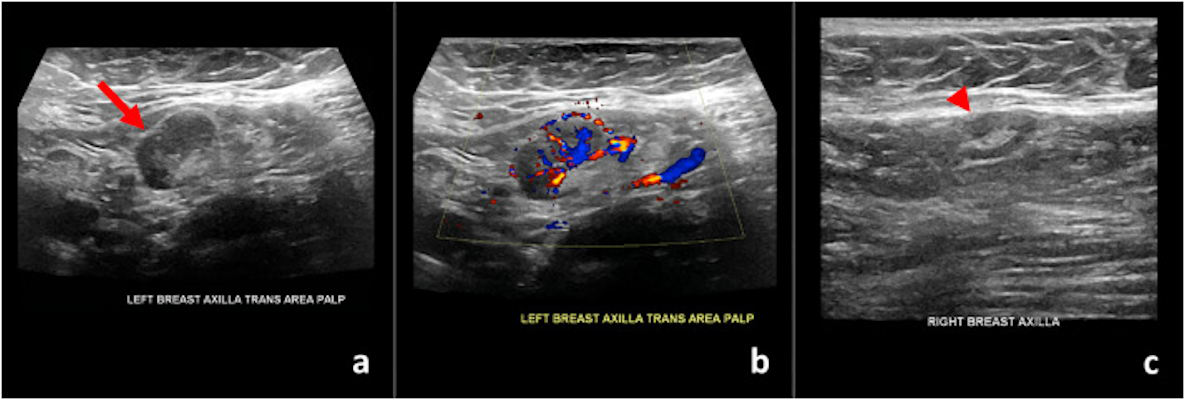
Ultrasound images of a 59-year-old patient who found a palpable lump nine days after receiving the first dose of the Pfizer-BioNTech vaccine in her left arm
Ultrasound images of a 59-year-old patient who found a palpable lump nine days after receiving the first dose of the Pfizer-BioNTech vaccine in her left arm. (a) Gray-scale and (b) color Doppler images of an enlarged left axillary lymph node. (c) Normal right axillary lymph node. All images courtesy of Dr. Nishi Mehta / Clinical Imaging.

This appeared to be the case for four women who presented for breast imaging at NewYork-Presbyterian. The patients had no preexisting conditions and were all frontline workers who qualified for the first round of Moderna or Pfizer-BioNTech vaccines.

In one case, a 59-year-old woman with no personal history of breast cancer came to the department after finding a palpable lump near her left armpit. Her mammogram was unremarkable, but targeted sonography showed her left axillary lymph node had uniform cortical thickening of 0.7 cm in an area corresponding to the patient's concern.

When asked about her history, the patient said she had recently received the Pfizer-BioNTech COVID-19 vaccine and noticed the lump nine days after receiving her first vaccine dose.

Ultrasound images of a 42-year-old patient who presented for sonographic follow-up of probably benign bilateral breast masses
Ultrasound images of a 42-year-old patient who presented for sonographic follow-up of probably benign bilateral breast masses. The patient had received the first dose of the Moderna vaccine in her left arm 13 days prior to imaging. (a) Gray-scale and (b) color Doppler images of an enlarged left axillary lymph node. (c) Unremarkable right axilla.

The authors reported similar findings in two women who presented for routine breast ultrasound screening, as well as one woman who underwent ultrasound follow-up of probably benign bilateral breast masses. All three of these women had diffuse cortical thickening in at least one axillary lymph node within 13 days of receiving a first or second vaccine dose.

In all four cases, the authors recommended follow-up imaging with targeted ultrasound. NewYork-Presbyterian is currently recommending clinicians delay follow-up ultrasound imaging until four to 12 weeks after patients receive their second COVID-19 vaccine dose. If the adenopathy still persists at this time, then the authors would recommend biopsy to rule out cancer.

While the report focused on breast imaging, Mehta said breast imagers aren't the only types of radiologists who may come across axillary adenopathy.

"Thoracic imagers will likely see similar findings of unilateral axillary adenopathy and cervical adenopathy on chest imaging studies and should therefore be similarly aware of the diagnosis," she told AuntMinnie.com.

Axillary adenopathy may also be seen on mammography, noted Mehta. Axillary adenopathy after vaccination has also been documented as increased FDG uptake on PET and CT, she added.

"We expect similar findings post COVID-19 vaccination," she said.

Thứ Hai, 18 tháng 1, 2021

POCUS group launches MSK certification

 By AuntMinnie.com staff writers


January 18, 2021 -- The Point-of-Care Ultrasound (POCUS) Certification Academy has launched a new certification program for healthcare providers that use ultrasound in the provision of physical therapy and other disciplines

The academy's POCUS Musculoskeletal (MSK) Certification supports a new measure of proficiency with POCUS that raises the standard of musculoskeletal imaging by testing on best practices of clinical applications.

The certification is designed to support professionals in the fields of physical therapy, physiatry, athletic training, sports medicine, and orthopedics, among others.

Thứ Năm, 14 tháng 1, 2021

Thyroid cancer screening hits the right note


By Theresa Pablos, AuntMinnie staff writer

January 14, 2021

The ultrasound technique works because singing causes the elasticity of surrounding tissues to increase, making it easier for clinicians to pinpoint abnormally stiff areas. Singing during thyroid ultrasonography also has the potential to identify more thyroid tumors while simultaneously putting patients at ease.

"Developing noninvasive methods would reduce the stress of patients during their medical exams," stated lead author Steve Beuve, a doctoral student specializing in medical imaging at University of Tours, in a press release. "Having to sing during a medical exam can perhaps help release some of the nervous tension even more."

The authors called their new technique vocal passive elastography. The method was inspired by passive elastography, which detects changes in elasticity from natural vibrations caused by blood pulsing, heart rhythms, and other natural biological changes.

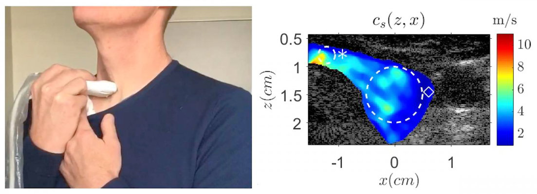
During vocal passive elastography, a clinician places a linear ultrasound probe at the front of a patient's neck. The patient then sings and maintains the "eee" tone, trying to match their tone to a 150-Hz sound playing from a speaker.

The singing vibrates the patient's trachea, which in turn produces vibrations throughout the thyroid. This allows the clinician to use an ultrafast frame rate to track changes in shear-wave velocity.

"The propagation of shear waves gives us information about mechanical properties of soft tissues," Beuve stated.

For their experiment, the team validated the concept by calculating shear-wave elastography (SWE) measurements as patients sang, then superimposing a map of that data onto B-mode ultrasound images. They created their own algorithm to reconstruct the raw image data and tested their algorithm on two different ultrasound systems.

The vocal passive elastography method succeeded in identifying particle tissue displacement induced by singing on both systems. The values also aligned well with SWE metrics, although the measurements were more similar on the left (thinner) side of the thyroid than on the right (thicker) side.

A volunteer holds a linear ultrasound probe to help prove the feasibility of vocal passive elastography
Left: A volunteer holds a linear ultrasound probe to help prove the feasibility of vocal passive elastography. Right: 2D shear wave speed as calculated by the new technique mapped onto an anatomical brightness mode ultrasound image. Image courtesy of Steve Beuve.

Overall, vocal passive elastography looked like a promising alternative to fine-needle aspiration biopsy, which misses up to 95% of thyroid cancers, according to the authors. It also drastically improved the thyroid signal-to-noise ratio by drowning out natural background noise from nearby organs.

The researchers are already conducting new experiments to further refine the technique, including investigating the benefits of asking patients to match different bandwidths. They are also interested in expanding the use of vocal passive elastography to areas outside of the vocal tract, such as the brain.

"We want to cooperate with physicians to propose protocols to verify the relevance of elasticity as a biomarker of pathogens," Beuve stated