Tổng số lượt xem trang

Thứ Ba, 15 tháng 2, 2022

US finds link between long COVID, viral effect on vagus nerve


By Amerigo Allegretto, AuntMinnie.com staff writer

February 15, 2022 -- Ultrasound found links between symptoms of long COVID-19 disease and the effects of the SARS-CoV-2 virus on the vagus nerve, according to research that will be presented at the European Congress of Clinical Microbiology & Infectious Diseases in April. A team led by Dr. Gemma Lladós from Germans Trias i Pujol Hospital in Badalona, Spain found that COVID-19 patients with symptoms of vagus nerve dysfunction mediated by SARS-CoV-2 also had nerve thickening, decreased esophageal and diaphragmatic mobility, and reduced maximum inspiratory pressures values.

"We need to improve our understanding of the mechanisms that may cause the persistence of symptoms to help patients and find interventions that can prevent and treat vagus nerve dysfunction," Lladós told AuntMinnie.com.

Stretching from the brain to the torso and into the heart, the vagus nerve plays an important role in bodily functions. These include controlling heart rate, speech, the gag reflex, transferring food from the mouth to the stomach, moving food through the intestines, and sweating, among others.

Post-COVID syndrome, also commonly known as long COVID, affects 10% to 15% of COVID-19 survivors, researchers wrote. Symptoms include persistent voice problems, difficulty in swallowing, dizziness, tachycardia, low blood pressure, and diarrhea.

Ultrasound images show longitudinal section of the left vagus at the cervical level
Spanish research has found possible links between long COVID and vagus nerve dysfunction in patients. Ultrasound images show (Above) longitudinal section of the left vagus at the cervical level, where there is a slight fusiform thickening, and (Below) an axial cut of left cervical vagus showing hyperechogenic perineural thickening. Images courtesy of Dr. Gemma Lladós et al.
an axial cut of left cervical vagus showing hyperechogenic perineural thickening

However, the researchers said the mechanism that causes long COVID is currently unknown. Therefore, Lladós et al wanted to determine the morphological and functional activities of the vagus nerve in their pilot study of long-COVID patients with dysfunction, using ultrasound and functional tests.

They looked at data from 22 patients who were selected from a cohort of 228 individuals who had at least one symptom suggestive of vagus nerve dysfunction. Patients had a median age of 44 years and presented between March and June 2021. Out of these, 20 were women.

Most common vagus nerve dysfunction symptoms in patients presenting with long COVID
Diarrhea73%
Tachycardia59%
Dizziness45%
Difficulty swallowing45%
Persistent voice problems45%

The study authors found that 19 patients had at least three dysfunction-related symptoms, and the median prior duration of symptoms was 14 months.

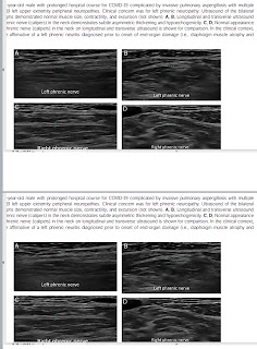
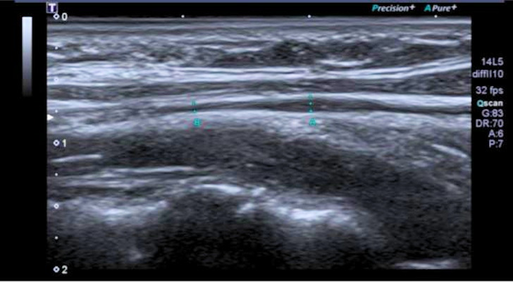
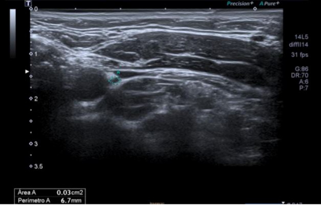
Six patients had mild vagus nerve thickening and higher echo on neck ultrasound scans. Thoracic ultrasound, meanwhile, showed flattened diaphragmatic curves in 10 patients. Ten out of 16 patients also showed reduced maximum inspiratory pressures.

"Radiologists may find by ultrasound in the lateral cervical area some images over the vagus nerve reflecting mild reactive inflammatory changes," Lladós said. "In addition, a flattened diaphragmatic curve visualized by chest ultrasound could translate into decreased diaphragmatic mobility during respiration."

The team also found eating and digestive dysfunction in patients, with 13 out of 18 having indication of trouble swallowing. Eight out of 19 patients also reported having their ability to deliver food to the stomach impaired, two of whom had difficulty swallowing. Acid reflux was also seen in nine of 19 patients, four of whom reported difficulty delivering food to the stomach and three having hiatal hernia.

The researchers also found abnormalities in voice function for eight out of 17 patients, seven of these reporting dysphonia, or persistent voice problems.

Lladós told AuntMinnie.com that the team wants to compare their results with healthy controls and with patients infected by SARS-CoV-2 without the persistence of symptoms. He said the aim is to know if there is more morphological and functional involvement of the vagus nerve in infected patients.

The team's findings will be presented at the European Congress of Clinical Microbiology & Infectious Diseases in Lisbon, Portugal, which will be held April 23-26.

expert reaction to conference abstract looking at long COVID and the vagus nerve 

A conference abstract presented at the European Congress of Clinical Microbiology & Infectious Diseases (ECCMID) Annual Meeting looks at vagus  nerve dysfunction in long COVID.


Dr David Strain, Chair of BMA Board of Science and Clinical senior lecturer and honorary consultant, University of Exeter Medical School, said:

“This is a conference abstract. The full paper is not available and therefore it is not possible to properly critique the data. It has also not undergone peer-review.

“The study appears to add to a growing collection of data suggesting at least some of the symptoms of long COVID is mediated through a direct impact on the nervous system. There is a well-established knowledge base that associates the vagus nerve with some of the cardinal features of long COVID such as palpitations, night sweats and gastrointestinal disturbances. Establishing vagal nerve damage is useful information, as there are recognised, albeit not perfect, treatments for other causes of vagal nerve dysfunction that may be extrapolated to be beneficial for people with this type of Long COVID.

“It is important to caveat that not all symptoms of long Covid may be associated with the direct impact of vagal dysfunction and there are many other presentations that will need further investigation. Further, we must remember that association is not necessarily causation. The Vagus nerve (so named because of its vagrant nature as it wanders through the body) is one of the longest nerves in the body and the longest and most complex of the cranial nerves. It is entirely possible that people susceptibility to nerve damage are at the greatest risk of developing long Covid; in this case this observation tells us little more than  nerve damage is an result of COVID, this is greater in those with Long Covid, and the damage is easy to detect in a long, complex nerve. Indeed, it is unlikely that vagal nerve damage could account for many other symptoms such as brain fog or muscle aches and pains. 

“Treatment with agents that have been demonstrated to be efficacious against vagal nerve dysfunction may provide relief for patients, however are unlikely to treat the underlying diagnosis, and thus patients would be at risk of rebound symptoms once treatment stops. Randomised Controlled trials into a Long Covid treatments will soon be commencing considering treatments for both underlying causes and symptomatic control I’m sure we will be able to evaluate these agents will only know when we respectively start with the symptoms whether reversal of this damage.”


Conference abstract: ‘Vagus nerve dysfunction in post-COVID-19 condition’ by G. Lladós et al was presented at the European Congress of Clinical Microbiology & Infectious Diseases (ECCMID) Annual Meeting to be held in Lisbon from 23-26 April.

The full study is not available and this research has not undergone peer-review. 


All our previous output on this subject can be seen at this weblink:

www.sciencemediacentre.org/tag/covid-19


Thứ Năm, 3 tháng 2, 2022

POCUS can help rule out pulmonary embolism (PE) in critically ill COVID-19 patients


By Amerigo Allegretto, AuntMinnie.com staff writer

February 3, 2022 -- Point-of-care ultrasound (POCUS) can help rule out pulmonary embolism (PE) in critically ill COVID-19 patients when imaging multiple organs, according to Dutch research published February 2 in the Journal of Critical CareA team led by Dr. Arthur Lieveld from Amsterdam University Medical Centers in the Netherlands wrote that POCUS could help determine which COVID-19 patients should undergo CT pulmonary angiography.

"In addition, finding right ventricular strain can make pulmonary embolism more likely in this setting, while a deep venous thrombosis would preclude the need for a CT pulmonary angiogram," Lieveld and colleagues wrote.

Critically ill COVID-19 patients have an increased risk of developing pulmonary embolism. While CT pulmonary angiography is the standard for diagnosing this, transporting patients for imaging carries health risks. CT also requires more time for decontamination and more medical personnel, which reduces time that could be dedicated toward patient care.

POCUS has been explored for imaging COVID-19 patients due to its portability and use for bedside imaging. While evidence is limited in detecting pulmonary embolism, some recent statements recommend using POCUS for critically ill patients.

Lieveld and colleagues wanted to find out how accurate POCUS is in diagnosing PE in these patients by looking at multiple organs together and separately. These include the lung, deep veins, and the heart.

The researchers looked at data from 70 COVID-19 patients in the intensive care unit with an average age of 67.5 years, 23 patients of whom had a pulmonary embolism. They also wrote that the median scan time with POCUS was 14 minutes.

The team found that imaging multiple organs together yielded higher accuracy than when single organs were imaged. Multiorgan POCUS had a sensitivity of 87.5% and a specificity of 25%.

POCUS' diagnostic accuracy for subpleural consolidations meanwhile was 42.9%, 75.6% for deep venous thrombosis, and 74.4% for right ventricular strain. The sensitivity and specificity for deep venous thrombosis was 24% and 88.8%, while sensitivity and specificity for right ventricular strain were 40% and 83%, respectively.

The researchers wrote that based on these results, subpleural consolidations are "hardly useful" in detecting pulmonary embolisms. However, they added that the high sensitivity displayed by multiorgan POCUS, which includes these consolidations, means this method may have a role in ruling out pulmonary embolism.

"The main driver of the good sensitivity seems to be subpleural consolidations, which ... seem to be ubiquitous in critically ill COVID-19 patients," the study authors wrote.

They also wrote that POCUS could remove some socioeconomic barriers as an affordable, easy-to-use tool for timely imaging.

"Moreover, it is easy to learn for [para]medical personnel," researchers added. "In our study, all POCUS scans were performed by intensive care unit residents. Considering that nonspecialist physicians see and treat the bulk of COVID-19 patients, this underscores the applicability of POCUS in a real-life setting."


HÌNH ẢNH SIÊU ÂM PHỔI SAU NHIỄM COVID-19 3 THÁNG

TỔNG QUAN

Hiện chưa có bài báo nào về khám kiểm tra siêu âm phổi [LUS] sau nhiễm COVID-19  tại thành phố HCM và y văn nước ngoài. Trong giai đoạn hậu nhiễm COVID-19, chúng tôi đã triển khai việc theo dõi và ghi nhận lại kết quả LUS sau nhiễm 3 tháng COVID-19 tại MEDIC Hòa Hảo.

MỤC TIÊU

Ghi nhận tổn thương phổi tồn tại bằng siêu âm phổi (LUS) dựa theo đề nghị của Rouby bằng cách khảo sát 12 vùng của 2 phổi P và T và sau đó tính điểm LUS Score total.

PHƯƠNG PHÁP

Khảo sát tiền cứu mô tả và cắt ngang những bệnh nhân và gia đình có nhiễm COVID-19 nay đã có xét nghiệm RT-PCR âm tính, tự đến khám tại MEDIC Hòa Hảo.

Thực hiện khám LUS chuẩn hóa với tinh chỉnh hệ số mechanical index MI <0,7, tắt tất cả các bộ lọc  như Harmonic và S-Harmonic, THI… tùy theo hãng máy siêu âm có tại MEDIC.


Phối hợp với X-Quang và CT nếu có yêu cầu của bệnh nhân và bác sĩ lâm sàng.

Thống kê nhân thân và bệnh sử theo MedCalc v. 20. Hồ sơ được lưu trữ điện toán hóa và truy cập qua mã QR code của MEDIC cho từng bệnh nhân.


KẾT QUẢ


Từ cuối tháng 8 năm 2021 đến cuối tháng 12/2021, báo cáo này gồm 70 người [48 nam và 22 nữ], tuổi từ 34-60, trung bình  sau nhiễm COVID-19 khoảng 90 ngày, trong đó có 11 nhân viên nữ của khoa siêu âm. Tất cả đều có thời gian cách ly và điều trị trong bệnh viện, đều có khó thở mức độ trung bình. Số người nhiễm COVID-19  sau chích ngừa mũi 2 # 20 trường hợp.


Hình ảnh LUS  sau nhiễm COVID-19 của nghiên cứu này gồm còn tồn tại dày màng phổi có gián đoạn [70/70 ca], lightbeam [20/70ca], subpleural consolidation nhỏ dưới 20mm [38/70 ca]. Đa số phát hiện tồn tại tổn thương ở vùng phổi sau bên [posterior lateral] cả 2 phổi. Phần lớn thương tổn thuộc  nhóm C và D theo phân loại thống nhất hiện nay, có LUS Score total bình quân # 11.

 Lúc khám LUS các bệnh nhân đều than phiền còn khó thở, mệt ít. Nhiều  bệnh nhân khai báo bị tê vùng lưng và đau cơ, rụng tóc [nữ 40%], nổi mày đay, nổi mụn, viêm nang lông, sọc gờ móng tay. Tất cả đều trong quá trình hồi phục dần.


BÀN LUẬN


Hình ảnh LUS trong nhiễm COVID-19  đã được mô tả bởi nhiều tác giả, được hệ thống hóa vùng khám, và định lượng bằng cách cho điểm theo phân loại thống nhất (4 loại A B C D) và thang điểm 0 1 2 3, như đã đề cập. Có tác giả nhận xét rằng hình ảnh siêu âm LUS trong nhiễm COVID-19 rõ nhất khoảng 2 tuần sau khởi phát rồi kém rõ dần. 

Trong nhóm bệnh sau nhiễm COVID-19 tại MEDIC, chúng tôi  dựa theo phân loại và thang điểm định lượng trên. Tất cả dều được khám LUS trước khi làm xét nghiệm cận lâm sàng và chẩn đoán hình ảnh khác.

Sau nhiễm COVID-19  hình ảnh siêu âm phổi gồm những tổn thương tồn tại nhỏ như dày màng phổi, lightbeam, small subpleural consolidation thường ở vùng phổi đáy sau bên [posterior lateral] cả 2 phổi. LUS Score total #11 trong đa số 65/70 ca cho thấy mức độ các thương tổn tồn tại này không cần can thiệp và không ảnh hưởng nhiều đến chức năng sống sau nhiễm COVID-19.

Dày màng phổi và dày màng phổi gián đoạn [dotted] thường gặp trong viêm phổi COVID-19  và một số bệnh lý khác. Dày lá thành và cả lá tạng màng phổi. LUS xác định dày màng phổi với bề dày <10mm. Giá trị cut-off của xơ hoá phổi nhẹ là dưới 2,4 mm theo Manelescu 2020.  B-lines và lightbeam xuất hiện cùng với dày màng phổi trong viêm phổi mô kẽ, qua giai đoạn cấp từ 10-12 ngày ít gặp lightbeam hơn.

Đông đặc phổi (consolidation) trong COVID-19  thường ở cạnh [subpleural nodule] màng phổi dày, với dạng nốt nhỏ tròn echo kém, có khi xếp thành dãy, ít gặp hơn trong giai đoạn post COVID-19  và không giới hạn rõ ở giai đọan sau viêm phổi. Khi phổi viêm, vùng đông đặc có dạng 3 cạnh, echo kém và tương đương với tổn thương kính mờ trên CT. Những đông đặc nhỏ dạng nốt trong giai đoạn cấp dường như có liên quan đến vi huyết khối trong nhu mô phổi khi thực nghiệm trên sinh vật.  Trong báo cáo này có 2 trường hợp nhầm với tổn thương u phổi và u nấm phổi, vì chúng không ở sát ngay dưới màng phổi bị dày.

Những đông đặc nhỏ dạng nốt, thường ở ngay dưới màng phổi, echo kém, đường viền không rõ trong giai đoạn sau nhiễm COVID-19 kèm theo dày màng phổi gián đoạn ở vùng đáy phổi sau [vùng 5,6].

Tuy nhiên LUS không thể phát hiện những tổn thương phổi sâu hơn 10 cm.

 Dù vậy bệnh nhân vẫn than phiền còn khó thở nhất là khi gắng sức; cần tìm thêm cách xác minh. Các nghiên cứu nước ngoài giải thích là do tự kháng thể tấn công vào phổi giống như bệnh tự miễn, nên diễn tiến tổn thương thầm lặng dần dẩn đến hoá xơ. Thường bệnh nhân có phản ứng miễn dịch mạnh thì phản ứng xơ hóa sẽ mạnh.

Một số bệnh nhân trong nghiên cứu này có chụp X-quang [31 ca] và CT [09 ca]. Tuy nhiên số kết quả tương hợp với LUS  ghi nhận được là thấp.

Có 09/70 ca LUS có chụp CT, 06 ca phù hợp tổn thương phổi do COVID-19 [06/70 ca].


Kết quả CT

COVID-19

TB

K phổi

Khác [u nấm phổi]

Tổng cộng =   09      

06

01

01

01


Có 31/70 ca LUS có chụp X-quang phổi, 26 ca không ghi nhận có tổn thương phổi COVID-19,  04 ca viêm phổi trong đó 01 ca có tràn dịch màng phổi, ca này CT xác nhận thuyên tắc phổi thùy dưới 2 bên do nhiễm COVID-19 và D-dimer tăng cao. Chỉ có 02/31 ca X-quang  phổi nghi COVID-19.


Kết quả X-quang

COVID 19 

VP + TDMP

TB xơ thâm nhiễm thùy trên 2 bên

KHÁC [U NẤM ĐỈNH PHỔI]

Âm tính COVID19

 Tổng cộng= 31

02

02

01

26


Tại Medic, kết quả LUS và X-quang phổi cũng như CT chưa có đồng thuận cao; y văn cho biết X-quang phổi COVID-19 chỉ nhạy khoảng 17% so với LUS. Do bản chất siêu âm tùy thuộc người làm LUS nên cũng ghi nhận đây như một yếu tố giúp giải thích sự thiếu tương hợp giữa LUS với X-quang.


Cần chờ đợi kết quả của những nghiên cứu khác post COVID-19 với cỡ mẫu lớn hơn.


KẾT LUẬN


Với 70 ca  sau nhiễm COVID-19 đến khám tại MEDIC, chúng tôi ghi nhận có tồn tại tổn thương phổi mức độ nhẹ, đa số có LUS Score total #11, sau 3 tháng nhiễm COVID-19 các tổn thương phổi này [như dày màng phổi và đông đặc nhỏ] trở nên không rõ và thu nhỏ dần. Hình ảnh siêu âm LUS chưa tương hợp với CT và X-quang phổi là điều nên khảo sát thêm với số liệu lớn hơn.

Giới hạn của chúng tôi là một sơ kết cục bộ với số liệu còn ít và chưa đồng thuận với các phương tiện chẩn đoán hình ảnh khác.

Cần tiếp tục khám LUS ở các mốc 3,6,12 tháng cho những bệnh nhân có khó thở trong giai đoạn cấp và hồi phục và đối chiếu với các nghiên cứu lớn, đa trung tâm để đánh giá đầy đủ khả năng của LUS trong viêm phổi COVID-19 và hậu COVID-19.


TÀI LIỆU THAM KHẢO CHÍNH

Jaime Gil-Rodríguez et al: Ultrasound findings of lung ultrasonography in COVID-19: A systematic review,  Europeen Journal of  Radiology, Review Article,| Volume 148110156MARCH 01, 2022. Available online 20 January 2022 0720-048X/© 2022 Elsevier B.V. All rights reserved. 

Markus H. Lerchbaumer et al: Point‑of‑care lung ultrasound in COVID‑19 patients: inter‑ and intra‑observer agreement in a prospective observational study, Scientifc Reports | (2021) 11:10678 | https://doi.org/10.1038/s41598-021-90153-2.

 
Giovannettia G et al: Lung ultrasonography for long-term follow-up of COVID-19 survivors compared to chest CT scan,  Respiratory Medicine 181, March 2021 https://doi.org/10.1016/j.rmed.2021.106384

McDermott C et al: Combatting COVID-19: is ultrasound an important piece in the diagnostic puzzle? Emerg Med J, 2020 Oct;37(10):644-649. doi: 10.1136/emermed-2020-209721. Epub 2020 Sep 9.   

Vetrugno et al : The “pandemic” increase in lung ultrasound use in response to Covid-19: can we complement computed tomography findings? A narrative review, Ultrasound J (2020) 12:39  https://doi.org/10.1186/s13089-020-00185-4.

Thứ Sáu, 21 tháng 1, 2022

POCUS on par with x-ray for COVID-19 diagnosis, management


By Amerigo Allegretto, AuntMinnie.com staff writer

January 21, 2022 -- Point-of-care ultrasound (POCUS) for lung imaging is noninferior to chest x-ray when it comes to diagnosing COVID-19, according to an Italian study published January 18 in the Journal of Ultrasound in Medicine.

A team led by Dr. Costantino Caroselli from the National Institute of Rest and Care for the Elderly in Ancona found that ultrasound characteristics seen in COVID-positive patients included B-lines, irregular pleural lines, and small consolidation.

"Lung ultrasound holds the promise of an accurate, radiation-free, and affordable diagnostic and monitoring tool in COVID-19 pneumonia," Caroselli and colleagues wrote.

Lung imaging is an important tool in helping diagnose and manage COVID-19 in patients. Previous studies have shown ultrasound's promise in determining early lung involvement in patients while preventing radiation exposure.

However, the researchers said that the lack of a unified approach for POCUS among clinicians is keeping this method from wider use. They added this is caused by a lack of data on patient outcomes and lung ultrasound having a lower specificity than chest x-ray.

Caroselli's team wanted to show how useful ultrasound is in diagnosing lung disease early in COVID-positive patients and compare their results to findings on chest x-ray. They looked at data from 479 patients, 82.7% of whom were diagnosed with COVID-19 by molecular testing.

Lung POCUS was performed by trained emergency physicians on patients presenting for possible COVID-19 evaluation before they underwent conventional radiologic examination or while they were waiting for the report.

Most common findings on lung POCUS in COVID patients
B-lines80.2%
Irregular pleural lines59.3%
Small subpleural consolidations55.3%

Chest x-rays were found to be normal, meanwhile, in 17.9% of all cases.

The team also tested the prognostic capabilities of lung POCUS. Pleural effusion, small consolidation, male sex, and clinical history were associated with hospital admission rate. However, x-ray outcomes were not associated with this rate.

Both POCUS and x-ray have significant roles in determining the need for orotracheal intubation for patients, the study authors wrote. Lung ultrasound small consolidation, x-ray ground glass, and x-ray interstitial pattern were found to be linked to patients needing invasive mechanical ventilation.

Meanwhile, pleural effusion found on ultrasound and x-ray small consolidation were the only predictors of mortality.

The researchers wrote that lung POCUS could help clinicians rapidly evaluate and triage patients as coronavirus surges persist.

"Our analysis confirmed that, as well as supporting COVID-19 diagnosis, lung ultrasound was useful in prognostic stratification relating to the need for hospitalization, orotracheal intubation, and mortality among the patients studied," they wrote.

Chủ Nhật, 2 tháng 1, 2022

Siêu âm, người phục vụ toàn bộ dân số sau đại dịch COVID-19

 Ultraschall, der Handwerker im Dienste der gesamten Bevölkerung nach der COVID-19-Pandemie

Fabio Piscaglia, Bernardo Stefanini,Vito Cantisani


Kể từ khi công nghệ siêu âm B-mode ra đời khoảng 40 năm trước, phương pháp siêu âm đã phát triển nhiều nhất và đã vượt qua sự phát triển của các phương pháp chẩn đoán hình ảnh khác như CT hoặc MRI . Siêu âm hiện nay được sử dụng thường xuyên, đáp ứng nhu cầu của toàn bộ bệnh nhân. Các bài báo được xuất bản trong số này của tạp chí là một ví dụ minh họa rất rõ ràng về tính linh hoạt đó. Ví dụ, nếu chúng ta xem xét độ tuổi của dân số, những bài báo này cho thấy siêu âm được sử dụng thường xuyên với lợi ích to lớn ra sao tại bất kỳ thời điểm nào của cuộc đời con người. Việc sử dụng siêu âm khám từ giai đoạn rất sớm, trong tử cung trước khi sinh [ 1 ], chuyển sang thời kỳ chu sinh [ 2 ], thời kỳ trưởng thành [3 ] [ 4 ], thời gian mà CT hoặc MRI ít được sử dụng và cuối cùng là bệnh của người cao tuổi, chẳng hạn như chứng mất trí nhớ Alzheimer [ 5 ]. Không chỉ các ứng dụng của siêu âm bao gồm toàn bộ cuộc sống con người về mặt thời gian, mà kích thước và giá cả của máy siêu âm còn từ các máy cao cấp nhất, [tương đối lớn chỉ có thể di chuyển trên bánh xe, dùng công nghệ tiên tiến nhất [ 3 ] và có giá hơn 100 000 Euro,] đến máy siêu âm cỡ bỏ túi, được sử dụng thuận tiện tại giường [ 6] và dễ dàng cầm bằng một tay. Các công nghệ gần đây  còn thêm khả năng kết nối đầu dò siêu âm hoạt động bằng pin với một số điện thoại thông minh  có hiệu quả. Công nghệ này bỏ qua nhu cầu của máy siêu âm chuyên dụng và có thể giữ giá đầu vào thấp, làm cho giá mua giữa máy siêu âm mắc nhất và rẻ nhất chênh lệch nhau hơn 100 lần. 

CT hoặc MRI thì không thể có khác biệt này của máy siêu âm [về kích thước, khả năng và giá cả]. 

Hơn nữa, siêu âm không chỉ có khả năng hiển thị thông tin hình thái mà còn hiển thị về chức năng: ví dụ như sự hiện diện và vận tốc của dòng máu trong các mạch máu với Doppler, độ cứng của mô với kỹ thuật đàn hồi [ 7 ], tưới máu mô với định lượng siêu âm cản âm [8 ] và gần đây là phép đo siêu âm mô phân tán / độ nhớt hoặc định lượng hàm lượng mỡ gan [ 3 ] [ 9 ] [ 10 ] [ 11] hầu như tất cả đều sẵn có trong cùng một thiết bị. 

Bên cạnh đó, đại dịch COVID-19 đã thực tế sử dụng siêu âm cho nhiều vùng cơ thể và môi trường sống. Siêu âm có thể khảo sát chính xác các cấu trúc ngoài da bằng đầu dò tần số siêu cao cũng như các vùng sâu cơ thể như tim hoặc mạch máu sau phúc mạc. 

Ngoài ra, siêu âm  phổi đã được khẳng định là có hiệu quả, nhưng thường chỉ giới hạn ở các đơn vị chăm sóc đặc biệt, chưa được sử dụng rộng rãi. Sự cần thiết phải đánh giá những bệnh nhân bị nhiễm SARS-Cov-2, thường bị tổn thương chủ yếu ở phổi  vẫn còn hạn chế cho những người có nhu cầu đến bệnh viện để tránh tình trạng quá tải các cơ sở y tế . Trên thực tế, siêu âm phổi đã nhanh chóng trở thành một phần của việc đánh giá những bệnh nhân như vậy, và đạt được thành công lớn [12 ] [ 13 ] [ 14 ]. Cách khám siêu âm phổi tương đối dễ học, làm cho các bác sĩ tin tưởng hơn trong quyết định giữ bệnh nhân ở nhà hoặc chuyển họ đến bệnh viện và đã được tích hợp liền mạch trong đánh giá siêu âm lâm sàng của những bệnh nhân đó. Trong số tạp chí này, tiềm năng của siêu âm phổi đã được đề cập [ 15]. 

Tuy nhiên,  việc tích hợp siêu âm vào đánh giá lâm sàng của bệnh nhân là điều đáng chú ý, khi siêu âm được sử dụng bởi các bác sĩ không chỉ là người khám siêu âm mà còn  chủ yếu chịu trách nhiệm về các quyết định lâm sàng. Đây là khái niệm mới nổi lên về siêu âm tại điểm chăm sóc [PocUS] hoặc  siêu âm hỗ trợ thăm khám cho bệnh nhân. Trong đó dùng siêu âm  như phần mở rộng kiểm tra sức khỏe, giúp bác sĩ đưa ra quyết định nhất quán hơn khi lập kế hoạch  chẩn đoán và điều trị tốt nhất . Điều này đã được định nghĩa là Kiểm tra Y tế Hỗ trợ Siêu âm (SAME) [ 16 ] [ 17 ]. Một ví dụ về khái niệm này cũng được minh họa trong một bài báo của số báo hiện tại [4 ]. Do đó, cũng từ quan điểm này, siêu âm đang cho thấy phạm vi ứng dụng rộng rãi nhất  từ khoa X-quang với siêu âm đa thông số  [multiparametric], đến các khoa lâm sàng,  khoa cấp cứu , các đơn vị chăm sóc đặc biệt cho đến  dịch vụ tại chỗ, trên xe cứu thương và đến tận nhà bệnh nhân.

Nhìn chung, sau đại dịch COVID-19, siêu âm dường như đã trở nên mở rộng thường xuyên hơn trong hoạt động y tế tốt nhất phục vụ cộng đồng .