Tổng số lượt xem trang

Thứ Hai, 23 tháng 1, 2023

The Enormous Potential of Ultrasound:

 Making a Difference in Patients’ Lives

The use of ultrasound in medicine began during World War II in several countries around the world. The work of Dr. Karl Theodore Dussik in Austria in 1942 using ultrasonic waves to detect brain tumors was one of the first indications that this noninvasive technology had tremendous potential. Although many others in the USA, Japan and Europe are also considered pioneers, the work of Professor Ian Donald and his colleagues in Scotland in the 1950s did much to facilitate practical technology and applications, leading to the wider use of ultrasound in medical practice we see today.

ULS Modes of Operation.png

For many parents, their first experience with ultrasound shows them their unborn baby’s little hands and tiny spines — a grainy glimpse into their family’s future. But ultrasound has vast applications beyond assessing the health of a growing baby. It is used to look closely at the heart after a patient’s heart attack, evaluate lung function in a patient stricken by COVID-19 and assess why a patient brought in by ambulance is in shock. Having clear 2D, 3D and 4D real-time images and clips that accurately capture tissues, fluids and structures inside the body — while being safe by not exposing the patient to any ionizing radiation — is powerful for diagnosis, treatment and improved patient outcomes.

Ultrasound allows physicians to see what’s going on in the human body by sending high-frequency sound waves and receiving echoes, which convert into images that physicians can visualize, attain measurements from and use in diagnostics. Ultrasound images and clips are processed in real time today, so there is no delay in the ability to understand a patient’s condition. In fact, through advocacy, training and technological development, ultrasound has developed into the second-most-used diagnostic imaging technology (after X-ray) and produces billions of diagnostic images each year.[1]

Now, with support from digital solutions and artificial intelligence, caregivers, physicians and surgeons are using ultrasound in real time for precision care at the bedsides of patients, in the operating room and the traditional doctor’s office, at home or wherever the patient happens to be.

Vscan Air Lung Pleural Line Ambulance.jpg

Ultrasound in Surgery

GE Healthcare, with its long history of innovation in ultrasound and a quest for healthcare to be smarter and more efficient, is bringing ultrasound to interventional suites and operating rooms. With our 2021 acquisition of BK Medical, we can use our global scale to bring active imaging technology — which helps surgeons visualize anatomy and navigate inside the body — to more customers around the world. Adding the fast-growing and relatively new field of real-time surgical visualization to GE Healthcare’s pre- and post-operative ultrasound capabilities will allow surgeons to provide a continuum of care from diagnosis through minimally invasive or robotic surgery and beyond.

BK Medical.png

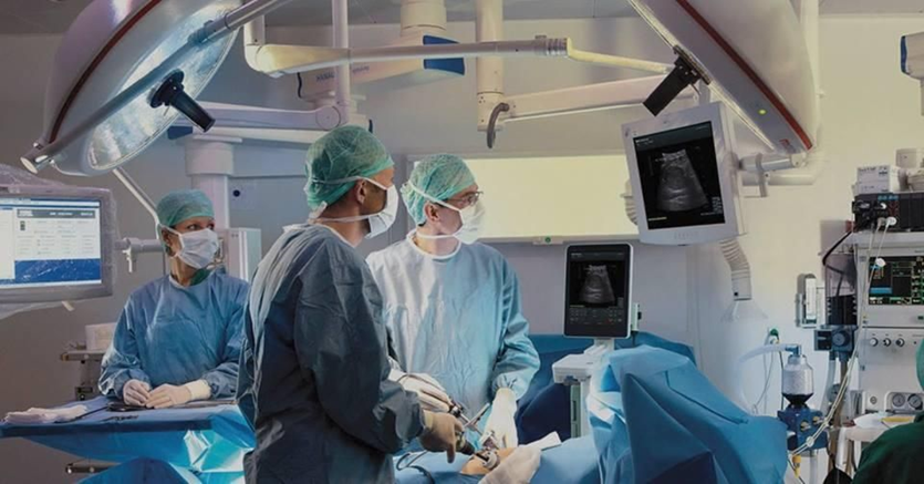
With precision health, ultrasound has evolved to address procedure-specific needs in disciplines ultrasound didn’t have a presence in previously: neurosurgery, spine and general surgery, and urology. That means when a patient undergoes neurosurgery to remove a tumor, for example, instead of the physician relying on static images from a CT scan, the surgeon can see onscreen exactly where the tumor is in real time and determine the best course for removal.

Imagine a surgeon able to operate on certain fetal anomalies in weeks 11-12 — something that until just recently was possible only in week 20 — and achieve a better outcome for the unborn baby. Or a physician who is performing a lumpectomy on a breast cancer patient and can see exactly where the tumor is and excise it precisely with clean margins, ensuring the patient’s successful breast cancer treatment.

Adding real-time surgical intervention to our pre- and post-operative ultrasound capabilities not only gives us entry into the operating room but improves surgeons’ decision-making, efficiency, accuracy and healthcare outcomes for millions of people.

Bringing Ultrasound to the Masses

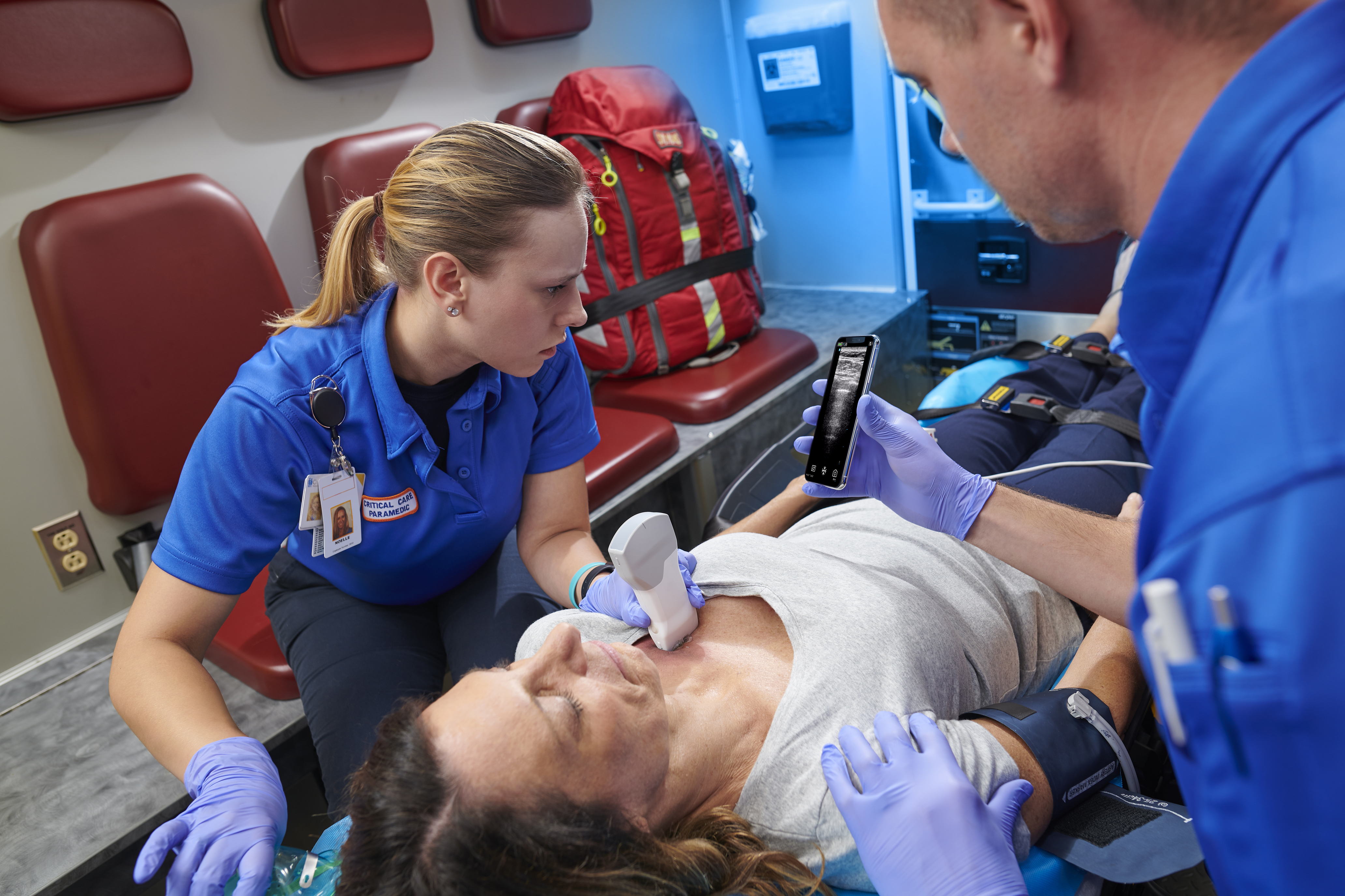
In 2010, GE Healthcare pioneered its first Vscan portable ultrasound system, which can fit in a physician’s pocket. In 2021, a decade later and with more than 50,000 units of Vscan sold globally, the third-generation handheld Vscan Air has been released. This pocket-size ultrasound, with crystal-clear image capability, is advantageous to sonographers who traditionally must train extensively and practice for years in their field. The device wirelessly pairs with any modern smartphone and is simple and easy to use, with an app that guides the user with one-click imaging. Those images can then be sent to the cloud, where a healthcare provider can view the image. Technology and AI are enabling clinicians to spend more of their time engaging with patients, rather than fiddling with the device. Indeed, the Vscan Air was built to be inclusive and have applicability across a wide variety of clinical specialties, clinician types, education levels and clinical settings.

In Germany, for instance, primary care physicians use the device to improve the speed of diagnosis. In the U.S., it has been used on helicopters during patient air transports. In Japan, home-care workers use it to better care for aging patients, while in developing countries it’s used to provide images for expectant moms who might not have access to a maternity clinic. As a global player in the industry, we understand the potential to one day put ultrasound technology such as the Vscan Air into the hands of not just healthcare providers, but patients.

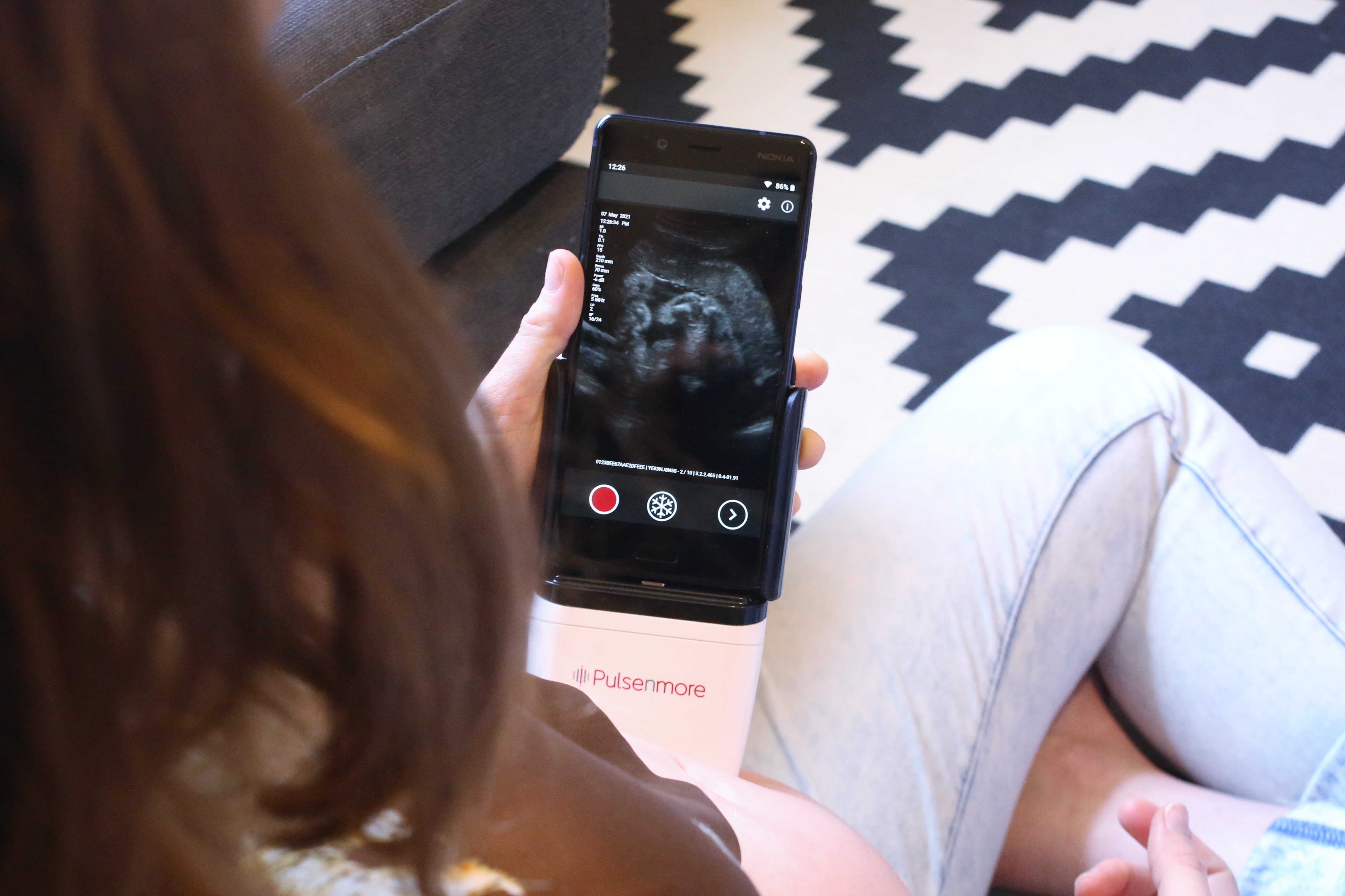
Healthcare providers today are predicting a significant shift of care services from traditional facilities to the home by 2025, without a reduction in quality or access. In-home use could create tremendous value for healthcare facilities and physician groups, care-at-home providers, technology companies and investors, and could improve patients’ quality of care and experience. That is precisely why we announced a strategic investment in Israeli home-use ultrasound startup Pulsenmore. Their novel self-operated prenatal home ultrasound solution, combined with a smartphone, enables pregnant women to self-scan for remote clinical assessment by their healthcare provider.

9N7A6304-1.jpg

With so many potential applications for this technology in development, including follicular monitoring for women undergoing in-vitro fertilization and remote monitoring for chronic heart failure and end-stage renal disease, we look to ultrasound as one day soon becoming the standard of care.

The Transformative Benefits of AI

At its simplest, artificial intelligence (AI) is just a tool — albeit a very powerful, industry-changing one that helps healthcare providers become better, faster and more efficient at what they were already doing. AI built directly into devices essentially enables clinicians to spend more of their time engaging with patients, rather than fiddling with the device or technology. AI algorithms help identify diseases more efficiently and with greater accuracy — meaning critical cases can be prioritized and treatment mapped out quickly.

Take, for example, doctors getting a handle on potential complications from COVID-19. Typically, physicians determine whether a heart is pumping enough blood through the primary arteries to organs by taking an ultrasound measurement called velocity time integral (VTI). Our hardware with AI-enabled features can get the VTI reading in seconds — dramatically reducing the number of keystrokes and the time it would take to calculate the flow rate manually.

We believe that “AI will eventually be what we think of software development today: the basis for every application in healthcare, for every solution, that we learn to harness as a tool to drive healthcare forward.”

Por-Rott-W-A-22-3-2021 10 Kopie (2) copy.jpg

Roland Rott, Ultrasound President and CEO, GE Healthcare

A Passion for Purpose

Healthcare enterprises are demanding tools that allow them to operate their fleets of devices efficiently and securely, from end to end. Small practices and individual clinicians are asking for the right tools that allow them to maximize workflow efficiencies, and collaboration to allow more time to focus on patient care and outcomes. Ultrasound innovation is deep in our DNA. As a market leader in ultrasound and a pioneer in the space, it is this focus and passion for purpose that drives our commitment to innovate and deliver better tools to physicians so they can simplify decision-making and provide faster, more personalized care to patients.

We continue to invest heavily in a digital future that allows all clinicians to collaborate seamlessly with peers, practice within an optimized department and provide their best — and most efficient — care to patients at every single site.

Whether treating a patient with COVID-19 using ultrasound to determine lung function at the point of care or using ultrasound to diagnose (and then assist in) an emergency appendectomy, or even using 4D ultrasound technology in interventional cardiology procedures, ultrasound provides an extra set of “eyes” to both physicians and surgeons, making them more efficient and creating better outcomes for their patients. We couldn’t be more excited about the future of ultrasound.

 
REFERENCES

[1] Amar Bhidé and Srikant Datar, “Case Histories of Significant Medical Advances: Development of Ultrasound Scanning,” Harvard Business School Working Paper 20-003 (2019-2021), https://www.hbs.edu/ris/Publication%20Files/20-003_7d51bf0d-d94d-44de-b08f-e12ff8bc02e0.pdf.


Emerging Trends in Ultrasound Imaging

 By Karen Koblan, Ultrasound Solutions Corp.

We are close to a new era in ultrasound technology. From helping healthcare specialists detect several diseases such as cancerous cells to showing real-time images inside the mother’s womb, ultrasound technology is a go-to way for various specialists to deal with a wide range of diseases and tasks.

Let us take a closer look at how the emerging technologies of ultra-compact ultrasound, 3D and 4D ultrasound, artificial intelligence (AI), tissue harmonic imaging, and volumetric ultrasound are impacting the future of ultrasound imaging.

1. Ultra-Compact Ultrasound

Ultra-compact or portable ultrasound machines have taken imaging technology by storm. Previously, medical professionals had to use large, bulky, and complicated ultrasound machines for treating patients. Now, healthcare and clinical laboratories are opting for these ultra-compact imaging machines due to their portability and ease of use.

Portable ultrasound machines are also being used by healthcare specialists. In particular, portable ultrasound machines are useful in detecting UTIs. These machines offer a number of advantages over standard methods of UTI detection, such as computed tomography (CT) or magnetic resonance imaging (MRI). Portable ultrasound machines provide superior imaging quality and allow for real-time image guidance. These machines are perfect for primary care settings.

2. 3D And 4D Real-Time Ultrasound Imaging

The use of three-dimensional (3D) real-time imaging ultrasound technology is being driven by the demand for more accurate diagnostic images. This type of imaging provides a clear picture of the internal organs and can be used to detect abnormalities such as tumors. It is becoming more popular because it gives a better view of what is going on inside the body.

3D real-time imaging is becoming more popular for fetal ultrasound. This technology gives a more detailed view of the baby. This technology is new, and there is no standard protocol for its use yet. However, more hospitals are expected to start using this technology in the near future.

Further, this technology helps determine diseases with ease. It helps to achieve better visuals of human body organs. Compared to 2D imaging technology, 3D ultrasound imaging technology also takes less time.

Four-dimensional (4D) ultrasound imaging technology is even more convenient for healthcare specialists such as gynecologists. Compared to 3D ultrasound technology, 4D ultrasound shows live motion with the help of several images. With this technology, gynecologists can observe the live movement of the baby in the mother’s womb.

4D real-time ultrasound imaging provides a lot of benefits that traditional two-dimensional imaging does not, especially to gynecologists. This type of imaging gives a more complete view of the fetus, as well as showing how the fetus is developing over time. This technology can also be used to monitor multiple fetuses simultaneously. This is beneficial for high-risk pregnancies.

3. Artificial Intelligence

AI or artificial intelligence has a lot of potential in the medtech and imaging industry, including in the area of ultrasound technology. Ultrasound waves are used to create images of the inside of the body. This technology has been used for diagnostic purposes for many years. However, interpreting these images can be tricky, even for experienced radiologists. This is where AI comes in to help.

AI-enabled ultrasound machines can quickly and accurately interpret images. This can help doctors diagnose and treat patients faster. Additionally, AI can help identify patterns that human observers may miss. For example, AI can help identify early signs of several serious diseases, including cancer, heart disease, and stroke. This technology has the potential to save lives by providing earlier diagnosis and treatment.

4. Tissue Harmonic Imaging

Tissue harmonic imaging (THI) is another new emerging technology that's rapidly changing the use of standard ultrasound techniques. THI is an advanced technology that produces images with greater clarity than standard ultrasound. This means that clinicians can make more accurate diagnoses using THI, and this makes it particularly well-suited for use in cardiac imaging.

In addition, THI technology requires less power and can be performed more quickly, making it more convenient for both patients and clinicians. Further, THI is less likely than standard ultrasound to produce artifacts, which can often lead to inaccurate diagnoses

5. Volumetric Ultrasound

In general, volumetric imaging creates images of objects in space by combining multiple 2D images taken from different angles. This allows for a more complete view of an object than would be possible with just a single image. Now, the same concept is being used for medical diagnosis purposes.

Volumetric ultrasound provides 3D images of the body by steering a 2D array transducer in a scan format, using sound waves and computer algorithms to create images of the inside of the body. This imaging modality can be helpful in identifying cancer cells, tumor cells, and other abnormalities, as well as diagnosing various conditions, such as various heart diseases. Further, volumetric ultrasound is often used to help guide procedures such as biopsies and needle injections.

It is often used to image the fetus during pregnancy. It can also be used to image other organs and structures, as well as to assess relationships between different structures of human body organs. It is a great way to see small structures and is very clear.

Final Thought

Ultra-compact ultrasound, 3D and 4D ultrasound, artificial intelligence (AI), tissue harmonic imaging, and volumetric ultrasound are impacting – and will continue to impact -- the future of ultrasound technology, benefiting both healthcare providers and patients.

Education is the catalyst to increased adoption of handheld ultrasound

By Mustafa Hassan, AuntMinnie.com contributing writer


January 20, 2023

Handheld ultrasound has not yet reached mainstream adoption, but the worldwide market is still projected to reach over $500 million by 2026, according to Signify Research's newly published Handheld Ultrasound Deep Dive Report 2022.

In addition, market revenues are estimated to have grown by approximately 30% in 2021, driven by strong growth in the U.S., the biggest market for handheld ultrasound. Despite the global challenges for handheld ultrasound vendors in 2022, such as rising inflation and supply chain disruptions, the handheld ultrasound market is expected to experience double-digit growth, and this is forecast to continue through to 2026.

Most of the market growth will be fueled by increased adoption of handheld devices by new users of ultrasound, such as primary care physicians, nurses, emergency medical technicians (EMTs), and midwives. The key market trends are discussed below.

AI

The steep learning curve and subsequent ultrasound skills shortage are two of the biggest barriers to the wider use of ultrasound. These challenges are exacerbated in handheld ultrasound, where there is a higher proportion of new and less experienced users compared with cart and compact ultrasound.

As the expansion of the handheld ultrasound market in the coming years is expected to be strongly driven by new user groups, this barrier will be greater than ever before. These barriers can be partially addressed by artificial intelligence (AI) solutions that guide users with positioning and moving the ultrasound probe.

To date, most image guidance AI solutions, such as those from Caption Health, UltraSight, and DESKi, are for cardiac scans. AI image guidance solutions will have a bigger impact when they can also be used for other clinical applications, making it easier for users such as nurses to use ultrasound during their rounds. Though this is starting to happen -- with image guidance AI solutions for the thyroid (developed by Medo.AI), women's health (developed by Pulsenmore), and vascular applications (developed by ThinkSono) -- it will take time, capital, and data to develop image capture support for other body areas. In the interim, we expect to see more anatomy labeling solutions to assist with image capture.

In addition to AI for image capture support, solutions for image analysis will make ultrasound more efficient for users. These applications need to be embraced by physicians, some of whom may be wary about the performance and limitations of AI.

For AI to be more widely used in handheld ultrasound, one of the main challenges that will need to be overcome is how the AI applications are paid for. The solutions need to be affordable to the customer, in line with the price paid for the scanner, yet still enable AI vendors and potential OEM partners to make a profit. Another challenge is validating the AI solutions, which can be costly. This is one of the main reasons for the lack of ultrasound AI adoption in China.

Teleultrasound

With education being the most significant barrier preventing the adoption of handheld ultrasound, teleultrasound can have a positive influence by connecting novice users with experienced experts. With ultrasound increasingly being taught as part of the curriculum at medical schools, a teleultrasound platform enables newly trained physicians to connect with an ultrasound expert to help them become more comfortable in using ultrasound in real-life cases and to get a second opinion on a diagnosis.

With the current backlog in patients requiring imaging and changes in reimbursement, there is a drive to move imaging to out-of-hospital settings, and teleultrasound platforms, along with other digital solutions, can play a role in facilitating this shift.

Table 1 “Teleultrasound solutions available in the handheld ultrasound market"

Signify table


Diverging Business Models

During the early years of handheld ultrasound, most devices were purchased with a one-time payment (capital expenditure). This payment model is familiar and accepted by healthcare providers and enables vendors with a broad range of ultrasound systems to sell handheld scanners to their existing customer base as an "add on" with bundled deals.

More recently, the new breed of dedicated handheld ultrasound vendors, like Butterfly Network, Vave, and Clarius Mobile Health, have introduced subscription payment models. Due to the low cost of handheld ultrasound devices, with the global average selling price now down to around $4,000, vendors need large sales volumes to make a profit. This is a challenge as handheld ultrasound has not yet achieved mainstream adoption. As such, most dedicated handheld ultrasound vendors are not yet profitable.

Vendors are now seeking subscription revenue streams, either to replace the upfront payment for the device or to supplement the initial device purchase, to generate additional and recurring revenue. Vendors are now launching digital solutions that are chargeable add-on services, such as teleultrasound and AI applications. Moreover, the subscription model enables vendors to lower the initial cost of the device, opening the market to cost-sensitive customers, such as new users of ultrasound.


Competitive Analysis

Signify marketshare


The leading vendors of handheld ultrasound are Philips, Butterfly Network, GE HealthCare, and Clarius. The largest Chinese handheld ultrasound vendors are SonoStar, Youkey, and Stork Healthcare. These vendors are mostly focused on their domestic market, except for SonoStar which has had some limited success in Western Europe and the U.S.

While the market was initially dominated by vendors such as GE HealthCare and Philips with a full ultrasound product portfolio, dedicated handheld vendors are becoming increasingly prominent. Initially competing with other ultrasound system types, notably low-end compact systems, handheld ultrasound vendors are increasingly developing customer bases distinct from users of compact systems. This trend will increase as handheld ultrasound becomes more mainstream and is adopted by increasingly newer users of ultrasound in new use cases such as in plastic surgery and medical aesthetics.

Although the majority of handheld ultrasound sales are wired devices, accounting for around 75% of handheld ultrasound sales revenue in 2021, wireless handheld ultrasound devices are expected to dominate the market in the future. The current product mix of wired versus wireless devices in the market is due more to the supplier mix than customer need, as most vendors currently offer wired devices. Among the larger handheld ultrasound vendors, Butterfly Network and Philips only offer wired devices and GE Healthcare launched its first wireless device in 2021.

The Signify view

The outlook for the handheld ultrasound market is promising, with a projected 2021-2026 compound annual growth rate of 24.7%. Growth will be driven by existing ultrasound users, either to replace compact systems or as an adjunct to existing systems, as well as new users of ultrasound such as nurses, primary care physicians, and midwives. Some vendors are also promoting the use of handheld ultrasound to enable patients to self-scan in the home setting.

Primary care is forecast to be the fastest growing clinical application for handheld ultrasound, with growth propelled by increased adoption of handheld ultrasound by primary care physicians. It is estimated that the percentage of primary care physicians that use ultrasound is in the single digits in most countries. The lower cost and ultraportability of handheld scanners are expected to notably increase the use of ultrasound in primary care.

The use of ultrasound in plastic surgery and medical aesthetics to improve procedure safety, is expected to be one of the fastest emerging applications of handheld ultrasound, especially in Western Europe and the U.S. Ultrasound will increasingly be used by plastic surgeons to differentiate their practice and reassure customers of the safety of cosmetic procedures.

"Handheld Ultrasound Market Deep Dive" presents market dynamics, estimates, forecasts, competitive analysis and major drivers for the handheld ultrasound equipment market. It segments the market into 30 countries/regions and into the five main clinical applications; radiology, cardiology, women's health, point of care, and specialty markets, with these markets subsegmented into 24 clinical applications overall.

Mustafa Hassan is an analyst at Signify Research, an independent supplier of market intelligence and consultancy to the global healthcare technology industry. Signify's major coverage areas are healthcare IT, medical imaging, and digital health


Chủ Nhật, 8 tháng 1, 2023

Model separates malignant from benign thyroid nodules on ultrasound


By Amerigo Allegretto, AuntMinnie.com staff writer

December 27, 2022 -- A deep-learning model developed by Chinese researchers can identify thyroid malignancies on ultrasound images, a study published December 21 in Computers in Biology and Medicine found.

Researchers led by Zhenggang Yu from Huaqiao University in Quanzhou found that by using an attention mechanism, their model can distinguish malignant from benign thyroid nodules with a high level of accuracy. They also highlighted the model's high speed.

"Young physicians and small hospitals with limited resources can benefit from using this method to assist with thyroid ultrasound examination diagnosis," Yu and co-authors wrote.

Early diagnosis of thyroid cancer with the aid of ultrasound imaging can lead to better treatment outcomes. However, this is dependent on the experience of the sonographer.

The researchers noted that while computer-aided diagnosis (CADx) techniques are available, their ability to differentiate between features seen in benign and malignant nodules is still limited.

Yu and fellow researchers wanted to propose another such method, but with an attentional mechanism with lightweight properties. They hypothesized that this could help identify nodules and distinguish malignant from benign findings by using a bounding box. They also highlighted that the model could do this without using much hardware while still being cost-effective.

The team tested the algorithm on data collected in 2020 and 2021 from a total of 1,921 thyroid ultrasound images from 1,357 patients. They looked at mean average precision (mAP) to evaluate performance and also assessed different intersection-over-union (IoU) thresholds, which measure the accuracy of an object detector on a particular dataset.

The researchers reported that the model showed good diagnostic results for benign and malignant thyroid nodules, especially for malignant nodules.

Performance of AI model for characterizing thyroid nodules on ultrasound
All nodulesBenignMalignant
Precision0.8850.8290.941
Recall0.8510.8380.863
mAP (0.5 IoU)0.890.8680.911
mAP (0.5 – 0.95 IoU)0.5010.4780.523

The proposed model's mAP (with 0.5 IoU threshold) of 0.89 and an average speed of seven milliseconds, suggests high accuracy and rapid detection, according to the authors. They also highlighted that the model performs well with nodules of different sizes.

"In the clinical setting, it is possible to achieve a direct diagnosis of thyroid cancer once ultrasound images have been acquired, thus making the method practical," they added.

The study authors suggested that their model can aid inexperienced sonographers in this setting and can help them to free up their workloads. They added that it can also help avoid unnecessary invasive fine-needle aspiration.

However, the authors also called for better sample balance in testing the model. They wrote that their study was imbalanced between high- and low-risk nodules, which could result in poorer learning for the model.

Elastography helps detect autoimmune thyroid diseases in children


By Amerigo Allegretto, AuntMinnie.com staff writer

December 13, 2022


Although the study findings are promising for identifying autoimmune thyroid diseases in children, authors Dr. Eda Celebi Bitkin and Dr. Nursen Toprak from Van Yüzüncü Yıl University wrote, although they noted that elastography by itself is not enough to differentiate between the two diseases.

"If the clinical aspect is poor or thyroid-stimulating hormone receptor antibodies are missing, the combined use of conventional ultrasound and acoustic radiation force impulse may be helpful in distinguishing between the two diseases," the authors wrote.

Hashimoto's thyroiditis and Graves' disease are autoimmune diseases that affect the thyroid gland. Ultrasound findings and autoantibody elevation are typically used to diagnose either disease. In rare cases, ultrasound findings may be normal in both diseases, but previous research has suggested that shear-wave velocity values from elastography are higher in autoimmune thyroiditis than in healthy thyroid glands.

Bitkin and Toprak explored the use of elastography in 88 pediatric patients with Hashimoto's thyroiditis and Graves' disease. Out of these, 28 had Hashimoto's thyroiditis, 20 had Graves' disease, and the remaining 40 were healthy controls. All patients underwent thyroid elastography and ultrasound.

The duo found that shear-wave velocity values of patients with either disease showed higher thyroid tissue stiffness than healthy controls, but when such values were compared between both diseases, the team found no significant difference (p = 0.73). As for conventional ultrasound, patients with Hashimoto's thyroiditis had a hypoechoic echo pattern similar to those with Graves' disease (p < 0.05).

"These ultrasound imaging findings reflect longer-term inflammation and may not be found in the early stages of thyroiditis," Bitkin and Toprak wrote.

The researchers did not track disease stage in their study but suggested that the degree of tissue stiffness may change as the diseases progress. They called for future studies to consider disease staging, especially for patients with Hashimoto's thyroiditis.

Thứ Sáu, 6 tháng 1, 2023

FATTY LIVER DISEASE AREA

 Abstract

Liver steatosis is a chronic liver disease that is becoming one of the most important global health problems, due to its direct connection with metabolic syndrome, its significant impact on patients’ socioeconomic status and frailty, and the occurrence of advanced chronic liver disease. In recent years, there has been rapid technological progress in the ultrasound-based diagnostics field that can help us to quantitatively assess liver steatosis, including continuous attenuation parameters in A and B ultrasound modes, backscatter coefficients (e.g., speed of sound) and ultrasound envelope statistic parametric imaging. The methods used in this field are widely available, have favorable time and financial profiles, and are well accepted by patients. Less is known about their reliability in defining the presence and degree of liver steatosis. Numerous study reports have shown the methods’ favorable negative and positive predictive values in comparison with reference investigations (liver biopsy and MRI). Important research has also evaluated the role of these methods in diagnosing and monitoring non-alcoholic fatty liver disease (NAFLD). Since NAFLD is becoming the dominant global cause of liver cirrhosis, and due to the close but complex interplay of liver steatosis with the coexistence of liver fibrosis, knowledge regarding NAFLD’s influence on the progression of liver fibrosis is of crucial importance. Study findings, therefore, indicate the possibility of using these same diagnostic methods to evaluate the impact of NAFLD on the patient’s liver fibrosis progression risk, metabolic risk factors, cardiovascular complications, and the occurrence of hepatocellular carcinoma. The mentioned areas are particularly important in light of the fact that most of the known chronic liver disease etiologies are increasingly intertwined with the simultaneous presence of NAFLD.


Thứ Ba, 3 tháng 1, 2023

JMU Vol 49-4 2022


Download from THU VIEN MEDIC 

https://drive.google.com/file/d/11i8Xbukp263jeNWc1fRrHdfifCTOaJnj/view?usp=sharing


Thứ Bảy, 31 tháng 12, 2022

Liver in Advance, US, CT, and MRI Moving toward the Future


ABSTRACT


Over the past two decades, the epidemiology of chronic liver disease has changed with an increase in the prevalence of nonalcoholic fatty liver disease in parallel to the advent of curative treatments for hepatitis C. Recent developments provided new tools for diagnosis and monitoring of liver diseases based on ultrasound (US), computed tomography (CT), and magnetic resonance imaging (MRI), as applied for assessing steatosis, fibrosis, and focal lesions. This narrative review aims to discuss the emerging approaches for qualitative and quantitative liver imaging, focusing on those expected to become adopted in clinical practice in the next 5 to 10 years. While radiomics is an emerging tool for many of these applications, dedicated techniques have been investigated for US (controlled attenuation parameter, backscatter coefficient, elastography methods such as point shear wave elastography [pSWE] and transient elastography [TE], novel Doppler techniques, and three-dimensional contrast-enhanced ultrasound [3D-CEUS]), CT (dual-energy, spectral photon counting, extracellular volume fraction, perfusion, and surface nodularity), and MRI (proton density fat fraction [PDFF], elastography [MRE], contrast enhancement index, relative enhancement, T1 mapping on the hepatobiliary phase, perfusion). Concurrently, the advent of abbreviated MRI protocols will help fulfill an increasing number of examination requests in an era of healthcare resource constraints.

Key points

  • Technical advances in liver imaging have been observed for ultrasound, computed tomography, and magnetic resonance imaging (MRI).

  • Quantitative liver imaging biomarkers are promising to measure disease severity and reduce interoperator variability.

  • Quantitative liver imaging biomarkers have the potential to be increasingly adopted in clinical practice.

  • Abbreviated MRI protocols will help fulfill an increasing number of examination requests.

Thứ Tư, 28 tháng 12, 2022

TI-RADS helps classify thyroid lesions on ultrasound

By Amerigo Allegretto, AuntMinnie.com staff writer


May 3, 2022

Researchers in three separate but related presentations discussed existing and new research that shows the effectiveness of TI-RADS in thyroid imaging with ultrasound.

"It provides descriptive features that are associated with increased rates of malignancy," said Dr. Michelle Melany from Cedars-Sinai Medical Center in Los Angeles in her keynote speech. "It helps categorize nodules and increases the number of reports with unambiguous management recommendations."

The American College of Radiology (ACR) in 2017 developed a standardized TI-RADS risk-stratification system to inform practitioners about which thyroid nodules require biopsy. This was in response to costly interventions for nodules that turned out to be benign on biopsy.

TI-RADS uses a scoring scale that goes from 0 to 5. A score of 0 indicates a benign lesion, while 5 represents a highly suspicious finding.

Benefits of TI-RADS include having higher sensitivity than risk stratification systems, ensuring all nodules can be classified, and reducing unnecessary biopsies, among others.


However, in her ARRS 2022 talk, Melany also listed some challenges the system faces. These include having a higher interobserver variability, varying appearances, and dependence on scanning parameters.

"In most of our practices, we're super busy. If only the ultrasound technician scans, then we're really dependent on what's captured and recorded on still images or cinematic clips," Melany said.

In an abstract presentation following Melany's keynote address, Dr. Logan Haug from the Mayo Clinic in Arizona showed research he co-led assessing whether adjusting TI-RADS could further reduce including benign nodules while keeping all malignant nodules. The current TI-RADS assign a score of two points for any lobulation found on ultrasound.

The team retrospectively looked at all ultrasound and pathology records to identify 516 thyroid nodules that underwent fine-needle aspiration (FNA) and had 58 lobulated margins in a two-year period. Images were divided into two groups showing "only macrolobulated" or "microlobulations present." Microlobulations were defined as being less than 2.5 mm in circumference.

Out of the lobulated margins, 14 were found to be malignant. A total of 37 lobulated margins were solely macrolobulated (two malignant), while 21 were microlobulated (12 malignant) (p < 0.0001).

Of nodules greater than or equal to 10 mm, 53 were lobulated, including 10 malignancies. The researchers found that under current TI-RADS standards, all 53 nodules met the criteria for FNA. However, under the team's microlobulation TI-RADS standards, 38 met the criteria for FNA.

In a second abstract presentation, Dr. Meredith Bara from the University of Alberta in Canada talked about research she co-led that looked at interreader agreement after teaching sessions for residents.

The study authors used 50 test nodules scored independently by three radiology residents with no formal TI-RADS training. Bara said the residents underwent a one-hour teaching session after initial scoring, which included reviewing sonographic features of nodules and 30 training cases.

The team found that the residents showed "significant" improvement in agreement after training, with a pooled specificity between 76.6% and 96.8%. They showed the strongest agreement on nodular shape and composition.

Melany in her keynote listed some potential future modifications to TI-RADS that could further improve its effects. These include using active surveillance, changing points for certain features, incorporating nodule location, using artificial intelligence, and using newer ultrasonic methods such as contrast-enhanced ultrasound and elastography.