Tổng số lượt xem trang

Thứ Bảy, 21 tháng 4, 2018

CẮT TÚI MẬT NỘI SOI: TIÊU CHUẨN SIÊU ÂM TIÊN LƯỢNG KHÓ


Abstract

Purpose: Laparoscopic cholecystectomy (LC) has become the treatment of choice for cholelithiasis. Still some patients required conversion to open cholecystectomy (OC). Our aim was to develop a standardized Ultrasound based scoring system for preoperative prediction of difficult LC.

 Methods and materials: Ultrasound findings of 300 patients who underwent LC were reviewed retrospectively. Four parameters (time taken, biliary leakage, duct or arterial injury, and conversion) were analyzed to classify LC as easy or difficult. The following ultrasound findings were analyzed: GB wall thickness, pericholecystic collection, distended GB, impacted stones, multiple stones, CBD diameter and liver size. Out of seven parameters, four were statistically significant in our study. A score of 2 was assigned for the presence of each significant finding and a score of 1 was assigned for the remaining parameters to a total score of 11. A cut-off value of 5 was taken to predict easy and difficult LC.
 Results: 66 out of 83 cases of difficult LC and 199 out of 217 cases of easy LC were correctly predicted on the basis of scoring system. A score of >5 had sensitivity 80.7% and specificity 91.7% for correctly identifying difficult LC. Prediction came true in 78.8% difficult and 92.6% easy cases. US findings of GB wall thickness, distended GB, impacted stones and dilated CBD were found statistically significant.
Conclusion: This indigenous scoring system is effective in predicting conversion risk of LC to OC. Patients having high risk may be informed and scheduled appropriately and decision to convert to OC in case of anticipated difficulty may be taken earlier.

ª 2017, Elsevier Taiwan LLC and the Chinese Taipei Society of Ultrasound in Medicine. This is an open access article under the CC BY-NC-ND license (http://creativecommons.org/licenses/ by-nc-nd/4.0/).

TI-RADS REDUCES BIOPSY

Thyroid Imaging Reporting and Data System Reduces Biopsies

Apr 18, 2018
Criteria from the American College of Radiology (ACR) Thyroid Imaging Reporting and Data System (TI-RADS) offers a meaningful reduction in the number of thyroid nodules recommended for biopsy, according to a study published in the journal Radiology
Researchers from several states performed a retrospective study to compare the biopsy rate and diagnostic accuracy before and after applying ACR TI-RADS criteria for thyroid nodule evaluation. Eight radiologists with three to 32 years of experience in thyroid ultrasonography were asked to review the ultrasound features of 100 thyroid nodules that were cytologically proven and/or pathologically proven. Nodules evaluated in five US categories and biopsy recommendations were provided based on the radiologists’ practice patterns without knowledge of ACR TI-RADS criteria. Three other expert radiologists were reference standard readers for the imaging findings. ACR TI-RADS criteria were retrospectively applied to the features assigned by the eight radiologists to produce biopsy recommendations. Comparison was made for biopsy rate, sensitivity, specificity, and accuracy.
The results showed 15 of the 100 nodules (15 percent) were malignant. The mean number of nodules recommended for biopsy by the eight radiologists was 80 ± 16 (standard deviation) based on their own practice patterns and 57 ± 11 with retrospective application of ACR TI-RADS criteria.
Without ACR TI-RADS criteria:
Sensitivity  95 percent
Specificity  20 percent
Accuracy  28 percent
With ACR TI-RADS criteria:
Sensitivity 92 percent
Specificity 44 percent
Accuracy 52 percent
Expert consensus:
Sensitivity 87 percent
Specificity 51 percent
Accuracy 56 percent

The researchers noted that although fewer malignancies were recommended for biopsy with ACR TI-RADS criteria, the majority met the criteria for follow-up US. Only three of 120 (2.5 percent) malignancy encounters required no follow-up or biopsy. Expert consensus recommended biopsy in 55 of 100 nodules with ACR TI-RADS criteria.
Not only did the ACR TI-RADS criteria offer a meaningful reduction in the number of thyroid nodules recommended for biopsy, the researchers wrote, they significantly improve the accuracy of recommendations for nodule management.

Thứ Tư, 18 tháng 4, 2018

SUBCUTANEOUS FAT MEASUREMENT by US


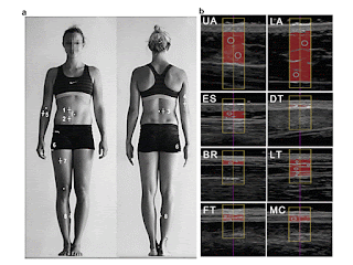
Abstract—A recently standardized ultrasound technique for measuring subcutaneous adipose tissue (SAT) was applied to normal-weight, overweight and obese persons. Eight measurement sites were used: upper abdomen, lower abdomen, erector spinae, distal triceps, brachioradialis, lateral thigh, front thigh and medial calf. Fat compression was avoided. Fat patterning in 38 participants (body mass index: 18.6–40.3 kgm22 ; SAT thickness sums from eight sites: 12–245 mm) was evaluated using a software specifically designed for semi-automatic multiple thickness measurements in SAT (sound speed: 1450 m/s) that also quantifies embedded fibrous structures. With respect to ultrasound intra-observer results, the correlation coefficient r 5 0.999 (p < 0.01), standard error of the estimate 5 1.1 mm and 95% of measurements were within ±2.2 mm. For the normal-weight subgroup, the median measurement deviation was 0.43 mm (1.1% of mean thickness), and for the obese/overweight subgroup it was 0.89 mm (0.5%). The eight sites used here are suggested to represent inter-individual differences in SAT patterning. High measurement accuracy and reliability can be obtained in all groups, from lean to overweight and obese, provided that measurers are trained appropriately.
Fig. 1. B-Mode ultrasound measurement of uncompressed subcutaneous fat. (a) Sites for ultrasound measurement of subcutaneous adipose tissue (SAT) patterning: upper abdomen (1, UA), lower abdomen (2, LA), erector spinae (3, ES), distal triceps (4, DT), brachioradialis (5, BR), lateral thigh (6, LT), front thigh (7, FT) and medial calf (8, MC). Body height was used as the reference length for all distances. A detailed description of the standardized marking and of the ultrasound measurement technique can be found in M€uller et al. (2016). (b) Ultrasound images and evaluations of SAT thicknesses. Participant A: male, body mass index 5 25.5, body mass 5 80.7 kg, stature 5 1.78 m. (c) Survey plot of SAT patterning according to (b). The columns represent the mean values of the semi-automatic multiple thickness measurements for the eight ultrasound sites. The mean thickness value of the SAT thickness in a given ultrasound image (within the region of interest) is termed dINCL (gray) when fibrous structures are included and dEXCL (black) when fibrous structures are subtracted. Sum of the eight thicknesses DINCL 5 44.3 mm (DEXCL 5 39.8 mm). (d) Survey plot of a participant B with similar body mass index. Body mass index 5 25.4 kgm22 ; body mass 5 75.5 kg, stature 5 1.72 m. The SAT thickness sum was 86.9 mm (77.9 mm), 96% higher than in participant A.


(E-mail: wolfram.mueller@medunigraz.at) 2016 The Authors. Published by Elsevier Inc. on behalf of World Federation for Ultrasound in Medicine & Biology. This is an open access article under the CC BY-NC-ND license (http://creativecommons.org/licenses/by-nc-nd/4.0/).

Key Words: Body composition, Subcutaneous adipose tissue, Overweight, Obesity, Ultrasound measurement precision.