Tổng số lượt xem trang

Thứ Sáu, 5 tháng 10, 2012

Different Sonographic Features of Triple-Negative Breast Cancer and Non-TNBC

Abstract

Objectives—Triple-negative breast cancer (TNBC) is known to have unique molecular, clinical, and pathologic characteristics. The growth pattern of this cancer may also affect its appearance on sonography. Our study evaluated the sonographic features of TNBC according to the American College of Radiology Breast Imaging Reporting and Data System sonographic classification system and compared these features with those of non-TNBC.
Methods—Data from 315 consecutive breast cancer cases were collected. The images were reevaluated by an examiner blinded to the patients’ characteristics and histologic results according to the Breast Imaging Reporting and Data System. The sonographic features of TNBC (n = 33) and non-TNBC (n = 282) were compared.
 
Conclusions—Triple-negative breast cancer and non-TNBC have different sonographic features. This finding can be explained by the pathologic profile of this breast cancer subtype. Some of the distinct sonographic criteria for TNBC are more likely to be associated with benign masses. Knowledge of the distinct sonographic features of TNBC would help the examiner avoid false-negative classification of this tumor type.

         
Discussion

Early Detection of TNBC
Triple-negative breast cancer is known to be an especially aggressive subtype of breast cancer. It is characterized by the complete absence of ER, PR, and HER2 over expression. However, this definition only tackles the phenotype and merely scratches the surface of the underlying processes within breast cancer cells.
Nowadays, we have a much deeper insight into the molecular backgrounds of the different subtypes of breast cancer. From gene expression profiles, we have learned that there are at least 5 different intrinsic subtypes of breast cancer (luminal A, luminal B, claudin low, HER2 enriched, and basal like).57 Basal-like breast cancer is not synonymous with TNBC, although there is overlap, and most basal-like breast cancers are triple negative and vice versa. There is also a strong association with BRCA gene mutations in both TNBC and basal-like breast cancer.24 Although we are just starting to determine the molecular basis of TNBC, this type of breast cancer remains an extraordinary challenge for the clinicians who treat patients with TNBC in primary care.

Triple-negative breast cancer is known to occur in younger woman, as shown in our study. The overall outcome is poor because patients with TNBC are at higher risk of early relapse and distant visceral metastases. Endocrine treatment for endocrine-responsive disease and HER2-directed treatment in cases of HER2 overexpression provide the most effective available treatment for suitable patients with breast cancer.25,26 Unfortunately, these treatment options do not exist for patients with TNBC. Therefore, chemotherapy remains the only systemic option for improving the prognosis and is usually necessary for patients with TNBC, as illustrated in our data set. New therapeutic agents, such as poly(ADP-ribose) polymerase inhibitors, are under development but have not yet been approved for the treatment of TNBC.27 As shown in neoadjuvant trials, TNBC responds relatively well to different chemotherapeutic agents, but paradoxically, the overall prognosis remains poor, and whenever a pathologically complete response cannot be achieved, the prognosis is even worse.2830

Therefore, early detection of this aggressive subtype of breast cancer has an important prognostic relevance. Against this background, breast sonography plays an essential and specific role by adding more diagnostic accuracy to the mammogram. Lesion evaluation with sonography thus plays a crucial role in the diagnostic and therapeutic pathway of patients with breast cancer. False-negative results in evaluations of breast cancers may result in a delayed diagnosis and a worse outcome.


Sonographic Features of TNBC in the Literature

In the literature, we found 3 related publications describing sonographic characteristics of TNBC, although to our knowledge, there has never been a systematic evaluation according to the BI-RADS sonographic classification and a comparison to non-TNBC. Dogan et al31 reviewed 44 patients with TNBC. They noted that TNBC may be occult on mammography and sonography and frequently has benign features. In contrast, magnetic resonance imaging identified all 44 cases correctly and showed features that had a high positive predictive value for malignancy. Ko et al32 retrospectively reviewed mammographic and sonographic findings of 245 patients from January 2007 to October 2008. Triple-negative breast cancers occurred in 35.5% of the cases (n = 87), and these cancers were more likely to have circumscribed margins, more likely to be markedly hypoechoic, and less likely to show posterior shadowing. Kojima and Tsunoda33 reported on 88 patients with TNBC. In their retrospective analysis of cases from January 2007 to April 2010, they observed that TNBC was more likely to present as a mass and less likely to show attenuating posterior echoes. These 3 studies already mentioned a few of the sonographic characteristics, and the results correlate well with our own findings.

Sonographic Features of TNBC in Our Study

The aim of our study was to scrutinize the distinct sonographic features of TNBC according to the BI-RADS sonographic classification, and our results show that TNBC is characterized by typical sonographic criteria that are to a certain degree different from those of non-TNBC (Table 4). The shape of TNBC was more likely to be oval or round, and the echogenic halo was observed significantly less often compared to non-TNBC. Furthermore, Cooper ligaments were displaced (rather than disrupted) significantly more often in TNBC. This finding can be explained by the typical growth pattern of TNBC, which is described as a “pushing border” in the absence of an infiltrating margin. This smooth appearance is usually associated with noninfiltrative processes of the breast but also occurs in TNBC as an expression of rapid tumor escalation. We believe that our observation on sonography is the macroscopic correlate of this histopathologic behavior. This kind of smooth mass margin has also been described for TNBC on magnetic resonance imaging.34
The pathologic phenomenon of a pushing margin at which the tumoredge shows a circumscribed boundary also explains the elevated rate of lobulated or microlobulated margins on sonography. The echogenic halo is an expression of a desmoplastic response and inflammation. Desmoplasia is thought to be a critical event in cancer progression and a prognostic indicator and is regularly found in TNBC.35 We were notable to find a sonographic-morphologic correlate for the desmoplastic reaction. On the contrary, an echogenic halo was observed significantly less often in our TNBC group. We suppose that the aforementioned rapid tumor progression dominates the appearance of the tumors on sonography. Hence, desmoplasia is actually present in triple-negative tumors but has no sonographic correlation.

The particularly disordered growth of malignant tumors leads to an increase in tissue layers with different impedances and consequently an increase in reflected, absorbed, and scattered echoes. In invasive ductal carcinoma, a trabecular appearance is common, and a syncytial growth pattern is rare. As a result, malignant tumors are usually associated with posterior acoustic shadowing. We found a significantly increased rate of posterior acoustic enhancement (instead of shadowing, unchanged echoes, or a mixed pattern) in our TNBC group. This feature is usually associated with benign nodules that are growing in a regular and controlled manner or with cystic lesions. As shown in recent studies, TNBC shows a syncytial growth pattern in about 56% of cases.36 In this pattern, tumor cells are closely apposed and nestled against one another and lack distinct cytoplasmic membranes, consequently forming a large syncytium. This growth pattern creates fewer layers than the trabecular pattern and may therefore explain the improved propagation of the ultrasonic waves.


Clinical Features of TNBC in Our Study

The prevalence of TNBC in our study cohort was 10.5% and was slightly lower than reported in the literature (11.2%–39.9% in certain ethnic groups).3,4 Because we did not select the cases and censoring due to missing data was a random event, this decreased prevalence can be considered a general effect due to the population that we serve in our community (97.6% European white, 1.7% Central and East Asian, 0.4% African, and 0.3% other). It might also be due to the strict definition of TNBC in our study, with a cutoff for ER and PR levels of 1% or greater for endocrine responsiveness, whereas other authors have used cutoffs of up to 10% or greater.4 However, our definition is consistent with the American Society of Clinical Oncology/College of American Pathologists recommendations.37

We observed a trend for higher breast density in the TNBC group. This result was most probably biased by the unbalanced age distribution, with the TNBC group being considerably younger than the non-TNBC group. Younger patients are more likely to have dense breast tissue. Yang et al1 reported no correlation between triple negativity and breast density in their age-adjusted collection of patients.

With regard to the final tumor stage, there was no difference in the T classification of the two groups. Regarding the N classification, we found a significantly higher rate of positive axillary lymph nodes in the TNBC group. As shown by previous studies, even small breast cancers tend to disseminate axillary lymph node metastases if a biologically unfavorable type of breast cancer is present. Maibenco et al38 reported an increasing rate of axillary lymph node metastases with higher histologic grades. Most cases of TNBC are reported to be grade 3, as found in our study. Furthermore, the intrinsic characteristics of TNBC that are determined by its unique molecular profile are known to determine its aggressive behavior, which may explain the increased lymph node involvement in the TNBC group.

Limitations of Our Study

The main limitation of our study was the relatively small sample size. Thus, although we were able to show significant differences for some sonographic features of TNBC and non-TNBC, for other features there was only a trend. We believe that significant differences for these features could be revealed with a larger sample size. Furthermore, we did not assess the combination of characteristics because that approach was not planned. Another limitation was the fact that only a single observer evaluated the sonograms. Data on interobserver concordance were not generated. This factor should be investigated further. An analogous study comparing other immunohistochemical subtypes of breast cancer (eg, HER2 positive versus HER2 negative) would also be beneficial. Investigation of the correlation with the intrinsic subtype would also be interesting. We were not able to determine the intrinsic subtype for our patients and thus were restricted to describing the TNBC phenotype.

Summary

We found that TNBC has distinct sonographic features compared to non-TNBC. These features can be explained by the typical growth pattern of this tumor type. Therefore, TNBC may lack some of the characteristic criteria for malignancy and may even mimic a benign lesion. Being familiar with this behavior would help the examiner avoid false-negative classification of TNBC in the future.
          FURTHER READING:


Study Divides Breast Cancer Into Four Distinct Types

By GINA KOLATA  The New York Times, Health
Published: September 23, 2012

In findings that are fundamentally reshaping the scientific understanding of breast cancer, researchers have identified four genetically distinct types of the cancer. And within those types, they found hallmark genetic changes that are driving many cancers.

These discoveries, published online on Sunday in the journal Nature, are expected to lead to new treatments with drugs already approved for cancers in other parts of the body and new ideas for more precise treatments aimed at genetic aberrations that now have no known treatment.

The study is the first comprehensive genetic analysis of breast cancer, which kills more than 35,000 women a year in the United States. The new paper, and several smaller recent studies, are electrifying the field.

“This is the road map for how we might cure breast cancer in the future,” said Dr. Matthew Ellis of Washington University, a researcher for the study.

Researchers and patient advocates caution that it will still take years to translate the new insights into transformative new treatments. Even within the four major types of breast cancer, individual tumors appear to be driven by their own sets of genetic changes. A wide variety of drugs will most likely need to be developed to tailor medicines to individual tumors.

“There are a lot of steps that turn basic science into clinically meaningful results,” said Karuna Jaggar, executive director of Breast Cancer Action, an advocacy group. “It is the ‘stay tuned’ story.”

The study is part of a large federal project, the Cancer Genome Atlas, to build maps of genetic changes in common cancers. Reports on similar studies of lung and colon cancer have been published recently. The breast cancer study was based on an analysis of tumors from 825 patients.

“There has never been a breast cancer genomics project on this scale,” said the atlas’s program director, Brad Ozenberger of the National Institutes of Health.

The investigators identified at least 40 genetic alterations that might be attacked by drugs. Many of them are already being developed for other types of cancer that have the same mutations. “We now have a good view of what goes wrong in breast cancer,” said Joe Gray, a genetic expert at Oregon Health & Science University, who was not involved in the study. “We haven’t had that before.”

The study focused on the most common types of breast cancer that are thought to arise in the milk duct. It concentrated on early breast cancers that had not yet spread to other parts of the body in order to find genetic changes that could be attacked, stopping a cancer before it metastasized.

The study’s biggest surprise involved a particularly deadly breast cancer whose tumor cells resemble basal cells of the skin and sweat glands, which are in the deepest layer of the skin. These breast cells form a scaffolding for milk duct cells. This type of cancer is often called triple negative and accounts for a small percentage of breast cancer.

But researchers found that this cancer was entirely different from the other types of breast cancer and much more resembles ovarian cancer and a type of lung cancer.

“It’s incredible,” said Dr. James Ingle of the Mayo Clinic, one of the study’s 348 authors, of the ovarian cancer connection. “It raises the possibility that there may be a common cause.”

There are immediate therapeutic implications. The study gives a biologic reason to try some routine treatments for ovarian cancer instead of a common class of drugs used in breast cancer known as anthracyclines. Anthracyclines, Dr. Ellis said, “are the drugs most breast cancer patients dread because they are associated with heart damage and leukemia.”

A new type of drug, PARP inhibitors, that seems to help squelch ovarian cancers, should also be tried in basal-like breast cancer, Dr. Ellis said.

Basal-like cancers are most prevalent in younger women, in African-Americans and in women with breast cancer genes BRCA1 and BRCA2.

Two other types of breast cancer, accounting for most cases of the disease, arise from the luminal cells that line milk ducts. These cancers have proteins on their surfaces that grab estrogen, fueling their growth. Just about everyone with estrogen-fueled cancer gets the same treatment. Some do well. Others do not.

The genetic analysis divided these cancers into two distinct types. Patients with luminal A cancer had good prognoses while those with luminal B did not, suggesting that perhaps patients with the first kind of tumor might do well with just hormonal therapy to block estrogen from spurring their cancers while those with the second kind might do better with chemotherapy in addition to hormonal therapy.

In some cases, genetic aberrations were so strongly associated with one or the other luminal subtype that they appeared to be the actual cause of the cancer, said Dr. Charles Perou of the University of North Carolina, who is the lead author of the study. And he called that “a stunning finding.”

“We are really getting at the roots of these cancers,” he said.

After basal-like cancers, and luminal A and B cancers, the fourth type of breast cancer is what the researchers called HER2-enriched. Breast cancers often have extra copies of a gene, HER2, that drives their growth. A drug, Herceptin, can block the gene and has changed the prognosis for these patients from one of the worst in breast cancer to one of the best.

Yet although Herceptin is approved for every breast cancer patient whose tumor makes too much HER2, the new analysis finds that not all of these tumors are alike. The HER2-enriched should respond readily to Herceptin; the other type might not.

The only way to know is to do a clinical trial, and one is already being planned. Herceptin is expensive and can occasionally damage the heart. “We absolutely only want to give it to patients who can benefit,” Dr. Perou said.

For now, despite the tantalizing possibilities, patients will have to wait for clinical trials to see whether drugs that block the genetic aberrations can stop the cancers. And it could be a vast undertaking to get all the drug testing done. Because there are so many different ways a breast cancer cell can go awry, there may have to be dozens of drug studies, each focusing on a different genetic change.

One of Dr. Ellis’s patients, Elizabeth Stark, 48, has a basal-type breast cancer. She has gone through three rounds of chemotherapy, surgery and radiation over the past four years. Her disease is stable now and Dr. Stark, a biochemist at Pfizer, says she knows it will take time for the explosion of genetic data to produce new treatments that might help her.

“In 10 years it will be different,” she said, adding emphatically, “I know I will be here in 10 years.”


Thứ Năm, 4 tháng 10, 2012

TĂNG NHÃN ÁP và LƯU LƯỢNG MÁU NHÃN CẦU


Abstract

Open-angle glaucoma (OAG) is a chronic progressive optic neuropathy that is increasing in prevalence worldwide. Currently, intraocular pressure is the only known modifiable risk factor. With lowering of intraocular pressure, the proportion of individuals who experience progression of visual field defects is reduced but continues to occur in some individuals. Many other risk factors have been identified, including decreased ocular perfusion pressure and decreased ocular blood flow. Various imaging methodologies have shown an association between OAG and altered blood flow in the various circulations: retrobulbar, retinal, optic nerve head and choroidal. In addition, different morphological alterations have been found to be associated with OAG. This review will cover the evidence that supports the association between altered ocular blood flow and glaucoma. Furthermore, it serves to describe the future methodologies that will assess ocular metabolism, which will strive to move the field closer to definitively understanding the effect of vascular changes on OAG.


Expert Commentary


The understanding of glaucoma has come a long way from the identification of IOP as a risk factor for glaucoma. Numerous other risk factors have been identified including decreased OPP, decreased OBF, circadian fluctuations in vascular parameters and vascular dysregulation. However, increased IOP continues to be the only modifiable risk factor for the progression of glaucoma. The association between altered OBF and glaucoma has been repeatedly defined; however, the pathophysiologic effect of altered blood flow on glaucomatous damage remains to be understood. Moreover, there are a lack of progression data for parameters such as OPP and OBF. Owing to a lack of large-scale longitudinal clinical trials, more evidence needs to be present before a recommendation can be made about measuring OBF in a clinical setting. Some of the aforementioned studies have small sample sizes and do not possess large statistical power, which could cause confusion due to reduced reproducibility of the studies. OBF measurements remain to be a research methodology to understand more about the pathophysiology of glaucoma. Nevertheless, the use of the methodologies of to assess OBF are continually providing more information that will be used to further understand the pathophysiology of glaucoma.

From the use of CDI for measuring retrobulbar vessels to SLO angiography measuring retinal and choroidal circulations, the blood flow methodologies are each able to assess a subset of the ocular circulation. However, none are comprehensive in their assessment and a combination of the various methodologies must be used to thoroughly analyze OBF. Furthermore, each methodology has its disadvantages, such as CDI’s inability to measure blood flow volume. The relatively new technologies of retinal oximetry and FD-OCT have shown promise to provide continued useful information, with retinal oximetry’s ability to more directly measure tissue metabolism and FD-OCT’s ability to provide accurate measurements of blood flow in absolute units. For further usefulness of OBF data, a comprehensive and standardized approach needs to be implemented.


Key Issues

  • Intraocular pressure is the only known treatable risk factor to decrease progression of open-angle glaucoma.
  • Sufficient evidence exists from clinical trials to conclude that ocular blood flow deficits are associated with glaucoma.
  • Recent evidence has shown that blood flow deficits lead to structural and functional damage.
  • In large population trials, decreased ocular perfusion pressure has been associated with the prevalence and progression of glaucoma.
  • Greater fluctuations in ocular blood flow and ocular perfusion pressure have been shown to be associated with the development of glaucoma and progression of visual field loss.
  • Currently, there is insufficient evidence to conclude that insufficient blood flow directly causes glaucoma progression.
  • Future studies will look at glaucoma progression as it relates to ocular blood flow parameters in longitudinal studies involving an increased number of patients, and more standardized methods.
  • Assessment of blood flow will need to move away from surrogate measurers of blood flow and more towards measurement of oxygenation and metabolism of ocular tissues.

Thứ Ba, 2 tháng 10, 2012

NHÂN CA K THỰC QUẢN DI CĂN GAN TẠI MEDIC: CHẨN ĐOÁN PHÂN BIỆT


Esophageal cancer is an important cause of cancer death, with an incidence of about 8-10/100 000 and 13 300 deaths  in the United States in 2004 [8]. About 35% of all patients already suffer from liver metastasis by the time of diagnosis of the primary tumor. Since patients are excluded from potentially curative resection of the tumor when metastases are present, the diagnosis of liver metastases plays a crucial role for further treatment. The prognosis of these patients is also highly dependant on the respectability of the tumor.

Distant metastases, especially liver metastasis, can be diagnosed by CT scan or MRI with high sensitivity and specificity[6]. Sensitivities of these diagnostic means range between 74% and 85%[9]. In these series, almost all false-negative results occurred when lesions were less than 1.5 cm in diameter. Therefore, non-invasive detection of small metastases can be diffi  cult or even impossible. When suspicious lesions are found by CT scan, further differentiation is possible by additional MRI imaging [5]. Differential diagnosis of liver metastases includes benign liver lesions, including hemangiomas, adenomas, von Meyenburg complexes or infectious lesions e.g. miliary tuberculosis [5].


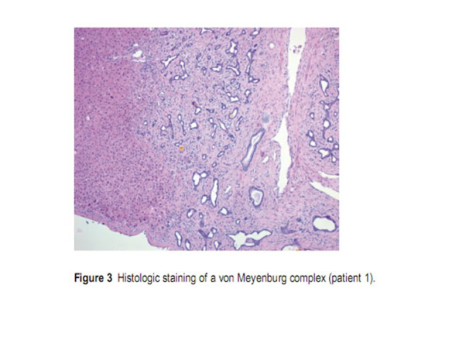
Bile duct hamartomas (von Meyenburg complexes) of the liver are usually detected during laparotomy or autopsy as an incidental finding. Multilocular occurrence is possible although they are rarely spread throughout the whole liver, as it was observed in our first patient. They may be found in normal liver tissue, but also in association with Caroli’s syndrome, congenital hepatic fibrosis (CHF) or autosomal dominant polycystic kindney disease (ADPKD) [10]. Histology of von Meyenburg complexes consists of a variable number of dilated small bile ducts, embedded in a fibrous, sometimes hyalinizing stroma (Figure 3).

If detected by CT scan or MRI, von Meyenburg complexes appear as small intrahepatic cystoid lesions. The lesions are frequently located adjacent to the portal veins, although the lesions can also be located everywhere else [5]. However, it remains difficult to differentiate between metastases and benign liver lesions. Moreover, small liver lesions with a diameter of less than 1.5 cm are often not detected by CT or MRI [9].

Since the treatment of metastatic disease is completely different from resectable esophageal cancer, liver lesions need to be identified and characterized as early as possible.


In our presented patients, the preoperative staging did not reveal any liver metastases. This underlines the importance of exact diagnostic measures in cases of unexpected intraoperative findings. Besides intraoperative ultrasound of the liver, frozen section is the gold standard for further differentiation of liver lesions of unknown origin.

Von Meyenburg complexes are defined as innocuous lesions. However, there are about 10 reported cases of neoplastic transformation of von Meyenburg complexes resulting in cholangiocarcinomas [2,3].

In conclusion, von Meyenburg complexes are an important differential diagnosis of hepatic metastases. As the existence of liver metastases is crucial for therapeutic decision making in malignant diseases, this differential diagnosis must be carefully clarified. Since von Meyenburg complexes are usually less than 5 mm in size, they can escape preoperative radiologic diagnostics. The macroscopic appearance of von Meyenburg complexes can mimic liver metastasis as demonstrated in our reported patients. This underlines the importance of intraoperative frozen sections to make the correct diagnosis.


Thứ Sáu, 28 tháng 9, 2012

SIÊU ÂM trong ICU


Discussion
Our study is unique in that it is a surveillance study of 125 consecutive patients whose evaluation entailed a global assessment encompassing clinical and ultrasound examination. Unlike previous works,2–6 it was performed to evaluate a protocol for head-to-toe ultrasound examination performed by a team of ICU physicians experienced in ultrasonography. Real-time ultrasound evaluation (within 12 h of admission) was done by the attending physician. The approach is based on the assumption that intensive care physicians with enough expertise can interpret sonographic images. Ultrasound training is not mandatory for ICU physicians in Italy, although proposals for graded competence have been advanced. Since 2000, the Society for Academic Emergency Medicine guidelines for physician training in emergency ultrasonography12 have informed the continuing medical education program of our ICU physicians. During their time in the course, physicians are required to perform 150 practice sections, including 60 heart, 10 chest, 60 abdomen, and 20 peripheral vein ultrasound examinations. After the course, the physicians receive 1 yr of tutored instruction. Drawn up in 2008, the ICU-sound protocol was tested for 6 months before entering daily clinical practice. Our data suggest that the protocol is not excessively time-consuming: a mean duration of 19.5 min to complete the scan is acceptable considering the protocol's complexity. Patient- and environment-related ultrasonography limitations had little influence on the majority of the ultrasound examinations in this study.
We used optic nerve sonography as an additional noninvasive diagnostic tool to detect increased intracranial pressure preceding emergency computed tomography or the decision to start invasive monitoring of intracranial pressure. Ultrasound examination revealed new findings of optic nerve sheath enlargement in six cases, in all of which computed tomography showed unknown intracranial hypertension, demonstrating the importance of this easy-to-perform ultrasound evaluation at admittance to a general ICU.
 
 
Image Tools
Two cases of unknown deep venous thrombosis were identified, one an internal jugular vein thrombosis (fig. 1). Upper extremity venous thrombosis is thought to be quite rare, but a recent report found that 18% of all cases involve upper extremity thrombi. Patients in the ICU setting are especially vulnerable to developing upper extremity thrombosis, and the clinical symptoms associated with an upper-extremity clot are frequently absent because of the extensive collateral network. A combined strategy using echocardiography and venous ultrasonography is a reliable method for diagnosing pulmonary embolism at the bedside.
 
Pleural effusion is often encountered in ICU patients; the diagnosis relies mostly on an anteroposterior chest radiograph obtained at the bedside. Pleural ultrasonography, an alternative imaging modality, has been validated against chest computed tomography, the accepted reference for diagnostic methods to identify pleural disease. The prevalence of significant pleural effusions in a medical ICU varies widely from 8.4 to 62%. In our study, effusions (small effusions less than 200 ml were not considered) were found in 35/125 patients (28%), 20 (57.1%) of whom underwent drainage and thoracenthesis. The estimated drainage volume was obtained using the formula proposed by Balik et al. It has been recently shown, however, that this formula can underestimate the volume, whereas a multiplane approach can increase the accuracy of quantifying small and moderate pleural effusion. Although more time-consuming (10 min), an accurate evaluation of effusion volume is a critical element in deciding whether to perform thoracenthesis. And it becomes even more relevant when weaning is considered, because drainage may reduce the work of breathing and increase respiratory muscle efficiency. The decision to drain was based on our clinical practice (reduced chest wall compliance, difficult weaning, refractory hypodynamic circulatory states) or when ultrasound suggested an infectious effusion (homogeneous echogenicity, septation, fibrin strands, nodular pleural changes). Pleural effusion can cause dissociation between effective preload and cardiac filling pressures. Drainage may be effective in patients with refractory hypodynamic circulatory states, particularly when there is evidence for diastolic chamber collapse.
Image Tools
Ultrasound examination was effective in evidencing the presence of anterior pneumothorax not detected by supine anteroposterior chest radiography (three cases). This finding holds clinical relevance, because during positive pressure ventilation a small pneumothorax may progress and cause hemodynamic instability. Ultrasound has proved to be more sensitive than anteroposterior chest radiography in the diagnosis of pneumothorax and can decrease the need for computed tomography for the diagnosis of occult pneumothorax. Lower lobe parenchymal consolidation without air bronchogram visualization can be difficult to distinguish from pleural effusion on an anteroposterior chest radiograph. Ultrasonography showing consolidation (with or without pleural effusion) can help in avoiding a possible mistake because of a misread chest radiograph (fig. 2).
 
 
Image Tools
Lung sonography is a useful aid in differentiating cardiogenic respiratory failure from acute airflow limitation, consolidation, pleural effusion, or pulmonary embolism. In our study, the diagnostic efficacy of lung ultrasound to differentiate dyspneic patients is well represented. Lung ultrasound pointed out 55 new findings, enabling us to differentiate the etiologic diagnosis in patients with a generic admitting diagnosis of acute respiratory insufficiency. Combining the data from lung sonography and echocardiography can enhance the diagnostic accuracy in differentiating respiratory insufficiency (fig. 3).
 
Image Tools
In a previous study, transthoracic echocardiography performed by intensivists provided new information and changed management in 37% of critical patients and added useful information to an additional 47% of patients, but did not alter immediate treatment. In our study, echocardiography revealed 53 new findings, leading to changes in therapy in 11/125 patients. These data confirm the importance of transthoracic echocardiography in ICU patients.
 
Abdominal examination evidenced 20 new pathologic findings and induced changes in therapy in 3/125 cases. The presence of the classic signs of acute acalculous cholecystitis at ultrasound examination, together with clinical data, enabled us to identify the origin of sepsis of unknown etiology (fig. 4). In our study, new ultrasound abnormalities were most often detected in patients with septic shock, followed by those with acute cardiac decompensation. This is not surprising, as these patients have multiple organ failure. ICU mortality was higher, but not statistically significant, in patients with two or more pathologic findings compared with patients without any findings. Mortality generally depends on the specific type of the most life-threatening abnormality, along with many other factors unrelated to ultrasound findings. Of note is that patients with no ultrasonographic pathologic findings had a better prognosis.
 
We plan to extend the ICU-sound protocol to include other diagnostic tools: transcranial Doppler and positive end-expiratory pressure-induced lung recruitment. Recent reports have shown, in fact, that lung reaeration can be assessed by ultrasound. It is rapid and repeatable at the bedside, does not require sedation and paralysis, and can be used to analyze dependent and not-dependent lung regions. Patients with acute lung injury, the adult respiratory distress syndrome, and pulmonary edema may benefit from ultrasound monitoring of positive end-expiratory pressure-induced reaeration; however, a limitation is that lung hyperinflation cannot be assessed.
 
Study Limitations
 
There is a potential for bias, as the operators in this study were not blinded to the patient's clinical picture, which is difficult to entirely eliminate in any use of ultrasound. The study relied only on transthoracic echocardiography; therefore, the true prevalence of cardiac ultrasound abnormalities may have been underestimated. Also, the prevalence of pleural effusion may have been underestimated with the use of a formula, which has been shown inadequate to quantify small and moderate effusions. An evaluation of the usefulness of a head-to-toe versus a focused examination was beyond the scope of this study. The major finding of this study is the discovery of a high prevalence of unsuspected clinical abnormalities in ICU patients. Ultrasound examination permitted us to modify the diagnosis and to improve the treatment, with prompt changes in therapeutic strategy. Patient- and environment-related sonography limitations had little influence on the majority of the examinations. The test can be performed by the attending ICU physician, with minimal risk of overuse or misinterpretation. Moreover, ultrasound use in the ICU could be optimized by making ultrasonography a routine part of intensive care training during residency. In experienced hands, rapid global assessment of critical patients at ICU admittance under this ultrasound protocol holds potential for improving healthcare quality.


Thứ Năm, 27 tháng 9, 2012

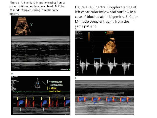
M-mode SONOGRAPHY and FETAL ARRHYTHMIAS


o        © 2012 by the American Institute of Ultrasound in Medicine

SIÊU ÂM ĐIỀU TRỊ VIÊM GÂN VÔI HOÁ CHÓP XOAY


Sonographically Guided Lavage Aspiration Technique
 

The patient is positioned such that the calcification to be treated is well visualized and amenable to needle placement and that both the patient and physician will be comfortable throughout the procedure. Depending on the

calcification location, particularly within the supraspinatus tendon and deep to the overlying acromion process of the scapula, it may be necessary to experiment with various armpositions of the patient to achieve this goal. Generally, the procedure is performed with the patient in the lateral decubitus or supine position for supraspinatus and infraspinatus calcification and in the supine position if the subscapularis tendon is the target.


A high-resolution linear probe (10–17 MHz) is used for this procedure, as the target calcification is usually very superficial. A 25-gauge needle is used for local anesthesia, and a 16- to 18-gauge needle is generally used for the lavage aspiration. Although some authors have advocated a smaller needle size (22 gauge), 20 in this author’s experience, a larger needle allows a faster andmore complete evacuation of calcium. Although others also advocate a 2-needle approach, and excellent results have been achieved with this method, this author has not found that approach to substantially improve calcium removal or to decrease the procedure time.
 
A recent study of 462 patients suggests that warming the lavage fluid may help improve dissolution of calcium and shorten the procedure time, which may prove to further optimize this technique.

However, this large study did not show a significant difference in patient outcomes as determined by visual analog scale scores between the two groups.

In preparation for the lavage aspiration, a series of syringes are prepared (3–6, depending on the size of the calcification) containing a mixture of saline and an anesthetic. This author favors a blend of 70% sterile saline and 30% lidocaine, 1%. A syringe containing an additional anesthetic (0.25% bupivacaine HCl) and cortisone (triamcinolone acetonide or methylprednisolone acetate) is also prepared for the subacromial-subdeltoid bursal injection that concludes the procedure, with the bupivacaine providing relief of postprocedure discomfort for several hours.

Continuous sonographic visualization of the calcification and needle is necessary throughout the procedure.

A liberal amount of anesthetic is administered subcutaneously, within the deeper soft tissues and within the subacromial-subdeltoid bursa, being sure that no air is introduced into the soft tissues or adjacent subacromial-subdeltoid bursa, extending from the skin entry site to themargin of the calcification along the expected path of lavage needle placement. If air is injected and is superficial to the target calcification, particularly within the subacromial-subdeltoid bursa, the calcification may be entirely obscured, and the proceduremay need to be postponed until the air is resorbed, which may take several days. With appropriate local anesthetic administration, the procedure is generally well tolerated with only mild discomfort. With continuous sonographic visualization, the 16- or 18-gauge needle is advanced into the epicenter of the calcific focus with a single puncture (Figure 3). Using the syringes filled with the anesthetic/saline  mixture noted above, intermittent plunger pressure and release are performed until a cavity forms within the focus of calcification (Figure 4). At this point, swirling of echogenic material (calcium) will be seen within the cavity, and with plunger release, this calcific material will decompress into the syringe. If more than a single puncture is made into the lesion, the lavage material may decompress through this additional hole in the calcific focus, and the yield of calcium removed will be diminished.
 

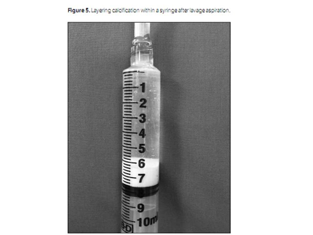
As large amounts of calcium fill the syringe, exchange is made for new clear syringes until no further calcification may be removed. The calcific material removed will be seen to layer within the dependent portion of the syringe (Figure 5). At this point in the procedure, any remaining calcification along the wall of the original focus is fenestrated using the needle. If additional foci of calcification are present, these are treated in the same manner. At the conclusion of this process, any remaining calcium fragments too small for lavage are also fenestrated with the needle. Finally, the mixture of anesthetic and corticosteroid described above is injected into the adjacent subacromial-subdeltoid bursa, which will provide considerable pain relief over the next several weeks to months as additional calcific material decompresses into the bursa from the involved rotator cuff tendon.


Although follow-up radiographs are not routinely obtained, they may show a rapid and marked decrease in the amount of calcification remaining within the tendon. In patients with recurrent or residual pain after therapy, subsequent sonographic examinations may be performed to assess the degree of calcification within the tendon or subacromial-subdeltoid bursa. Patients in this group may often be effectively treated with a sonographically guided bursal cortisone and anesthetic injection.


Conclusions

Calcific tendinosis of the rotator cuff is a commonly diagnosed entity that is responsible for a great deal of patient pain and limitation of mobility. Although radiographs remain the mainstay of initial calcium visualization and diagnosis, sonography can localize the calcification to the specific tendon involved, assess the entire rotator cuff for tears or tendinosis, and also evaluate the adjacent biceps tendon and subacromial-subdeltoid bursa for concomitant abnormalities. Finally, diagnostic sonography provides the means by which this condition can be safely treated by the percutaneous technique described above. This technique quickly removes and fragments the problematic calcification with a low incidence of complications, and multiple studies have shown an excellent clinical response in most patients. Lavage aspiration with sonographic guidance has thus become the optimal modality for effective treatment of this painful condition.

Thứ Hai, 24 tháng 9, 2012

NHÂN CA VỠ TÁ TRÀNG tại MEDIC: VAI TRÒ CHẨN ĐOÁN và THEO DÕI của SIÊU ÂM


A traumatic duodenal hematoma (DH) is an unusual event, occurring mainly in children and young individuals, with a male predominance in both age groups. Furthermore, it can be a diagnostic challenge because of unreliable history, nonspecific signs and symptoms, delayed appearance, and the duodenum’s retroperitoneal location.1,2
Sonography is considered a reliable screening tool for blunt abdominal trauma (BAT)3,4; however, since the beginning of the last decade, only a small number of reported DH cases5–,8 have been described by sonography.

Discussion

Accurate diagnosis is essential for proper treatment of a DH. The clinical appearance and findings including abdominal pain, vomiting, tenderness, and a palpable mass can be nonspecific, accompanied by unremarkable laboratory test results.6,8
Blunt abdominal trauma, sometimes minor, is the leading cause of DHs, which occur in approximately four fifths of patients.9,10 Bleeding disorders, Henoch-Schönlein purpura, anticoagulation therapy, alcoholism, pancreatitis, tumors, duodenal ulcers, and local or iatrogenic factors are other implicative causes.7,10–,13

Most hematomas resolve spontaneously without permanent changes. Treatment may be surgical or conservative using nasogastric suction and adequate parenteral nutrition. Expectant treatment of an isolated DH is generally preferred. Failure of conservative treatment is considered when there is no evidence of partial resolution after 5 days or complete resolution after 10 days or in cases of perforation, indicating surgical treatment.14

All pictures extracted from http://cai.md.chula.ac.th/lesson/atlas/T/page1t.html
 

An upper GI series was for many years the only diagnostic tool for DHs before the advent of CT, which has been established as the examination of choice for duodenal injuries, especially in disclosing complications such as perforation and abscesses.15 However, CT was found to be diagnostic in 60% of patients with duodenal perforation.1


Various sonographic patterns have been described in DHs: (1) a duodenal wall thickening with hypoechogenicity16; (2) a duodenum-related mass of variable echogenicity, depending on the age of the hematoma7; and (3) a prevertebral cystic lesion simulating a pancreatic pseudocyst.6 This variability may reflect the difficulty in distinguishing the origin of small retroperitoneal lesions proximal to the bowel wall in the upper abdomen because of the enteric gas component and also the different characteristics of a hematoma depending on its age. Color-coded imaging has been shown to be helpful in differentiating a spontaneous DH from an intestinal mass.8
Sonography may be the first examination performed in a patient with epigastric abdominal pain or a palpable abdominal mass,8 and it is useful to be familiar with this uncommon entity. In BAT, sonography can additionally show associated lesions, including pancreatic traumatic pseudocysts and parenchymal lacerations, or a small amount of ascites caused by peritoneal blood or pancreatic fluid.17

In conclusion, sonography may play a primary role, both in the diagnosis and the monitoring of DHs, when conservative treatment is attempted. Computed tomography may be reserved for inconclusive cases.

*          © 2004 by the American Institute of Ultrasound in Medicine