Tổng số lượt xem trang

Thứ Ba, 16 tháng 10, 2012

FIRST US TOOL for DENSE BREAST


Clinical Context

The National Cancer Institute estimates that approximately 40% of women undergoing screening mammography have dense breast tissue. High breast density hinders mammographic interpretation, which may delay detection of breast cancer until it is at a more advanced and difficult-to-treat stage.
Less-dense breasts have a high amount of fatty tissue, whereas dense breasts have a high amount of connective and fibroglandular tissue. Breast cancer and fibroglandular breast tissue both appear as solid white areas on mammograms, rendering interpretation more difficult.

Study Synopsis and Perspective

The US Food and Drug Administration (FDA) approved the first ultrasound device for use in combination with mammography in women with dense breast tissue.
The device, known as the somo-v Automated Breast Ultrasound System (ABUS), provides clinicians with an additional resource in screening women with dense breasts. The indication is limited to use in women who have a negative mammogram result and no symptoms of breast cancer.
Mammograms of dense breasts can be difficult to interpret, the FDA points out in its announcement of the approval.
"A physician may recommend additional screening using ultrasound, for women with dense breast tissue and a negative mammogram," said Alberto Gutierrez, PhD, director of the Office of In Vitro Diagnostic Device Evaluation and Safety at the FDA's Center for Devices and Radiological Health. "The somo-v ABUS is a safe and effective breast ultrasound tool when such screening is recommended."
Approximately 40% of women undergoing screening mammography have dense breasts, according to National Cancer Institute estimates. These women have an increased risk for breast cancer, with detection usually at a more advanced and difficult-to-treat stage, the FDA said.
In April, an expert advisory committee of the FDA voted unanimously to recommend the expanded use of the ultrasound device as a screening tool for women with dense breast tissue.
However, as reported by Medscape Medical News, some of the panel members had concerns about the device because of its automated nature. In contrast to handheld ultrasound, medical specialists who operate an automated ultrasound do not have to be imaging experts, Robert Faulk, MD, from Medical Imaging Consultants in Omaha, Nebraska, noted during the meeting. "For instance, obstetricians and gynecologists could do ABUS and interpret the results themselves, along with mammography findings," Dr. Faulk said. "That could have a deleterious effect on healthcare for many women," he added.
"ABUS as a screening tool could potentially be applied to 40 million women in the United States [with dense breasts], and if it were used by nonimaging specialists, the false-positive rates could go through the ceiling," said Daniel Kopans, MD, from Harvard Medical School in Boston, Massachusetts. Dr. Kopans commented that postmarket surveys of the false-positive rate from the new ultrasound device could provide important data on the effect of its use as a screening tool in women with dense breasts.
Dense breasts have a high amount of connective and fibroglandular tissue compared with less-dense breasts, which have a high amount of fatty tissue, the FDA explained.
Fibroglandular breast tissue and tumors both appear as solid white areas on mammograms, which can complicate interpretation. Dense breast tissue may obscure smaller tumors, potentially delaying detection of breast cancer, according to the FDA.
Ultrasound imaging has been proven capable of detecting small masses in dense breasts.
A clinical study has shown a statistically significant increase in breast cancer detection when images generated by this new device were reviewed in conjunction with mammograms compared with mammograms alone. The study involved board-certified radiologists who reviewed mammograms alone or in conjunction with device-generated images for 200 women with dense breasts and negative mammograms.
As part of the approval, the FDA requires that the manufacturer train clinicians and technologists using the new ultrasound device, and that the manufacturer provide each facility with a manual clearly defining system tests required for initial, periodic, and yearly quality-control measures.
The ultrasound works via a transducer that directs high-frequency sound waves at the breast. The specially shaped transducer of the device can automatically scan the entire breast in approximately 1 minute to produce several images for review, according to the FDA press materials.
More information on the new ultrasound device is available on the FDA Web site.


Clinical Implications

  • The FDA has approved a new automated ultrasound device to be used in combination with mammography in women with dense breast tissue. This is the first ultrasound device approved for this indication. The specially shaped transducer of the device directs high-frequency sound waves at the breast to scan the entire breast in approximately 1 minute, automatically producing several images for review.
  • The FDA has approved the ultrasound device for use only in women with negative mammography results and no symptoms of breast cancer. In these women, the device is a safe and effective breast ultrasound tool. Compared with mammograms alone, mammograms plus images generated by the new device interpreted by board-certified radiologists were associated with a statistically significant increase in breast cancer detection.
  • Unlike handheld ultrasound, automated ultrasound does not have to be operated by an imaging expert. This may be a potential drawback in that medical practitioners without specialized radiographic training could use the new device and interpret the results themselves, along with mammography findings. This could dramatically increase false-positive rates. The FDA is requiring that the manufacturer train clinicians and technologists using the new ultrasound device, and that each facility is given a manual clearly defining system tests required for quality control.


    Abstract: The idea of an automated whole breast ultrasound was developed three decades ago. We present our initial experiences with the latest technical advance in this technique, the automated breast volume scanner (ABVS) ACUSON S2000TM. Volume data sets were collected from 50 patients and a database containing 23 women with no detectable lesions in conventional ultrasound (BI-RADS®-US 1), 13 women with clearly benign lesions (BI-RADS®-US 2), and 14 women with known breast cancer (BI-RADS®-US 5) was created. An independent examiner evaluated the ABVS data on a separate workstation without any prior knowledge of the patients’ histories. The diagnostic accuracy for the experimental ABVS was 66.0% (95% confidence interval [CI]: 52.9–79.1). The independent examiner detected all breast cancers in the volume data resulting in a calculated sensitivity of 100% in the described setting (95% CI: 73.2%–100%). After the ABVS examination, there were a high number of requests for second-look ultrasounds in 47% (95% CI: 30.9–63.5) of the healthy women (with either a clearly benign lesion or no breast lesions at all in conventional handheld ultrasound). Therefore, the specificity remained at 52.8% (95% CI: 35.7–69.2). When comparing the concordance of the ABVS with the gold standard (conventional handheld ultrasound), Cohen’s Kappa value as an estimation of the inter-rater reliability was κ = 0.37, indicating fair agreement. In conclusion, the ABVS must still be regarded as an experimental technique for breast ultrasound, which definitely needs to undergo further evaluation studies.

    Keywords: breast cancer, automated breast ultrasound, automated breast volume scanner, ABVS

Chủ Nhật, 14 tháng 10, 2012

NHÂN CA OVARIAN TERATOMA tại MEDIC


Figure 4d.  Mature cystic teratoma of the right ovary in a 19-year-old pregnant woman. (d) Photograph of the gross specimen shows yellowish, pasty sebaceous material (black arrowhead) and hair (white arrowheads) within the cyst cavity, findings that account for the fat echogenicity and signal intensity seen at US and MR imaging. Two molar teeth are also evident (arrows).
 
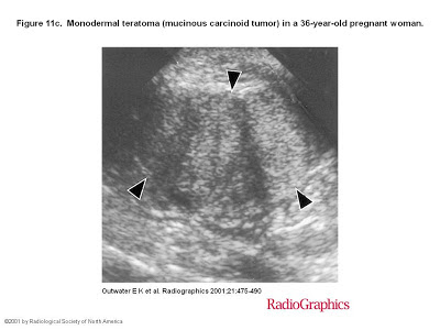
Figure 4(a) Sagittal transabdominal US image shows an echogenic mass with sound attenuation (arrows). 
 
Abstract
Ovarian teratomas include mature cystic teratomas (dermoid cysts), immature teratomas, and monodermal teratomas (eg, struma ovarii, carcinoid tumors, neural tumors). Most mature cystic teratomas can be diagnosed at ultrasonography (US) but may have a variety of appearances, characterized by echogenic sebaceous material and calcification.


At computed tomography (CT), fat attenuation within a cyst is diagnostic. At magnetic resonance (MR) imaging, the sebaceous component is specifically identified with fat-saturation techniques. The US appearances of immature teratoma are nonspecific, although the tumors are typically heterogeneous, partially solid lesions, usually with scattered calcifications.


At CT and MR imaging, immature teratomas characteristically have a large, irregular solid component containing coarse calcifications. Small foci of fat help identify these tumors. The US features of struma ovarii are also nonspecific, but a heterogeneous, predominantly solid mass may be seen. On T1- and T2-weighted images, the cystic spaces demonstrate both high and low signal intensity. Familiarity with the US, CT, and MR imaging features of ovarian teratomas can aid in differentiation and diagnosis.
.....
Most mature cystic teratomas can be diagnosed at US. However, the US diagnosis is complicated by the fact that these tumors may have a variety of appearances. Three manifestations occur most commonly. The most common manifestation is a cystic lesion with a densely echogenic tubercle (Rokitansky nodule) projecting into the cyst lumen (16). The second manifestation is a diffusely or partially echogenic mass with the echogenic area usually demonstrating sound attenuation owing to sebaceous material and hair within the cyst cavity (Fig 4) (17),(18). The third manifestation consists of multiple thin, echogenic bands caused by hair in the cyst cavity (Fig 3). Pure sebum within the cyst may be hypoechoic or anechoic (19). Fluid-fluid levels result from sebum floating above aqueous fluid, which appears more echogenic than the sebum layer (18). The dermoid plug is echogenic, with shadowing due to adipose tissue or calcifications within the plug or to hair arising from it. Diffuse echogenicity in these tumors is caused by hair mixed with the cyst fluid (Fig 4).
 
In a prospective US study that made use of these criteria, Mais et al (20) found a sensitivity of 58% and a specificity of 99% in the diagnosis of mature cystic teratoma. Numerous pitfalls have been described in the US diagnosis of mature cystic teratoma (21). Blood clot within a hemorrhagic cyst can appear echogenic, although a mature cystic teratoma usually demonstrates sound attenuation rather than increased through-transmission. Hemorrhagic cysts or blood clots typically demonstrate increased through-transmission. Echogenic bowel can frequently be mistaken for diffusely echogenic mature cystic teratoma and vice versa (21). Perforated appendix with appendicolith and fibrous lesions such as cystadenofibromas have also been described as false-positive findings (21),(22).
 © RSNA, 2001

Thứ Tư, 10 tháng 10, 2012

NHÂN CA RÒ ĐỘNG TĨNH MẠCH VÙNG CHẬU tại MEDIC


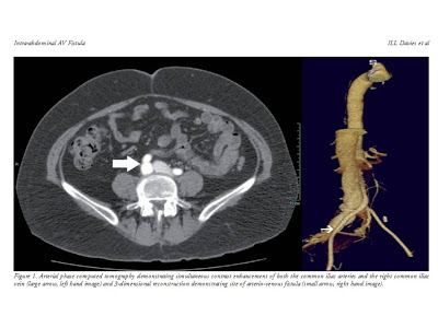
The AV fistula had the origine of right internal iliac artery and drains off blood to right internal iliac vein.  

DISCUSSION

Arteriovenous malformations (AVMs) of the female pelvis are uncommon. These may either be congenital or acquired. Congenital pelvic AVMs are characterised by a large number of arterial feeding branches. The arterial feeders are considered undifferentiated vascular structures following arrested embryonic development at various stages [1]. Acquired pelvic AVMs on the other hand, develop following spontaneous rupture of an atherosclerotic aneurysm into the adjacent veins or following penetrating trauma (less than 20%), e.g. gunshot wound or stabbings, or after lumbar disc surgery [2]. Therefore, acquired AVMs are most commonly arteriovenous fistulas (AVF). The AVFs are most commonly aorto-caval fistula, followed by ilio-iliac [3] and aorto-iliac. However, the etiology, clinical features, pathophysiology, principles of management and postoperative care for these fistulas are similar. Approximately 3 to 4% of all patients undergoing surgery for ruptured aorto-iliac aneurysm are found to have AVF [3]. These carry a better prognosis than intraperitoneal, retroperitoneal or enteric rupture of aorto-iliac aneurysms [5]. The disorder has a marked male preponderance [4]. Other rarer causes of acquired AVFs include Marfan’s syndrome, Ehler-Danlos syndrome, syphilis, Takayasu’s arteritis, invasion by malignant tumour [2] and in some the cause is never ascertained.


Pelvic AVMs may manifest with symptoms of pain, haemorrhage, haematuria, dyspareunia, or congestive heart failure, or symptoms secondary to mass effect on adjacent pelvic structures. Acquired AVF, secondary to surgery or trauma, tend to occur in younger patients as trauma or surgery tends to  affect younger patients. In contrast, spontaneous perforation of atherosclerotic aneurysm into adjacent veins tends to occur in the older population. Two large series report mean ages of 67.3 [6] and 69.7 years. The time of onset of symptoms is usually earlier, from hours to weeks [3]. The initial diagnosis is often that of an abdominal aortic aneurysm, AVF is often not suspected and often the diagnosis is only made at surgery [5].

Iliac artery aneurysms and ilio-iliac fistulas are usually associated with abdominal aortic aneurysm, as demonstrated in this case, though they may occur as isolated entities. Rupture of atherosclerotic aneurysms into the iliac vein may have three different clinical manifestations: sudden onset of high output cardiac failure; pulsatile lower abdominal mass associated with bruit and thrill; or unilateral intermittent claudication or venous congestion. Our patient presented with the first two clinical manifestations and the tricuspid regurgitation was most likely secondary to grossly dilated ventricle from high output cardiac failure.


Ultrasound, CT or even MRI is almost always ordered for assessment of the abdominal/iliac aneurysms. Colour Doppler ultrasound has demonstrated the AV fistulas as areas of high velocity turbulent flow with aliasing of colour signal and also shows any associated thrombus at the aneurysm or fistula. Detection of ilio-iliac fistulas may however be difficult, as demonstrated by this case, as these tortuous and aneurysmal vessels lie deep within in the pelvis and obscured by overlying bowel gas. CT angiography is excellent in demonstrating the aneurysm and fistulous communications [6], especially with the advent of multi-detector CT and 3D software. Early contrast opacification of the iliac veins and inferior vena cava, and site of the fistula are clearly visualised.

Conventional angiography still remains the ‘gold standard’ for assessment of AVMs as well as assisting in assessing options for endovascular management. Endovascular treatment has gained considerable favour in the management of arteriovenous fistula especially for those with significant high-risk comorbid factors.  Options available include percutaneous endovascular treatment with covered self-expanding stent graft to cover the mouth of the fistula. However, this is not always feasible due to the tortuous iliac vessels. Transcatheter embolisation with coils or detachable balloons [7] is generally not recommended due to the large size of the fistula, high flow and short neck.

Surgical options for AVF consist of endo-aneurysmal repair of the fistula and prosthetic graft replacement of the aortoiliac aneurysm but these are associated with high morbidity and mortality (approaching 60%) due to the emergent nature of the procedure [4]. Thus early diagnosis and appropriate management is of paramount importance. Unlike acquired AVFs, surgical treatment of congenital AVMs is difficult due to the extensive nature and the large number of dysplastic feeder vessels with the potential for exsanguinating haemorrhage and damage to surrounding structures [8].  Accordingly, transcatheter embolisation has become the treatment of choice [8]. Preoperative embolisation of AVMs has also been used as an adjunct to decrease intraoperative blood loss. Small asymptomatic AVMs that do not increase in size may be safely observed.

AVFs between major abdominal vessels are uncommon complication of aortoiliac aneurysm. Ilio-iliac fistula in a female patient is even rarer and associated with high morbidity and mortality especially if the diagnosis is not suspected. In this patient, in the absence of history of trauma or surgery and the presence of extensive aneurysmal disease involving the aorta and iliac arteries, it is reasonable to believe that the AV fistula was secondary to perforation of the aneurysmal right iliac artery into the right iliac vein. In addition to the rapid onset, the presence of a single communication and older age group lend more support to this diagnosis.

There has only been a single reported case in the literature with an ilio-ilial fistula secondary to atherosclerotic disease [5]. In addition the CT angiographic appearances of ilio-ilial AVF have not been described.


Spontaneous major intra-abdominal arteriovenous fistulas: a report of several cases, Astarita D, Filippone DR, Cohn JD. Angiology1985,  Sep;36(9):656-61.

 Abstract

Most major intra-abdominal fistulas result from trauma or surgery. Spontaneous fistulas are rare with less than 100 reported cases since 1831. From a review of hospital records, five such spontaneous fistulas were identified among 215 cases of abdominal aortic aneurysm between 1975 and 1983. These cases are presented and supplemented by 73 similar cases collected from a literature review for discussion of the salient features of clinical presentation and management of spontaneous major fistulas. Major intra-abdominal arteriovenous fistulas usually present with a machinery bruit over a pulsatile mass, but may present more subtly with pain and otherwise unexplained hematuria. Because these fistulas lead to refractory heart failure, surgery should be expeditious. Closure should be performed from within the aneurysm with arterial and pulmonary artery pressure monitoring. Care must be taken to prevent pulmonary embolization.

Thứ Hai, 8 tháng 10, 2012

GIẢI NOBEL Y HỌC 2012


Hai nhà khoa học John B.Gurdon và Shinya Yamanaka đã đoạt giải Nobel Y học năm nay 2012 nhờ việc phát hiện tế bào trưởng thành có thể tái lập trình để trở thành tế bào vạn năng (pluripotent).


Theo thông báo từ Ủy ban Nobel, Gurdon và Yamanaka đã nhận thấy các tế bào trưởng thành, chuyên biệt hoá có thể được can thiệp và lập trình lại để trở thành các tế bào gốc, chưa trưởng thành vốn có khả năng phát triển thành tất cả các dạng mô trong cơ thể. Nghiên cứu này đã “cách mạng hóa” nhận thức về cơ chế phát triển của tế bào và cơ quan sinh học.

Năm 1962, ông Gurdon đã phát hiện tế bào có một khả năng đặc biệt là đảo chiều. Trong một thí nghiệm kinh điển, ông đã thay thế nhân tế bào chưa trưởng thành trong một tế bào trứng ếch bằng nhân của một tế bào ruột trưởng thành. Kết quả là tế bào trứng biến đổi vẫn phát triển thành một con nòng nọc bình thường. DNA của tế bào trưởng thành vẫn có tất cả các thông tin cần thiết để phát triển mọi tế bào trong cơ thể ếch.

Hơn 40 năm sau, năm 2006, Shinya Yamaka phát hiện các tế bào trưởng thành nguyên dạng ở chuột có thể được lập trình để trở thành các tế bào gốc chưa trưởng thành. Bằng cách can thiệp và biến đổi chỉ một vài gene, ông đã có thể tái lập trình các tế bào trưởng thành trở thành tế bào gốc vạn năng hay tế bào gốc đa hiệu (pluripotent stem cell). Đây là thuật ngữ chỉ những tế bào chưa trưởng thành có khả năng phát triển thành mọi dạng tế bào khác nhau trong cơ thể.

Những phát kiến mang tính đột phá này, theo Ủy ban Nobel, đã làm thay đổi hoàn toàn cách nhìn nhận của khoa học về sự phát triển cũng như chuyên biệt hóa ở cấp độ tế bào. “Tế bào trưởng thành không phải giam mình mãi mãi với một chức năng cụ thể và chuyên biệt nào. Bằng cách tái lập trình tế bào người, các nhà khoa học đã mở ra cơ hội mới để nghiên cứu nhiều loại bệnh cũng như phát triển phương pháp chẩn đoán và điều trị mới”.

Ứng dụng
 
Tế bào gốc có khả năng ứng dụng chữa trị nhiều bệnh từ ung thư, tiểu đường, để thay thế các mô và nội tạng bị hỏng của cơ thể nhờ khả năng phát triển thành mọi loại tế bào thay thế những tế bào bệnh tật. Nó có thể sửa chữa một trái tim sau cơn đột quỵ hoặc đảo ngược tiến trình phát triển của bệnh Alzheimer.
Nghiên cứu được trao giải Nobel y học năm nay đã mở ra một phương cách mới để có được tế bào gốc mà không phải dùng đến phôi thai, một vấn đề gây nhiều tranh cãi đạo đức. “Thiếu nội tạng để cấy ghép là một vấn đề lớn tại nhiều quốc gia hiện nay” - ông Yamanaka nhấn mạnh.
Một trong các ứng dụng thực tế khác của nghiên cứu này là khả năng nghiên cứu tận gốc rễ các căn bệnh thông qua tế bào của người mắc bệnh để tìm ra biện pháp chữa trị. “Tế bào da có thể được lấy từ người bệnh mắc nhiều căn bệnh khác nhau, tái lập trình và xem xét trong phòng thí nghiệm để xem chúng khác với những tế bào khỏe mạnh như thế nào”, Ủy ban Nobel cho biết.