Tổng số lượt xem trang

Thứ Sáu, 7 tháng 12, 2012

Shear Wave Elastography in the Diagnosis of Thyroid Nodules: Feasibility in the Case of Coexistent Chronic Autoimmune Hashimoto's Thyroiditis.


SUMMARY

 Objective ShearWave™ Elastography (SWE) is real-time, quantitative and user-independent technique, recently introduced in the diagnostic work-up of thyroid nodules. Hashimoto’s thyroiditis (HT), characterized by variable degrees of lymphocytic infiltration and fibrosis, might affect shear wave propagation. The aim of this study was to assess the feasibility of SWE in cytologically benign thyroid nodules within both Hashimoto’s and nonautoimmune thyroid glands. The effect of autoimmunity on the gland stiffness was also evaluated.

 Design longitudinal study in a single centre.

 Patients SWE was performed in 75 patients with a benign thyroid nodule at cytology: 33 with Hashimoto’s thyroiditis (HT group) and 42 with uni- or multi-nodular goitre, negative for thyroid autoimmunity (non-HT group).

 Results The elasticity index (EI) of the extra-nodular tissue was greater, though not statistically significant, in the HT than in the non-HT group (24·0 ± 10·5 kPa vs 20·8 ± 10·4 kPa; P = 0·206). However, the EI of extra-nodular tissue was related to the TPOAb titre in the HT group (P = 0·02) and was significantly higher in patients with HT receiving L-thyroxine than in the euthyroid subjects (P = 0·02). The EI of thyroid nodules was similar in HT and non-HT groups. In both groups, the stiffness of nodules was significantly higher than that of the embedding tissue.

 Conclusions Our data indicate that SWE correctly defines the elasticity of thyroid nodules independently from the coexistence of autoimmune thyroiditis, always being able to differentiate nodular tissue from the surrounding parenchyma. In HT, the stiffness of extra-nodular tissue increases in relation to both the thyroid antibody titre and the degree of impairment of thyroid function.


BÀN LUẬN
Dữ liệu của chúng tôi  cho thấy kỹ thuật sử dụng siêu âm đàn hồi sóng biến dạng [shear wave elastograpphy, SWE] này có thể  xác định một cách chính xác độ đàn hồi của các hạt giáp, độc lập với nền  tuyến giáp cứng như trong viêm giáp Hashimoto (HT). Một gradient độ cứng đã được quan sát thấy trong tất cả các bệnh nhân, với các hạt giáp là cứng hơn đáng kể  so với các mô xung quanh, cả trong HT và trong các nhóm không-HT.

Viêm giáp tự miễn mạn tính HT tự nó [per se]  và độ nặng của nó, theo đánh giá của hiệu giá TPOAb và cần có  liệu pháp thay thế với L-T4 cho suy giáp, làm tăng thêm chỉ số đàn hồi [elastic index, EI] nhu mô quanh hạt. Tuy nhiên, ngay cả trong viêm giáp Hashimoto, một gradient độ đàn hồi đáng kể được duy trì giữa các hạt lành tính và mô-ngoài-hạt, chỉ số EI của hạt luôn lớn hơn so với nhu mô tuyến giáp mà hạt nhúng vào. Trong chấp nhận và thống nhất với phát hiện này, thấy có  quan hệ trực tiếp giữa các chỉ số đàn hồi EI của mô quanh hạt  và chỉ số EI các hạt lành tính ở viêm giáp Hashimoto.
Siêu âm đàn hồi sóng biến dạng SWE là một công cụ mạnh mẽ cho việc chẩn đoán các hạt giáp ác tính, chỉ số EI của hạt ác tính cao hơn đáng kể so với hạt lành tính.Theo chúng tôi biết, đây là nghiên cứu  khảo sát đầu tiên ảnh hưởng của viêm giáp HT  trên tính năng đàn hồi hạt giáp. Cũng được biết rõ rằng viêm giáp tự miễn mạn tính HT, được đặc trưng bằng thâm nhiễm tế bào lympho  và xơ hóa, làm thay đổi siêu cấu trúc [ultrastructure] tuyến giáp  gây cứng mô tuyến. Trong bệnh to cực [acromegaly], một nguyên do đặc trưng khác gây  xơ hóa  tuyến giáp, siêu âm đàn hồi đã cho thấy là không ích lợi  trong việc dự đoán tính chất ác tính của các hạt giáp. Điều này do bệnh nhân bệnh to cực dường như có một tỷ lệ  hạt cứng lớn hơn, nhưng không phải ác tính trên mô bệnh học. Ở những bệnh nhân này, xơ hóa và kết quả là độ cứng có thể do tăng tổng hợp collagen và lắng đọng gây ra bởi GH và IGF1.  Mặc dù hạn chế bởi loạt bệnh nhân của chúng tôi không nhiêu, những phát hiện của nghiên cứu này cho thấy viêm giáp tự miễn mạn tính HT không làm giảm khả năng siêu âm đàn hồi  sóng biến dạng trong  đánh giá các hạt giáp ở viêm giáp tự miễn mạn tính HT.

Điều này có liên quan đặc biệt bởi vì: (i) HT ảnh hưởng đến ít nhất 5% dân số nói chung,  (ii) bệnh nhân với HT có nhiều khả năng để phát triển các hạt giáp, do, ít nhất là một phần, từ kích thích TSH mãn tính;  (iii) một số nghiên cứu cho thấy mối liên quan giữa HT và ung thư tuyến giáp loại nhú [papillary carcinoma];  và (iv) một tỷ lệ cao  các kết quả  FNAC đáng ngờ hoặc không xác định được báo cáo khi kiểm tra hạt trong viêm giáp tự miễn Hashimoto.  Theo quan điểm này, việc xác định độ đàn hồi chính xác trở nên quan trọng trong quy trình  chẩn đoán các  hạt trong viêm giáp tự miễn  Hashimoto.
Để  kết luận, dữ liệu của chúng tôi cho thấy siêu âm đàn hồi sóng biến dạng SWE xác định  chính xác độ đàn hồi của các hạt giáp độc lập với viêm tuyến giáp tự miễn mạn tính cùng tồn tại, luôn  có khả năng phân biệt mô hạt từ nhu mô xung quanh. Trong viêm giáp  HT, độ cứng  mô ngoài hạt tăng thêm  trong tương quan cả hiệu giá kháng thể tuyến giáp  [thyroid antibody] lẫn mức độ suy giảm chức năng tuyến giáp, cao hơn ở những bệnh nhân đòi hỏi phải điều trị  L-thyroxine.

 --------------------------------------------------------------------------------------------


INTRODUCTION

Elastography is an evolving technique aimed at differentiating benign from malignant thyroid nodules. This technique allows in vivo estimation of the tissue mechanical properties using a conventional ultrasound (US) system with modified software. Similarly to palpation, the rationale of elastography is that a cancerous nodule is stiffer, with less elastic deformation compared with the surrounding thyroid tissue or a benign thyroid nodule. This issue is of particular interest because thyroid nodules are commonly encountered in clinical practice, being detected by US in 19–67% of randomly selected individuals.1,2 As only a minority of these are malignant (5–15%3,4), selecting the malignant ones for thyroid surgery represents a major clinical issue.

Elastosonography, and in particular real-time US-elastography (US-E), was recently investigated as a tool for stratifying the presurgical risk of malignancy in nodules with indeterminate or nondiagnostic cytology.5 Rago et al.6 evaluated tissue stiffness by US-E in a large group of patients with thyroid nodules who underwent surgery for compressive symptoms or suspicion of malignancy at fine-needle aspiration cytology (FNAC). US-E displayed a sensitivity of 97%, a specificity of 100%, a positive predictive value of 100% and a negative predictive value of 98%, independently from nodule size. Recently, the same approach was applied to patients bearing nodules with indeterminate or nondiagnostic cytology, confirming the association of nodular stiffness with malignancy.6 Although promising, static US-E is biased by several factors, such as poor reproducibility, inter-observer variability and lack of quantitative information.7–9 Furthermore, US-E was mainly evaluated in highly selected patients, excluding those with cystic nodules and nodules with eggshell calcification.10,11 As the estimation of elasticity is altered by the presence of a nearby hard area, this technique is not reliable in the study of multi-nodular goitres, which represent about 40% of all nodular thyroid glands.12

Shear Wave™ Elastography (SWE) is a new real-time imaging modality, which uses a linear US transducer operated with no added pressure. Transient pulses are used to generate propagating shear waves in the body, and elasticity is directly calculated by measuring the wave propagation speed. Currently, SWE is the only technique producing real-time quantitative information on selected areas of thyroid tissue.13 It is also claimed to be relatively operator-independent and reproducible. Sebag et al.14 investigated the efficiency of this technique in predicting malignancy in solitary or multiple thyroid nodules. Using a cut-off level of 65 kPa of elasticity index (EI), they could predict malignancy with a sensitivity of 85·2%, a specificity of 93·9% and a positive predictive value of 92·3%. However, the accuracy of SWE might be impaired in patients with acromegaly, in whom GH/IGF I-induced fibrosis has been reported to increase nodule hardness, as assessed by US-E, independently from the malignant nature of the lesion.15 A more common, potentially confounding condition, yet to be evaluated, is chronic autoimmune Hashimoto’s thyroiditis (HT) coexistent with thyroid nodules. At histology, HT is characterized by lymphocytic infiltration of the thyroid parenchyma associated with variable degrees of fibrosis. The characteristic US pattern of Hashimoto’s glands consists of an array of tiny hypoechoic nodules that may become confluent, interspersed with echogenic fibrous bands. These pathological changes might affect shear wave propagation, thus hampering the diagnostic potential of SWE in thyroid nodules. This issue is of particular interest for two reasons. First, an increased prevalence of thyroid cancer has been reported in patients with HT.16,17 Second, the diagnostic accuracy of FNAC may be diminished in nodules within a Hashimoto’s gland, resulting in a higher rate of indeterminate or suspicious cytological reports.18

To answer the question of whether SWE might be feasible in nodules associated with HT, we investigated a series of benign thyroid nodules at cytology, which were harboured either within a Hashimoto’s gland or within a nonautoimmune thyroid.

PATIENTS and METHODS

Patients

The study enrolled 33 patients with HT harbouring one or more thyroid nodules (HT group) and 42 patients with uni-nodular or multi-nodular goitre, who had tested negative for thyroid antibodies and had a normal echo pattern at US (non-HT group).19 Inclusion criteria were a previous FNAC indicating that the nodule was benign and the absence of a dominant cystic component within the nodule.

The diagnosis of HT was made in the presence of a positive test for thyroglobulin antibody (TgAb) and/or thyroid peroxidase (TPOAb) antibody, and a hypoechoic pattern of the thyroid at US. Fifty-four patients were euthyroid without therapy, and 14, all of them belonging to the HT group, were euthyroid on L-thyroxine (L-T4) therapy. In patients with HT, the indication of LT4 treatment was a diagnosis of subclinical or overt hypothyroidism. Another seven patients, all with nodular goitre, were on L-T4 TSH-suppressive treatment. In patients in L-T4 replacement therapy and L-T4 TSH-suppressive therapy, the duration of L-T4 treatment ranged from 12 to 156 months (median 36·0 months).

All patients gave their informed consent to participate in the study.

Cytopathological diagnosis

Selection of nodules for FNAC was made according to the current guidelines.20,21 FNAC was performed, under US guidance, by a skilled endocrinologist using a 23-gauge needle attached to a 5-ml syringe.


Shear Wave Elastography

After a preliminary US evaluation and a cytological diagnosis of a benign lesion, thyroid nodules were evaluated by SWE. Briefly, SWE generates a remote radiation force by focused ultrasonic beams, a patented technology called ‘Sonic Touch’. Several of these so-called pushing beams at increasing depths are transmitted to generate a quasi-plane shear wave frame that propagates throughout the whole imaging area. Then, an ultrafast echographic imaging sequence (Ultrafast Imaging System) is performed to acquire successive raw radiofrequency dots at a very high frame rate (up to 20,000 frames per second). From shear wave propagation speed, tissue elasticity can be derived based on Young’s modulus formula (E = σ/ε, where σ is stress and ε is strain). This results in a colour-coded image, blue representing softer tissue and red stiffer tissue. Quantitative information is delivered as EI expressed in kilo-Pascal (kPa).

Shear wave elastographic measurement was performed with the Aixplorer, developed by SuperSonic Imagine (Les Jardins de la Duranne, Aix en Provence, France).


RESULTS

Patients in the HT and non-HT groups were matched for age (Table 1). The calcitonin concentration was in the normal range in all patients, confirming the benign nature of the nodule.

Conventional US

Table 1 summarizes the main US features of evaluated nodules. In the HT group, the volumes of the thyroid and of the nodules were significantly lower than in the non-HT group. The main US features, which may suggest malignancy, such as hypoechogenicity, microcalcifications, absence of halo and increased vascularization were equally distributed in the HT and non-HT glands.

Shear Wave Elastography

 Effect of chronic autoimmune thyroiditis on the elasticity of extra-nodular thyroid parenchyma. The mean (±SD) EI of the extra-nodular tissue was higher in the HT than in the non-HT group, but the difference did not reach statistical significance (24·0 ± 10·5 kPa vs 20·8 ± 10·4 kPa; P = 0·2; Fig. 1). However, the severity of chronic autoimmune thyroiditis significantly affected the elasticity of extra-nodular thyroid parenchyma, as assessed by a significant direct relationship between the EI of extra-nodular tissue and the serum titre of TPOAb (P = 0·02). Moreover, the mean EI of the extra-nodular tissue was significantly higher in the 14 patients with HT who were on L-T4 substitution for hypothyroidism as compared with the 54 untreated euthyroid patients (27·3 ± 9·0 kPa vs 20·9 ± 10·4 kPa, respectively, P = 0·02).


Figure 1. Mean (±SD) elasticity index of extra-nodular thyroid parenchyma and of benign nodules in Hashimoto’s thyroiditis (HT) and non-HT groups of patients. 

 Effect of chronic autoimmune thyroiditis on the gradient of elasticity between thyroid nodules and extra-nodular tissue. The median EI was calculated for the whole cohort of patients, both for thyroid parenchyma (20·4 KPa) and for nodules (29·5 KPa). The percentage of patients having an EI of the thyroid parenchyma higher than the calculated median one did not differ in HT (51·3%) as compared with the non-HT group (48·7%). Similarly, HT and non-HT patients were equally distributed above and below the median value of elasticity measured in nodular tissue (46% and 54%, respectively).

The mean (±SD) EI of thyroid nodules did not significantly differ in the HT as compared with the non-HT group (Fig. 1). In the HT group, the EI of the extra-nodular tissue was significantly related to the EI of the thyroid nodules (r2 = 0·196, P = 0·01). This association was absent in the non-HT group. The stiffness of thyroid nodules was always higher than that of the embedding tissue in both the HT and non-HT groups, and the difference between the two regions of interest was statistically significant (Fig. 2).


Figure 2. Mean (SD) elasticity index of benign nodules and extra-nodular thyroid parenchyma in Hashimoto’s thyroiditis (HT) and non HT groups of patients. 

DISCUSSION

Our data using shear wave elastography confirm the ability of this technique to correctly define the elasticity of thyroid nodules, independent from the presence of a stiffer gland as observed in HT. A gradient of stiffness was observed in all patients, with thyroid nodules being significantly harder than the surrounding tissue, both in HT and in the non-HT groups.

Chronic autoimmune HT per se and its severity, as assessed by TPOAb titres and by the need of replacement therapy with L-T4 for hypothyroidism, increases the EI of extra-nodular parenchyma. However, even in Hashimoto’s glands, a significant elasticity gradient is maintained between benign nodules and the extra-nodular tissue, the EI of nodules always being greater than that of the embedding thyroid parenchyma. In agreement with this finding, a significant direct relation was observed between the EI of the extra-nodular tissue and that of the benign nodules in Hashimoto’s glands.

Shear wave elastography has been reported to be a powerful tool for the diagnosis of malignant thyroid nodules, the EI being significantly greater in malignant as compared with benign nodules.14 To the best of our knowledge, this is the first study investigating the influence of HT on the elastographic features of thyroid nodules. It is well established that chronic autoimmune HT, being characterized by lymphocytic infiltration and fibrosis, modifies thyroid ultrastructure resulting in a stiffer gland. In acromegaly, another condition characterized by fibrosis of the thyroid, elastosonography was found to be useless in predicting the malignant nature of thyroid nodules. This is because patients with acromegaly seem to have a greater prevalence of hard nodules, which are not malignant at cytopathological examination.15 In these patients, fibrosis and consequent stiffness are probably due to increased collagen synthesis and deposition induced by GH and IGF1.24 Although limited by the fact that our series of patients was not large, the findings of the present study indicate that chronic autoimmune HT does not impair the ability of shear wave elastography to evaluate thyroid nodules even in chronic autoimmune HT. This is of particular relevance because: (i) HT affects at least 5% of the general population;25 (ii) patients with HT are more likely to develop thyroid nodules, due, at least in part, to a chronic TSH stimulation;26 (iii) several studies indicate an association between HT and thyroid cancer of the papillary type;16,27–32 and (iv) a higher prevalence of suspicious or indeterminate FNAC reports was reported when examining nodules harboured within a Hashimoto’s gland.33 In view of these considerations, correctly defining elasticity becomes critically important in the diagnostic work-up of nodules within Hashimoto’s glands.

In conclusion, our data indicate that shear wave elastography correctly defines the elasticity of thyroid nodules independently from the coexistence of chronic autoimmune thyroiditis, always being able to differentiate nodular tissue from surrounding parenchyma. In HT glands, the stiffness of extra-nodular tissue increases in relation to both the thyroid antibody titre and the degree of thyroid function impairment, being higher in patients requiring L-thyroxine therapy.

Thứ Tư, 5 tháng 12, 2012

ARFI Imaging for Differentiation of Thyroid Nodules


Abstract

Background

Acoustic Radiation Force Impulse (ARFI)-Imaging is an ultrasound-based elastography method enabling quantitative measurement of tissue stiffness. The aim of the present study was to evaluate sensitivity and specificity of ARFI-imaging for differentiation of thyroid nodules and to compare it to the well evaluated qualitative real-time elastography (RTE).

Methods

ARFI-imaging involves the mechanical excitation of tissue using acoustic pulses to generate localized displacements resulting in shear-wave propagation which is tracked using correlation-based methods and recorded in m/s. Inclusion criteria were: nodules ≥5 mm, and cytological/histological assessment. All patients received conventional ultrasound, real-time elastography (RTE) and ARFI-imaging.

Results

One-hundred-fifty-eight nodules in 138 patients were available for analysis. One-hundred-thirty-seven nodules were benign on cytology/histology, and twenty-one nodules were malignant. The median velocity of ARFI-imaging in the healthy thyroid tissue, as well as in benign and malignant thyroid nodules was 1.76 m/s, 1.90 m/s, and 2.69 m/s, respectively. While no significant difference in median velocity was found between healthy thyroid tissue and benign thyroid nodules, a significant difference was found between malignant thyroid nodules on the one hand and healthy thyroid tissue (p = 0.0019) or benign thyroid nodules (p = 0.0039) on the other hand. No significant difference of diagnostic accuracy for the diagnosis of malignant thyroid nodules was found between RTE and ARFI-imaging (0.74 vs. 0.69, p = 0.54). The combination of RTE with ARFI did not improve diagnostic accuracy.

Conclusions

ARFI can be used as an additional tool in the diagnostic work up of thyroid nodules with high negative predictive value and comparable results to RTE.


Acoustic Radiation Force Impulse (ARFI)-Imaging


Velocities of ARFI measured in thyroid nodules and tissue are shown in Table 3. In 5 patients no measurement in the healthy thyroid gland was possible due to multinodular goiter. While no significant difference in median velocity was found between healthy thyroid tissue and benign thyroid nodules (p =0.068), a significant difference was found between healthy thyroid tissue and malignant thyroid nodules (p =0.0019), as well as between benign and malignant thyroid nodules (p = 0.0039), respectively.

The median success-rate of ARFI-measurement (number of valid measurements divided by the number of all measurements performed) was 100% in the healthy thyroid (mean: 99+/-2%, range: 91–100%), and 100% in thyroid nodules (mean: 92+/-17%,range: 9–100%). The lower mean success-rate for nodules accounts for the upper measurement limit of 8.4 m/s above which values were displayed as ‘‘x.xxm/s’’ and therefore counted as unsuccessful measurement.

AUROC for ARFI of the thyroid nodule for the diagnosis of malignant thyroid nodules was 0.69 [95-CI: 0.53;0.85] (p = 0.0043). The optimal cut-off with the highest sum of sensitivity and specificity (Youden cut-off) for ARFI-measurement in thyroid nodules was 2.57 m/s (Table 2). AUROC for the ratio of ARFI in  the nodule and healthy thyroid tissue for the diagnosis of malignant thyroid nodules was 0.71 [95-CI: 0.56;0.85] (p = 0.0025). The optimal cut-off (Youden cut-off) for ARFI-ratio was 1.57 m/s (Table 2). No significant difference was found between AUROC of ARFI of the nodule and ARFI-ratio (p.0.20). Details are shown in Table 2 and Figure 3.
 

Intra-observer variability expressed as the mean standard deviation of 10 measurements at one location was 0.46 within all thyroid nodules, and 0.21 within healthy thyroid tissue. It was higher in malignant nodules with 0.94 as compared to benign nodules with 0.39.


Discussion

RTE has become a well evaluated clinical tool enabling the determination of tissue elasticity using ultrasound devices. RTE is a qualitative elastography method evaluating changes in ultrasound pattern during strain and stress of direct or indirect tissue compression. A recent meta-analysis reported a sensitivity and specificity for RTE for the diagnosis of malignant thyroid nodules of 92%, and 90%, respectively [12]. Methods to quantify the colour coded images revealed by RTE were developed using strain value and ratio and histograms with the aim of reducing intra- and interobserver variability [27][29]. Nevertheless, besides a lot of promising study results two recent studies have challenged the usefulness of RTE in clinical practice by reporting no additional value as compared to qualified B-mode ultrasound [13], [30].

Quantitative elastography was well evaluated for the diagnosis of liver fibrosis with most studies evaluating transient elastography (FibroScan, Echosens, Paris) [31]. Hereby, a mechanical wave is send into the liver and the velocity of shear waves within the liver is measured. However, it was only developed for measurement in liver tissue. Recently, other quantitative elastography methods were developed, which are integrated in conventional ultrasound systems and can be performed in all solid organs [32], [33]. These quantitative methods also send a mechanical or acoustic wave into the tissue and measure the velocity of shear waves; the stiffer the tissue is, the faster the shear waves propagate. Only one previous study with 146 nodules from 93 patients evaluated shear wave elastography with SuperSonic Imaging (Aixplorer, Aixen Provence, France) and reported a sensitivity of 85%, and a specificity of 94% for the diagnosis of malignant thyroid nodules [34]. In a recently published pilot study [14] the feasibility of Acoustic Radiation Force Impulse (ARFI)-imaging to measure thyroid tissue and thyroid nodules was shown. To our knowledge, no previous study has compared the well evaluated qualitative RTE and the novel quantitative elastography methods for the differentiation of thyroid nodules. The results of the present study show comparable results for RTE and ARFI-imaging for the differentiation of benign and malignant thyroid nodules. Sensitivity and specificity of RTE in the present study was lower than in the published meta-analysis on RTE [12] with sensitivity of 76% vs. 92%, and specificity of 72% vs. 90%. However, the results or RTE in the present study were higher than in the study of Moon et al. [30] evaluating 703 nodules in 676 patients with sensitivity of 75% vs. 65%, and specificity of 72% vs. 58%. The results and these discrepancies again might be explained by the qualitative and operator-depending procedure of RTE. The advantage of ARFI-imaging is that the same acoustic wave is send into the tissue independent of the examiner pressing the button to start measurement, while for RTE the examiner needs to perform small compressions to the tissue which may vary. In addition, RTE determines tissues elasticity in relation to surrounding tissue, whereas ARFI is a quantitative method measuring the velocity of shear waves within a ROI.

The combination of RTE with ARFI-imaging improved specificity for the diagnosis of malignant thyroid nodules from 72% (RTE alone) to 92% (combination of both), but reduced sensitivity from 76% to 48%, respectively. Both methods revealed an excellent negative predictive value for excluding malignant thyroid nodules with 95% for RTE alone, and 93% for ARFI alone. The combination of both methods did not further improve NPV.

A possible clinical algorithm could be to use primarily one elastography method in combination with FNAB to exclude malignancy of a thyroid nodule and perform follow-up examinations in patients with benign FNAB and benign criteria on RTE or ARFI. However, both methods might be useful in combination if FNAB reveals benign cytology, but one elastography method shows criteria of malignancy. If then both methods (RTE and ARFI) report values in the range of malignancy, than operation could be advised despite the benign cytology. Nevertheless, of course B-mode ultrasound criteria must be included in such an algorithm. Further larger studies are necessary to find an optimal algorithm of B-mode ultrasound, qualitative and quantitative elastography and FNAB to optimize the work up of thyroid nodules.

The present study has the following limitations:

The reference standard was cytology only in 94/158 (59.5%) nodules with benign cytology. However ultrasound examination after 6 months did not show growth of nodule size as a sign of benign lesions. Nevertheless, false-negative cytology may have existed. Histology was the only excepted reference method for the diagnosis of malignant thyroid nodules. The malignant nodules were predominantly papillary carcinoma which might limit the diagnostic utility to this entity.

Cystic lesions without at least 5×5 mm of solid parts of the nodules were excluded from the present study, since ARFI –ROI measures 5×5 mm and cystic lesions produce artefacts on RTE mimicking hard tissue. Therefore, the results of the present study cannot draw any conclusion concerning the value of ARFI for predominantly cystic lesions.

The guidelines for clinical practice for the diagnosis and management of thyroid nodules of the American Association of Clinical Endocrinologists (AACE), Associazione Medici Endocrinologi (AME) and the European Thyroid Association recommend that suspicious thyroid nodules smaller than 10 mm should be assessed by FNAB [5]. However, in the present study, only 22 nodules with 5–10 mm in size were included, which was too small to perform a subanalysis. A recent study demonstrated, that RTE can be performed in thyroid nodules of 3–10 mm in size and is suitable for the diagnosis of microcarcinoma of the thyroid gland [35]. Future studies should evaluate the value of ARFI and the combination of ARFI with RTE in thyroid nodules smaller than 10 mm.
The intra-observer variability expressed as the mean standard deviation of 10 measurements at one location was 0.46 within thyroid nodules, and 0.21 within healthy thyroid tissue. Especially in malignant thyroid nodules it was as high as 0.94. A reason might be that many measurements in malignant nodules resulted in “x.xx m/s” if the value exceeded the upper detection limit of 8.4 m/s. In these cases more than 10 measurement attempts were made to reach 10 numeric values. A software optimization increasing the velocity detection at velocities exceeding 8.4 m/s are needed to overcome this limitation.

In summary, the present study demonstrates comparable results for the novel quantitative Acoustic Radiation Force Impulse-Imaging as for the well evaluated qualitative real-time elastography for the differentiation of thyroid nodules. The combination of both methods did not significantly improve the diagnostic accuracy for the diagnosis of malignant thyroid nodules. Large multicenter studies are necessary to develop application algorithms for qualitative and quantitative elastography in clinical practice.


Thứ Hai, 3 tháng 12, 2012

AASLD 2012


Factors associated with failure of liver stiffness measurement using Supersonic Shear Imaging Elastography


AUTHORS/INSTITUTIONS: A. Jurchis, R. Sirli, I. Sporea, A. Popescu, S. Bota, O. Gradinaru, M. Szilaski, D. Suseanu, C. Ivascu, A. Martie, , University of Medicine and Pharmacy, Timisoara, ROMANIA|

ABSTRACT BODY: 
  Background and aim: Liver stiffness measurement (LSM) using Supersonic Shear Imaging Elastography (SSI) (Aixplorer ® device) is a novel rapid, non-invasive technique that evaluates liver fibrosis. In some cases, however, no elasticity measurements are obtained.

 The aim of this study was to assess the prevalence and factors associated with failure of LSM.

 Material and methods: Our study included 184 subjects with chronic liver disease of diverse etiologies or healthy volunteers. Failure was defined if 5 valid measurements (VM) could not be obtained. We analyzed the factors associated with failure.

 
Conclusion: Failure to obtain valid LSM by SSI was observed in 20.6% of the patients. Older age, higher BMI, higher weight were significantly associated with failure to obtain valid LSM. In over-weight and obese patients SSI is feasible in only approximately two thirds of the patients.

 
ARFI Elastography vs. Transient Elastography: which one is more influenced by high aminotransferases values


AUTHORS/INSTITUTIONS: S. Bota, I. Sporea, R. Sirli, A. Popescu, M. Danila, Gastroenterology and Hepatology,

Univerisity of Medicine and Pharmacy, Timisoara, ROMANIA|M. Peck-Radosavljevic, A. Ferlitsch, Gastroenterology

and Hepatology, Medical University, Vienna, AUSTRIA|H. Saito, H. Ebinuma, Internal Medicine, School of Medicine,

Keio University, Tokyo, JAPAN|M. Lupsor, R.I. Badea, IIIrd Medical Clinic, University of Medicine and Pharmacy, Cluj

Napoca, ROMANIA|M. Friedrich-Rust, C. Sarrazin, Internal Medicine 1, J.W. Goethe University, Frankfurt/Main,

GERMANY|F. Piscaglia, A. Borghi, Div. Internal Medicine, Dept. Clinical Medicine, University and General Hospital S.

Orsola-Malpighi, Bologna, ITALY|

ABSTRACT BODY:

Introduction: In the last years non-invasive methods for liver fibrosis evaluation became more and more popular, but it is not clear which method is better for fibrosis evaluation.

Aim: to compare the influence of elevated aminotransferases level on liver stiffness (LS) values assessed by means of Acoustic Radiation Force Impulse (ARFI) elastography and by Transient Elastography (TE) respectively.

Patients and methods: Our retrospective study included 512 patients (p), mean age 51.8±14.4 years, from 6 centers (5 countries) from Europe and Asia with chronic hepatitis: 357p (69.7%) with chronic hepatitis C, 82p (16.1%) with chronic hepatitis B, 2p (1.4%) with biviral infection and 71p (13.8%) with chronic hepatopathies of nonviral etiology.

We performed LB (evaluated according to the Metavir score), ARFI and TE measurements. We performed 10 valid ARFI and TE measurements in each patient and median values were calculated, expressed in meters/second (m/s) and kilopascals (kPa) respectively.


Conclusion:


In our study, the influence of elevated aminotransferases level was higher in case of TE as compared with ARFI elastography. ARFI measurements seem not to be influenced by elevated aminotransferases < 5 x ULN.

Thứ Bảy, 1 tháng 12, 2012

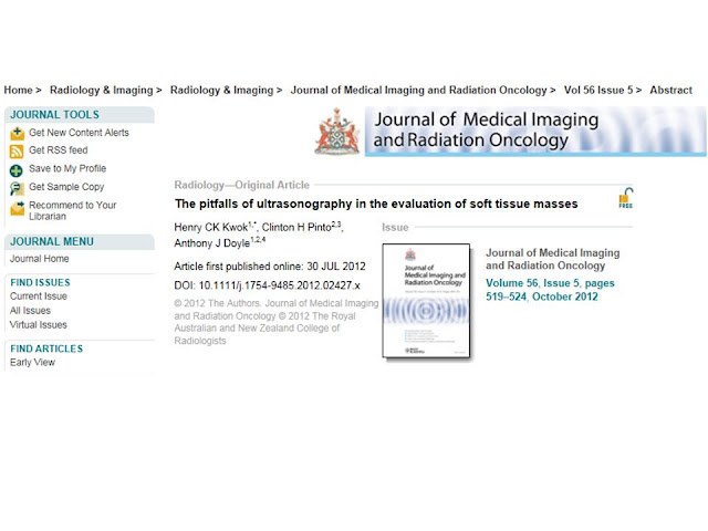
PITFALLS of ULTRASOUND on SOFT TISSUE MASSES


Abstract

Introduction: Ultrasonography is associated with a high error rate in the evaluation of soft tissue masses. The purposes of this study were to examine the nature of the diagnostic errors and to identify areas in which reporting could be improved.

Methods: Patients who had soft tissue tumours and received ultrasonography during a 10-year period (1999–2009) were identified from a local tumour registry. The sonographic and pathological diagnoses were categorised as either ‘benign’ or ‘non-benign’. The accuracy of ultrasonography was assessed by correlating the sonographic with the pathological diagnostic categories.
Recommendations from radiologists, where offered, were assessed for their appropriateness in the context of the pathological diagnosis.

Results: One hundred seventy-five patients received ultrasonography, of which 60 had ‘non-benign’ lesions and 115 had ‘benign’ lesions. Ultrasonography correctly diagnosed 35 and incorrectly diagnosed seven of the 60 ‘non-benign’ cases, and did not suggest a diagnosis in 18 cases. Most of the diagnostic errors related to misdiagnosing soft tissue tumours as haematomas (four out of seven). Recommendations for further management were offered by the radiologists in 144 cases, of which 52 had ‘non-benign’ pathology.There were eight ‘non-benign’ cases where no recommendation was offered, and the sonographic diagnosis was either incorrect or unavailable.

Conclusions: Ultrasonography lacks accuracy in the evaluation of soft tissue masses. Ongoing education is required to improve awareness of the limitations with its use. These limitations should be highlighted to the referrers, especially those who do not have specific training in this area.

Key words: diagnostic error; haematoma; neoplasm, connective and soft tissue; ultrasonography.


DISCUSSION

Ultrasonography lacks accuracy in the evaluation of soft tissue masses due to the non-specific nature of many imaging findings. The present study has reaffirmed our previous observations that ultrasonography has a high error rate in distinguishing non-benign from benign lesions. Despite our earlier experiences and the increased awareness of the limitations of ultrasonography, no significant improvement in error rates was observed between the current and the previous study periods.

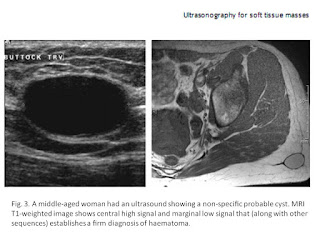
A common diagnostic error involves mistaking solid tumours for haematomas, sometimes resulting in diagnostic delay and suboptimal management. In a review of 31 cases of soft tissue tumours masquerading as haematoma, Ward etal. found that misdiagnosis was associated with diagnostic delays averaging 6.7months; furthermore, neither ultrasonography nor magnetic resonance could reliably differentiate soft tissue tumours and haematoma.[3] It may be useful to correlate with clinical history such as recent trauma and examination findings such as subcutaneous ecchymosis.[4] However, it is important to note that a history of trauma may be incidental, and ecchymosis can also occur with tumoural bleeding.[3]

Given these difficulties, it is not surprising that many radiologists err on the side of caution when confronted with a soft tissue mass on ultrasonography. Of the 115 patients with histologically benign lesions, ultrasonography suggested a suspicious diagnosis or recommended further evaluation in 92 cases (80%, positive likelihood ratio 1.1). A ‘positive’ ultrasonography result per se therefore adds little diagnostic value in the evaluation of patients with soft tissue masses. On the other hand, one should be cautious to assign a ‘negative’ result to a study without providing specific guidance or recommendation. Notably, diagnostic delays have been observed in false negative cases when such recommendation has not been explicitly made. This is particularly relevant in our local practice, where non-specialists (e.g. general practitioners) contribute up to 60% of the referrals for ultrasonography examination for soft tissue masses. In these circumstances, a short comment such as the following may help guide the referrers in appropriate cases:

The findings are non-specific. If the lesion does not resolve rapidly, or if the radiological diagnosis does not fit the clinical picture, a referral to a specialist surgeon is recommended and further imaging such as MRI may be appropriate.

Despite its limitations, ultrasonography may serve specific roles in the work-up of soft tissue masses. First, ultrasonography can confirm the presence of a mass, which can sometimes be difficult to ascertain clinically. Second, ultrasonography can differentiate cystic lesions from solid lesions. Third, ultrasonography can often reliably diagnose lesions with certain well-characterised sonographic features. For instance, a cyst adjacent to a tendon may suggest a ganglion, and a superficial well-defined echogenic mass may suggest a lipoma. It should be noted that the majority of these lesions are satisfactorily managed in the community without being referred to the registry, and are therefore excluded in this review. Fourth, ultrasonography may be used to guide biopsy of the lesions. This is particularly valuable in targeted biopsy of large, heterogeneous tumours.[5]

Ultrasonography has been utilised in tumour follow-up in the research setting. It has been used to detect tumour recurrence[6, 7] and monitor regression of tumour neovascularity induced by therapy for musculoskeletal sarcoma.[8] Ultrasonography may also be used in conjunction with MRI when susceptibility artefacts from orthopaedic hardware prevent evaluation of specific areas following surgery.[8] It has been suggested that colour Doppler flow imaging and spectral wave analysis may allow assessment of blood flow within soft tissue masses and, by inference, differentiate between malignant and benign tumours.[9]

In summary, ultrasonography has specific roles in the evaluation of soft tissue masses. However, aside from the recognisable entities of ganglion, superficial lipoma and obvious peripheral nerve sheath tumour, ultrasonography of soft tissue masses remains non-specific with respect to malignancy. Ongoing education is prudent to improve our understanding of its limitations and pitfalls. In addition, it may be important to highlight these limitations to the referrers, especially if they have no specific training in the management of soft tissue masses.

Thứ Sáu, 30 tháng 11, 2012

World’s First Wireless Ultrasound System


Siemens Showcases World’s First Wireless Ultrasound System at RSNA 2012

Wireless transducer technology will expand use of ultrasound into a variety of clinical settings


Press Release: Siemens Healthcare – Mon, Nov 26, 2012 10:00 AM EST

At the 98th Scientific Assembly and Annual Meeting of the Radiological Society of North America (RSNA), November 25-30 in Chicago, Siemens Healthcare (Booth #831, East Building/Lakeside Center at McCormick Place, Hall D) is introducing the ACUSON Freestyle™ ultrasound system that features wireless transducers, eliminating the impediment of cables in ultrasound imaging. To enable this pioneering technology, the system brings to the market a large number of innovations, including acoustics, system architecture, radio design, miniaturization, and image processing. The ACUSON Freestyle system will expand ultrasound’s use in interventional and therapeutic applications, where the technology provides numerous workflow and image quality advantages. The development of wireless ultrasound is in line with the objectives of the Healthcare Sector’s global initiative Agenda 2013 – specifically in the areas of innovation and accessibility.


For image acquisition and processing, the ACUSON Freestyle system employs advanced synthetic aperture imaging technology, an integration of proprietary hardware and software that was specifically developed for the wireless signal transmission of full-resolution digital image data at very high data rates. Focusing on each pixel in the image, this method produces excellent image quality throughout the field of view. This design reduces the transducer’s power requirements, increasing battery life. Wireless real-time ultrasound data transmission is further enabled through the proprietary development of a novel ultra-wideband radio technology, which, operating at a high frequency of 7.8 Gigahertz, is not susceptible to interference with other electronic equipment.


Three wireless transducers are available for the ACUSON Freestyle system, covering a range of general imaging, vascular, and high-frequency applications such as musculoskeletal and nerve imaging. The user can operate the transducers up to three meters away from the system, which includes an ergonomic interface that enables remote control of scanning parameters from within the sterile field. The ACUSON Freestyle system has a 38-centimeter, high-resolution LED display. The system console can be mounted easily on a lightweight cart and also operates on battery power.

The products mentioned here are not commercially available in all countries. Due to regulatory reasons the future availability in any country cannot be guaranteed. Further details are available from the local Siemens organizations.


Battery life and wireless specs

The transducer's rechargeable battery lasts for about 90 minutes of total scanning time. The system also comes with a spare battery pack that recharges at a charging station on the back of the system while the other one's working.

The development of the Acuson Freestyle was considered as a "huge technical challenge" as ultrasound is a real-time modality where the device must process a large amount of data fed from the transducer. The developers also had to make sure the wireless signals transmitted from the device wouldn't interfere with signals coming from all the other wireless systems in the hospital.

Ad Statistics


Times Displayed: 3165806
Times Visited: 1395


Thứ Tư, 28 tháng 11, 2012

NHÂN CA LOÉT DẠ DÀY @ MEDIC


Fig. 1. Case 1. A Transverse sonogram demonstrates an echogenic core surrounded by a thick hypoechoic halo lateral to head of the pancreas (arrow). B Ulcer and spasm at the apex of duodenal bulb (arrow).

Fig. 2. Case 2. A An irregular echogenic area surrounded by a hypoechoic rim between gallbladder and head of the pancreas (arrow). B Barium collection and bulber deformity due to duodenal ulcer (arrow).

Fig. 3. Case 3. Sonogram demonstrated a hypoechoic solid mass with a central echogenic area adjacent to gallbladder (arrow). Follow-up UGI series in the prone position showed a large ulcer in the postpyloric area.

Fig. 4. Case 4. A An echogenic area surrounded by a thick hypoechoic rim (arrow). B Severe deformity of duodenal bulb due to chronic peptic ulcer is visible on UGI exam (arrow).

Fig. 5. Case 5. A Linear echogenic appearance adjacent to gallbladder. Duodenal wall is thickened (arrow). After some water administration, linear echogeneity was seen in the anterior duodenal wall as a mucosal defect. B The x-ray demonstrates a linear ulcer in the duodenal bulb (arrow).

Discussion

Abdominal US is becoming more frequently used as a primary screening procedure for the evaluation of nonspecific abdominal complaints. Since barium is highly reflective to ultrasound, US is often performed before UGI series. Bowel lesions may therefore be encountered during the US examination. Occasionally, US will permit characterization of a bowel lesion even when it cannot be established radiologically.

The US patterns of normal bowel are variable, depending upon the intraluminal contents. Bowel may be either collapsed or contain varying quantities of fluid and gas. An US pattern consisting of a rounded mass with an echogenic core and a sonolucent halo (target or "bull's-eye" configuration) is commonly encountered on abdominal US arising from a collapsed mucus-filled bowel.

A similar configuration can be observed emanating from a variety of benign and malignant bowel lesions such as congenital pyloric stenosis, intramural hematomas, inflammatory bowel disease especially Crohn's, ileocecal tuberculosis, acute appendicitis, edema of the intestinal wall secondary to thrombosis of the mesenteric veins, Menetrier's disease, ischemia, diverticular disease, and bowel neoplasms [8-11]. However, the target configuration originating from bowel lesions has a hypoechoic halo that is abnormally thick and an echogenic core that is eccentrically located. This appearance has been given US descriptions: the cockade sign, the pseudokidney sign, or the abnormal target appearance [1, 8, 12]. Peristalsis will be absent or diminished, as observed with real-time scanning in affected bowel segment [12].

Echogenic cores in my cases are compatible with ulcer findings in x-rays, according to their shape, volume, and localization. This echogeneity might well result from necrotic fibrinoid debris covering the surface of the niche as a thin layer.

The hypoechoic halo around it might be a counterpart of wall edema and/or infiltration often produced during the active inflammatory phase of duodenal peptic ulcer. Epigastric pain that has been observed as the main complaint in all cases is a significant symptom supporting that the ulcer is in an active phase.

Relatively few reports have been published about US appearances of gastric and duodenal ulcers [2-7]. A case of large gastric ulcer presented with thickening of the gastric wall on US [1]. In another case of antral ulcer demonstrated by a UGI series, the hyperechoic core with an acoustic shadow surrounded by a hypoechoic halo was the US findings of an ulcer niche on the posterior antral wall [2]. Multiple gastric ulcers in a child were demonstrated as a mucosal defect on the thickened antral wall and their response to therapy was followed with US [3]. The appearances of my 5th case, which showed a mucosal defect on the thickened anterior wall of the duodenum, is very similar to this case.

Publications about US appearances of duodenal peptic ulcers are still few [7]. In one study, US appearances of 2 giant peptic ulcers are defined as cystic cavity. In 1 of these 2 cases, an echoic appearance surrounded by a hypoechoic rim, which is very similar to my cases, was defined as the thickening of the duodenal wall [6]. This appearance, however, may represent the ulcer while the cystic area adjacent to it might possibly be a pseudodiverticulum.

Gas in the lumen of the GI tract prevents US examination. When a duodenal peptic ulcer is in an acute inflammatory phase and accompanied with spasm, the collapse of the lumen eliminates gas accumulation. The use of excellent acoustic windows such as the liver and gallbladder is an important factor enabling us to study the duodenum without gas in the lumen. If an ulcer is located on the anterior wall of duodenum, even an open lumen will not prevent the demonstration of ulcer as in the 5th case. In conditions like acute appendicitis, intestinal wall thickening is easily recognized by ultrasonography. With probably more prominent wall thickening, mucosal changes of niche, and good acoustic windows, the acute duodenal ulcer should be expected to be more detectable than acute appendicitis.

In conclusion, the initial diagnosis of a duodenal peptic ulcer in the acute phase may first be indicated by the ultrasonologist. Further studies, especially correlated with duodenoscopy, are necessary to establish the role and importance of US in duodenal ulcer diagnosis.

Thứ Ba, 27 tháng 11, 2012

GUIDELINES for ULTRASOUND USE in RHEUMATOLOGY PRACTICE


The MSUS Committee presented recommendations for "reasonable" rather than "appropriate" use because the RAND analysis method used excludes cost consideration. The authors write, "Where risks of the procedure are minimal...and because costs are not considered, the analysis will inherently favor use of the procedure. Therefore, rather than use the term 'appropriate,' which we felt would be overstating the findings, we use the term 'reasonable' to mean that the evidence and/or consensus of the Talk Force Panel...supported the use of MSUS for the described scenario."

"Reasonable" includes use for:

  • articular pain, swelling, or mechanical symptoms without definitive diagnosis (glenohumeral, acromioclavicular, sternoclavicular, elbow, wrist, metacarpophalangeal, interphalangeal, hip, knee, ankle, and midfoot and metatarsophalangeal joints);
  • inflammatory arthritis and new or ongoing symptoms (glenohumeral, acromioclavicular, elbow, wrist, metacarpophalangeal, interphalangeal, hip, knee, ankle, midfoot and metatarsophalangeal, and entheseal joints);
  • shoulder pain or mechanical symptoms, but not adhesive capsulitis or as preparation for surgical intervention;
  • parotid and submandibular glands in suspected Sjogren's disease;
  • symptoms near a joint obscured by adipose tissue or soft tissue derangements (glenohumeral, acromioclavicular, elbow, wrist, hand, metacarpophalangeal, interphalangeal, hip, knee, ankle/foot, and metatarsophalangeal joints);
  • regional neuropathic pain to diagnose entrapment of the median nerve at the carpal tunnel, ulnar nerve at the cubital tunnel, and posterior tibial nerve at the tarsal tunnel; and
  • guiding articular and periarticular aspiration or injection at sites that include the synovial, tenosynovial, bursal, peritendinous, and perientheseal areas.

MSUS at the temporomandibular joint and costochondral joints was not considered reasonable because the interposition of bone often interferes with imaging in those areas.

The authors also emphasize that these recommendations apply to MSUS done as part of a thorough clinical evaluation in a rheumatology office. "It was not intended to include settings isolated from the rheumatologic assessment, such as might occur in a radiology department or operative setting, or other disciplines, such as podiatry or anesthesia," they write.
Arthritis Care Res. 2012;64:1625-1640.

Study Highlights

  • The ACR developed a summary of clinical scenarios achieving mainly positive recommendations for use of MSUS.
  • For patients with joint pain, swelling, or mechanical symptoms, without definitive clinical diagnosis, use of MSUS is reasonable at the glenohumeral, acromioclavicular, sternoclavicular, elbow, wrist, metacarpophalangeal, interphalangeal, hip, knee, ankle, midfoot, and metatarsophalangeal joints. However, use of MSUS is not reasonable at the temporomandibular joint and costochondral joints.
  • For a patient with present or previous monoarthralgia or oligoarthralgia but without a definitive clinical diagnosis, it is reasonable to use MSUS to look for subclinical inflammatory arthritis or enthesitis at asymptomatic glenohumeral, acromioclavicular, sternoclavicular, elbow, wrist, metacarpophalangeal, interphalangeal, hip, knee, ankle, midfoot, and metatarsophalangeal joints.
  • For a patient with diagnosed inflammatory arthritis and new or ongoing symptoms without a definitive clinical diagnosis, use of MSUS is reasonable to detect inflammation, structural damage, or an additional diagnosis at the glenohumeral, acromioclavicular, elbow, wrist, metacarpophalangeal, interphalangeal, hip, knee, ankle, midfoot, metatarsophalangeal, and entheseal sites.
  • For a patient with hip pain or mechanical symptoms without a definitive clinical diagnosis, use of MSUS is reasonable to assess effusion, intraarticular and periarticular lesions, and adjacent regional soft tissue structures.
  • For a patient with periarticular pain without a definitive clinical diagnosis, use of MSUS is reasonable to assess tendon and soft tissue disorders and adjacent swelling at the shoulder, elbow, hand, hip, knee, ankle, and forefoot.
  • For a patient with inflammatory-sounding entheseal, sacroiliac, or spinal pain, use of MSUS is reasonable to detect evidence of enthesopathy.
  • For a patient with shoulder pain or mechanical symptoms, without a definitive clinical diagnosis, use of MSUS is reasonable to detect underlying structural disorders. However, use of MSUS is not reasonable to detect adhesive capsulitis or to prepare for surgical intervention.
  • For a patient with regional mechanical symptoms but without a definitive clinical diagnosis, it is reasonable to use MSUS to detect inflammation, tendon, and soft tissue disorders at the shoulder, elbow, hand, wrist, hip, knee, ankle, and foot joints.
  • Use of MSUS is reasonable to assess the parotid and submandibular glands as part of an evaluation for Sjogren’s disease.
  • For a patient with symptoms near a joint surrounded by adipose or other local soft tissue abnormalities, use of MSUS is reasonable to facilitate clinical assessment at the glenohumeral, acromioclavicular, elbow, wrist, hand, metacarpophalangeal, interphalangeal, hip, knee, ankle/foot, and metatarsophalangeal joints.
  • For a patient with regional neuropathic pain without a definitive clinical diagnosis, use of MSUS is reasonable to diagnose entrapment of the median nerve at the carpal tunnel, the ulnar nerve at the cubital tunnel, and the posterior tibial nerve at the tarsal tunnel.
  • Use of MSUS is reasonable to guide articular and periarticular aspiration or injection at synovial, tenosynovial, bursal, peritendinous, and perientheseal sites.
  • Use of MSUS may be reasonable to guide synovial biopsy procedures.
  • Use of MSUS may be reasonable to monitor disease activity and structural progression at the glenohumeral, acromioclavicular, elbow, wrist, hand, metacarpophalangeal, interphalangeal, hip, knee, ankle, foot, and metatarsophalangeal sites in patients with inflammatory polyarthritis.