
Femoral hernias, Henry Robert Whalen, Gillian A Kidd, Patrick J O’Dwyer
BMJ2011;343doi:
http://dx.doi.org/10.1136/bmj.d7668(Published 8 December 2011)
Cite this as:BMJ2011;343:d7668
An overweight
65 year old woman visits her general practitioner with discomfort in her right
groin. On examination, the suggestion of a reducible groin lump is noted. She
is routinely referred to the surgical outpatient clinic with a possible
diagnosis of inguinal hernia. However, two weeks later and before her surgical
appointment, she again visits her general practitioner, this time with
vomiting, diarrhoea, and colicky abdominal pain. She is immediately referred to
the emergency department. An abdominal radiograph shows small bowel
obstruction. She is admitted to the surgical ward with a diagnosis of
obstructed femoral hernia and has a small bowel resection and emergency hernia
repair.
What is a femoral hernia?
A femoral
hernia is the protrusion of a peritoneal sac through the femoral ring into the
femoral canal, posterior and inferior to the inguinal ligament. The sac may
contain preperitoneal fat, omentum, small bowel, or other structures.
How common are femoral
hernias?
·
About 5000 femoral hernia repairs are carried out in the United Kingdom
each year
·
Femoral hernias account for a fifth of all groin hernias in females but
less than 1% of groin hernias in males
·
The 40% of femoral hernias that present acutely are associated with a
10-fold increased risk of mortality1 2
Why is a femoral hernia missed?
Evidence is
scarce as to the reason why femoral hernias are often missed and present as
emergencies. Patients may be aware of groin discomfort or a groin lump, but
they may not realise its clinical importance and may be reluctant to seek
medical help. Initially some patients present to primary care with vague
symptoms including groin discomfort that may be attributed to other disease
such as osteoarthritis. As femoral hernias are typically small, they may be
easily missed on examination, particularly in obese patients. Furthermore,
owing to the difficulty in clinically distinguishing groin hernias, femoral
hernias may be mistaken for inguinal hernias and referred for surgical opinion
on a non-urgent basis.3
In an
emergency, patients may present with signs of bowel obstruction, which include
colicky abdominal pain, vomiting, and abdominal distension. About a third of
patients do not complain of symptoms directly attributable to a hernia,4 and a groin lump is
not always present. Other diagnoses, such as gastroenteritis, enlarged groin
lymph node, diverticulitis, or constipation, may be made in error.⇓
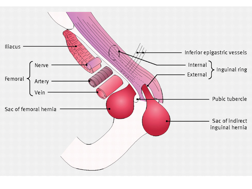
Inguinal
hernias are usually reducible and above the inguinal ligament. Femoral hernias
are often irreducible and below the inguinal ligament. Adapted with permission
from Ellis H. Clinical anatomy. 6th ed. Blackwell
Scientific, 1977
Retrospective
studies have observed that about 40% of hernias causing symptoms of acute bowel
obstruction are missed owing to a lack of groin examination.5 6 The researchers
concluded that female patients and all patients with femoral hernia were less
likely to have a groin examination, despite signs of bowel obstruction being
noted.5
Why does this matter?
Although
femoral hernias are less common than inguinal, they are associated with higher
rates of acute complication. The cumulative probability of strangulation for
femoral hernias is 22% three months after diagnosis, rising to 45% 21 months
after diagnosis, whereas the probability of strangulation for an inguinal
hernia is 3% and 4.5% respectively over the same time period.7
Several studies
have shown that acute femoral hernias and their subsequent complications are
associated with increased morbidity and mortality.1 2 8 9 10 Examples of
morbidity resulting from acute presentation include increased rates of bowel
resection, wound infection, and cardiovascular and respiratory complications.10 As elective femoral
hernia repair has been shown to be a relatively safe procedure (even in
patients aged over 80), it is generally accepted that femoral hernias should be
referred urgently and repaired electively.2 10 11 12
Missed femoral
hernia at emergency presentation delays time to surgery.5 One study has shown
an increased likelihood of bowel resection if surgery is undertaken more than
12 hours after the onset of acute symptoms.13 Preoperative delay
is clearly linked with an increase in bowel resection, and this is associated
with mortality rates that are about 20 times higher than those for patients
having elective hernia repair (which would not require a bowel resection).2
How is it diagnosed?
Clinical
Classically,
femoral hernias present as mildly painful, non-reducible groin lumps, located
inferolateral to the pubic tubercle. In contrast, inguinal hernias are found
superomedially. However, femoral hernias tend to move superiorly to a position
above the inguinal ligament, where they may be mistaken for an inguinal hernia.
Differentiation of groin hernias on clinical grounds is therefore unreliable,
irrespective of the experience of the examining doctor.14 In patients
presenting electively, only about 1% of groin hernias in males are likely to be
femoral, whereas the likelihood in females is about 20%.1 Clinical examination
alone is inaccurate in differentiating groin hernia.14 Therefore in
females, owing to the greater prevalence of femoral hernia, consider all groin
hernia to be femoral until proved otherwise.
Femoral hernias
may also present without a palpable lump and with only vague symptoms of
abdominal or groin pain. However, symptoms may vary and there is a lack of
evidence to predict the likelihood of a particular symptom indicating the
presence of a femoral hernia. Patients may present later with clinical features
of bowel obstruction. Undertake a detailed groin examination in all patients
presenting with bowel obstruction.
Investigations
Ultrasonography,
magnetic resonance imaging, and computed tomography (CT) have all been shown to
be accurate in detecting and differentiating groin hernias.
Ultrasonography
is widely available, non-invasive, and highly accurate in differentiating
inguinal from femoral hernia—with sensitivities and specificity of 100% being
reported in two studies.15 16 Its accuracy is,
however, operator dependent.
Magnetic
resonance imaging has been reported to be more accurate than ultrasonography in
detecting inguinal hernia.17 However, there is a lack
of evidence for whether magnetic resonance imaging is better than
ultrasonography in detecting and differentiating groin hernia. Therefore
ultrasonography should be the first choice for electively investigating
suspected groin hernia as it is more widely available, less costly, and
accurate.
CT scanning has
been shown to be accurate in differentiating groin hernias. One retrospective
study reports the correct identification of 74 of 75 hernias (28 femoral and 47
inguinal), which were later confirmed at operation.18 This is broadly
comparable with the non-invasive modalities outlined above, but as there is a
substantial radiation dose associated with CT scanning, it should not be used
electively for investigating suspected groin hernia. In the acute abdomen,
however, consider CT as the first choice for investigating suspected small
bowel obstruction in the presence of a negative clinical examination.
How is it managed?
In males, a
groin hernia suspected as being femoral on clinical examination requires urgent
referral, due to the risks of acute complications outlined above. All groin
hernia in females should be urgently referred for assessment.
Electively,
both open and laparoscopic repair using mesh have significantly lower
recurrence rates than repair using sutures only.1 Open repair has the
advantage that it can be performed under local anaesthetic. No evidence
suggests superiority of either method in the acute setting.
Some research
has suggested that femoral hernias may be overlooked during repair of suspected
inguinal hernias.19 So during surgical
repair of all groin hernias examine the femoral canal if an obvious inguinal
hernia is not observed.
Key points
·
Femoral hernias are more common in females and in people aged over 65
years and are associated with higher rates of complications such as
strangulation
·
Emergency surgery for femoral hernia is associated with a 10-fold
increased risk of mortality, which is further increased by preoperative delays
·
Clinical examination is unreliable in differentiating femoral from
inguinal hernia
·
Refer all females with groin hernia for urgent assessment and
management
·
Examine the groins of all patients presenting with signs of small bowel
obstruction
·
Ultrasound is the first line elective investigation for suspected
uncomplicated groin hernia, but in acute small bowel obstruction, CT scanning
is first choice


