Tổng số lượt xem trang

Thứ Sáu, 13 tháng 6, 2014

NHÂN CA NANG CỦA SỤN TUYẾN GIÁP TẠI MEDIC


Bệnh nhân nam 33 tuổi  khám kiểm tra vùng cổ, được bs Phạm thị Thanh Xuân ở phòng Siêu âm Trung tâm Medic, phát hiện có 1 nang nhỏ trong sụn giáp bên P, kích thước = 8x7mm.
Siêu âm đàn hồi ARFI xác  nhân là cấu trúc nang trong sụn giáp, với VTI= brighter color (mềm) và VTQ=1,5m/s.
Kết luận là nang sụn giáp P.
Y văn chưa thấy mô tả siêu âm nang sụn giáp, có bài về CT xác chẩn khối thanh quản là dystrophic ossifications của sụn giáp.


Thứ Tư, 11 tháng 6, 2014

DASH and Incisional Hernias

US Good Alternative to CT for Characterizing Incisional Hernias: Study
By James E. Barone MD
June 03, 2014

NEW YORK (Reuters Health) - Using dynamic abdominal sonography to measure incisional hernias is equivalent to using computerized tomography (CT), according to a new study.

As opposed to a CT scan, dynamic abdominal sonography for hernia (DASH) is a bedside procedure, its results are available immediately and it does not use ionizing radiation, researchers write in JAMA Surgery, online May 28.

"The DASH examination is a feasible and accurate method for evaluating hernias, particularly in those patients who have smaller hernias and who may have unclear physical exam findings," lead author Dr. Rebeccah B. Baucom from Vanderbilt University Medical Center in Nashville, Tennessee, told Reuters Health by email.

A previous paper by Dr. Baucom and colleagues showed that CT was better at detecting incisional hernias than physical examination by surgeons. In another study, the team found that DASH was as accurate as CT scan in diagnosing hernias.

To see if DASH could be used to plan operative procedures using measurements of hernia defect size, the researchers compared it with CT scans in 109 patients. One surgeon performed all of the DASH examinations, whereas one of three surgeons read the CT scans.

The mean patient BMI was 32.2, and 34% of the patients had at least one previous hernia repair.

As measured by CT, the mean surface area of hernias was 44.6 cm2, compared to 41.8 cm2 with DASH (p=0.82). The transverse dimensions were nearly identical at 5.20 cm for CT and 5.17 cm for DASH (p=0.71).

Measurements were comparably accurate for CT scan and DASH among the 14 morbidly obese patients (BMI>39.9), 49 who were obese (BMI 30 to 39.9), and 46 who had a BMI under 30.

The authors also looked at the subgroup of hernias that were 10 cm or larger in transverse dimension. Of the 15 patients in that category, the mean surface area of the hernias was 189.4 cm2 by CT and 171.3 cm2 by DASH, an insignificant difference (p=0.26).

"Several of our surgeons perform DASH examinations on their patients who present with suspected incisional hernias," said Dr. Baucom, adding that it is also being done more and more often at follow-ups after repair.

Dr. Michael Liang, who has studied ventral hernia repair but was not involved in the new work, said, "DASH is a safe and potentially effective modality for the surgeon to use to assess ventral incisional hernias."

But Dr. Liang, of the University of Texas Health Sciences Center at Houston, raised a number of concerns about the study. For example, he told Reuters Health, the authors did not use operative findings, the gold standard, to validate the ultrasound and CT results, nor did they adjust for the size of the hernias.

For recurrent, large, and complex ventral hernias, Dr. Liang said a CT scan will be obtained regardless of DASH availability because the former modality "is useful to assess defect size, peritoneal volume, lateral muscles, abdominal wall thickness and contraction, intra-abdominal organs, and old mesh."

According to Dr. Baucom and colleagues, however, DASH plus physical examination have been sufficient to evaluate most of these structures and relationships.

SOURCE: http://bit.ly/1nZk6EV

JAMA Surgery 2014.

Thứ Hai, 9 tháng 6, 2014

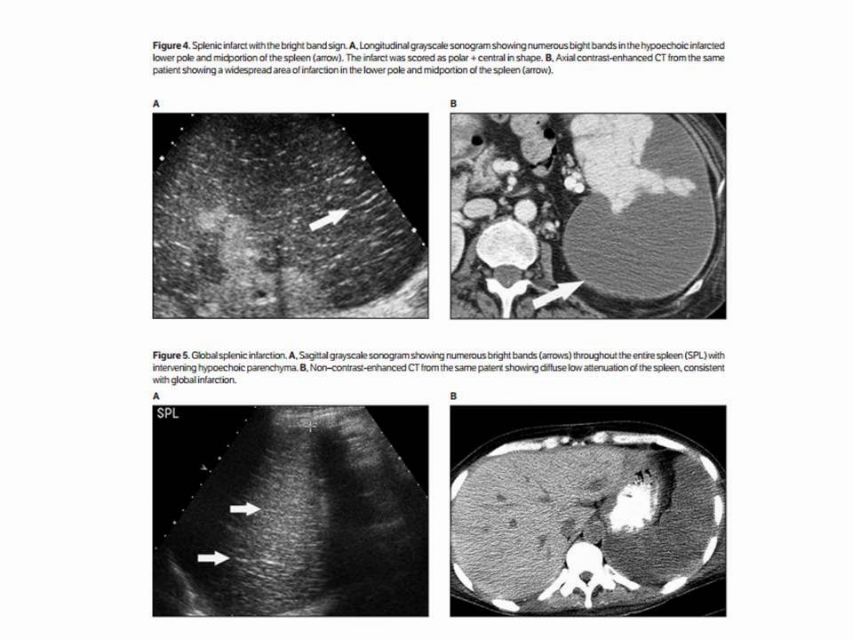
BRIGHT BAND SIGN in SPLENIC INFARCTION


In summary, the bright band sign was apparent in 91.9% of patients with splenic infarcts, including 95.7%
of those with nonclassic sonographic features of splenic infarction, was not present in a collection of 19 abnormal control patients, and was not seen in 100 normal control patients.
The bright band sign likely results from specular reflections returned from fibrous trabeculae that remain intact within infarcted regions while the neighboring splenic parenchyma undergoes necrosis. Although further work will be required to establish the role that the bright band sign may play inthe diagnosis of splenic infarction, the sign appears potentially useful, particularly for the many infarcts with nonclassic, nonspecific sonographic appearances.


Thứ Sáu, 6 tháng 6, 2014

ULTRASOUND GUIDELINES CATCHES INCIDENTAL THYROID CANCERS

In a retrospective 10-year review, a team led by Dr. Manisha Bahl from Duke University Medical Center found that adopting SRU guidelines for follow-up of incidental thyroid nodules would result in only 2% of thyroid cancers being missed. In addition, the missed tumors tended to be much smaller and more likely to be stage I than those found during workup recommended by the SRU guidelines.

"When the SRU criteria-negative incidental cancers are compared to SRU criteria-positive incidental cancers, they are smaller in size, more likely to be papillary carcinoma, and less likely to have nodal metastases," senior author Dr. Jenny Hoang told AuntMinnie.com. "In general, these characteristics belong to thyroid cancers that are more likely to have an indolent course and may never become symptomatic in the patient's lifetime."

A big problem

Incidental thyroid nodules spotted on ultrasound and other imaging modalities are a big healthcare problem in the U.S.: The nodules are common and workup can lead to patient anxiety and unnecessary and costly procedures or surgery, Hoang said.

Guidelines such as SRU's thyroid recommendations are important in reducing the growing number of biopsies being performed for incidental thyroid nodules. However, the SRU recommendations -- which were published in 2005 -- have not been widely adopted by clinicians and radiologists, including those at Duke, she said.

A previous Duke study found that using SRU recommendations could prevent 30% of biopsies. However, that study population consisted mostly of benign nodules "and did not adequately address the fear of missing more cancers in a larger cohort of patients with incidental thyroid nodules over time," she said. "In this paper we specifically addressed that fear: How many incidental cancers would be missed with the SRU recommendations over a decade?"

The group retrospectively reviewed data from thyroid surgery patients from January 1, 2003, through December 31, 2012. After evaluating imaging studies and reports for incidental thyroid nodules, the team categorized incidental nodules using the SRU criteria to ascertain the characteristics of malignant nodules that would and would not have been worked up (Radiology, June 2014, Vol. 271:3, pp. 888-894).

Of the 2,090 thyroid surgery patients included in the study, 680 were found to have thyroid cancer; 101 (15%) had incidental thyroid nodules detected on imaging. After the researchers applied the SRU recommendations to the 90 patients who had available ultrasound images or reports, they determined that 16 (18% of the 90 patients and 2% of all thyroid cancers) would have been missed using the criteria.

The tumors that would not have been worked up under the SRU criteria had a mean size of 1.1 cm (range, 0.9-1.4 cm), compared with a mean size of 2.5 cm (range, 1.0-7.6 cm) for those that had met the SRU recommendations. The difference was statistically significant (p < 0.001). Nearly all (15/16, 94%) were stage I, compared with 47 (64%) of the 74 incidental thyroid nodules that met the SRU criteria.

Fourteen (88%) of the 16 SRU criteria-negative cancers were papillary carcinoma; small papillary carcinoma has an extremely high survival rate. The other cases were a follicular carcinoma and a medullary carcinoma.


Transverse ultrasound image of an SRU criteria-negative thyroid cancer. Incidentally detected at CT, this 1.2-cm thyroid nodule is solid in composition. It does not meet the SRU recommendations for fine-needle aspiration biopsy. The postsurgical pathologic result was papillary cancer. Image courtesy of Dr. Manisha Bahl.
The researchers were surprised to find that patients with incidental thyroid cancers only represented 15% of patients who had surgery for thyroid cancer, Hoang said. The group's two prior studies had found that 29% of patients with incidental thyroid nodules underwent ultrasound-guided biopsy and 23% of patients had thyroid surgery for nodules.

"The overall lower proportion of incidental thyroid cancers among all thyroid cancer patients simply highlights the fact that benign incidental thyroid nodules are common, but cancer among the incidental nodules is uncommon," she said.

For practicing radiologists and clinicians, the clinical implication from this study and the group's prior studies is that SRU recommendations "can be used to better manage workup of incidental thyroid nodules and that SRU recommendations achieve the goal of ensuring that clinically important thyroid cancers would undergo biopsy, while avoiding unnecessary tests and surgery in patients with benign nodules," Hoang said.

Adoption of guidelines

In addition to SRU, the American Association of Clinical Endocrinologists (AACE) and the American Thyroid Association (ATA) have guidelines that recommend fine-needle aspiration (FNA) biopsy of thyroid nodules based on ultrasound findings. When speaking with a number of radiologists from different practice types over the past couple years, Hoang was most commonly told that the clinician decides which nodules receive biopsies.

"Most clinicians use a size cutoff of 1 cm for solid nodules which is part of the AACE and ATA guidelines," she said. "This is seemingly a small difference compared to the SRU 1.5-cm cutoff [for nodules without microcalcification], but another study from our group showed that one in four biopsies could be prevented with the SRU recommendations. Therefore, increasing the size cutoff by just 5 mm can substantially reduce the proportion of thyroid nodules that require biopsy."

Hoang said that she hopes this study will encourage more widespread adoption of the SRU recommendations.

"Radiologists and clinicians now have our study to allay fears of missing many and significant cancers by being more selective in the workup of incidental thyroid nodules," she said.

Hoang also noted that she is a member of an American College of Radiology (ACR) committee that will produce a white paper in the coming year on incidental thyroid nodules for all imaging modalities. Furthermore, Duke has developed a three-tier system for reporting incidental thyroid nodules found on CT, MRI, or PET/CT, she said.

Thứ Năm, 5 tháng 6, 2014

ULTRASOUND MATCHES CT for KIDNEY STONE IMAGING

Ultrasound Matches CT for Kidney Stone Imaging
Neil Osterweil
May 17, 2014

ORLANDO, Florida — Ultrasound was comparable to CT in its ability to discriminate between renal calculi and other causes of flank pain, according to the results of a new study.

"Routine utilization of CT as a knee-jerk response for someone presenting with acute colic is inappropriate, not only for cost but for radiation exposure," said Marshall L. Stoller, MD, professor and vice chair of urology at the University of California, San Francisco.

More important, this study shows that using ultrasound has no adverse impact on patient outcomes, said Dr. Stoller.

The results of the study were presented here at the American Urological Association 2014 Annual Scientific Meeting.

A Multicenter Study

Dr. Stoller and colleagues at 15 academic medical centers conducted a randomized trial in which adult patients presenting to an emergency department with suspected nephrolithiasis were randomly assigned to receive point-of-care ultrasound performed by an emergency physician, ultrasound performed by a radiologist, or abdominal CT. Additional tests were at the discretion of each patient's physician.

The investigators looked at the incidence of serious adverse events diagnosed within 30 days of imaging, cumulative radiation exposure, and cost. Follow-up with detailed, structured patient interviews was done at 3 and 7 days, and again at 1, 3, and 6 months.

The incidence of serious adverse events among patients assigned to CT was 11.1%, compared with 12.3% for patients who underwent ultrasound performed by an emergency physician and 10.6% for those whose ultrasound scans were performed by a radiologist. The differences were not significant.

Severe adverse events — including abdominal aortic aneurysm with rupture, pneumonia with sepsis, appendicitis with rupture, inflammatory bowel conditions and renal infarction, pyelonephritis with urosepsis, and ovarian torsion with necrosis — occurred in 5 of 908 patients (0.55%) assigned to emergency-physician-performed ultrasound, 3 of 893 patients (0.34%) assigned to radiologist-performed ultrasound, and 4 of 958 patients (0.42%) assigned to CT.

Not surprisingly, patients who underwent ultrasound had significantly lower average cumulative radiation exposures, at 10.5 mSv and 9.3 mSv for emergency-physician- and radiologist-performed ultrasound, respectively, compared with 17 mSv for patients who underwent CT (P < .0001).

There were no significant differences in hospital readmissions after discharge, average pain scores, pain resolution, or subsequent serious adverse events, the investigators found.

Average imaging costs were about 33% to 50% higher for CT (average, $300) than for ultrasound ($150 if done by an emergency physician, and $200 if done by a radiologist).

Concerns About Radiation

This study shows that emergency department physicians need not fear missing a diagnosis of kidney stones or other causes of acute pain if they choose ultrasound over CT as an initial imaging modality, said Margaret Pearle, MD, PhD, professor of urology at the University of Texas Southwestern Medical Center at Dallas and the Center for Mineral Metabolism and Clinical Research.

And patients can be confident that their chance of getting an accurate diagnosis is comparable, she added.

"I think in our institution, probably like the majority of institutions, if someone comes in with acute flank pain, the first test is a CT, although we're starting to see a little bit of a trend toward ultrasound," Dr. Pearle told Medscape Medical News.

The shift appears to be driven more by patient and clinician concerns about radiation exposure, rather than administrative worries over cost, she said.

"There's tremendous fear right now, particularly on the part of patients. They hear that radiation exposure is bad, and they don't [want to be exposed]," she said. "Stone disease is one of those conditions that is associated with repeated imaging studies because of repeated symptomatic episodes."

Dr. Pearle was not involved in the study.

Because of fragmentation and lack of coordination of healthcare, some patients get repeat exposure to ionizing radiation for the same episode of pain, said Dr. Stoller.

"It is not uncommon in my clinic that you'll see someone in their 20s coming in with 5 or 6 CTs from the past week or 2. That's a huge amount of radiation," he said.

The study was supported by the National Institutes of Health. Dr. Stoller and Dr. Pearle have reported no relevant financial relationships.

American Urological Association (AUA) 2014 Annual Scientific Meeting. Abstract PD4-03. Presented May 16, 2014.


Thứ Năm, 29 tháng 5, 2014

Dr. Birnholz on Ultrasound Part 15

In other news, Dr. Jason Birnholz is back with his latest Practice of Ultrasound installment. The article looks back at Dr. Birnholz's involvement in ultrasound from its origins, with the establishment of one of the first sonography clinical services at Massachusetts General Hospital, through today, with ultrasound becoming an increasingly common tool used throughout healthcare. He also ponders the rise of what he calls "checklist" ultrasound, in which the exam is performed by minimally trained healthcare personnel and ultrasound is used as a gateway to other imaging tools. This differs from the model of sonography performed by highly trained specialists who might be capable of rendering a diagnosis simply with the tools at hand. In today's era of point-of-care ultrasound and the "electronic stethoscope," it's clear that checklist imaging has won out. But that may not always be the case, especially with new ultrasound technology in development that could lead to major improvements in image quality.

Thứ Sáu, 23 tháng 5, 2014

Liver Cancer Ultrasound Screening

Liver Cancer Ultrasound Screening Can Improve Survival of Cirrhosis Patients

By Medimaging International staff writers
Posted on 20 May 2014


Image: Dr. Amit Singal, assistant professor of internal medicine and clinical sciences at the University of Texas Southwestern Medical Center – author of the paper “Early Detection, Curative Treatment, and Survival Rates for Hepatocellular Carcinoma Surveillance in Patients with Cirrhosis: A Meta-analysis” published in the Public Library of Science Medical Journal (Photo courtesy of the University of Texas Southwestern Medical Center).

Liver cancer survival rates could be improved if more individuals with cirrhosis are screened for tumors using inexpensive ultrasound scanning and blood tests, according to a recent review.

The meta-analysis of 47 studies involving more than 15,000 patients discovered that the three-year survival rate was much higher among patients who received liver cancer screening—51% for patients who were screened compared to 28% of unscreened patients. The review also found that cirrhosis patients who were screened for liver cancer were more likely to receive curative treatment instead of palliative care.

“Curative therapies, such as surgery or a liver transplant, are only available if patients are found to have liver cancer at an early stage. Unfortunately, right now, only a minority of patients’ cancers are found at an early stage,” said Dr. Amit Singal, an assistant professor of internal medicine and clinical sciences at the University of Texas (UT) Southwestern Medical Center (Dallas, USA), and medical director of the Liver Tumor Clinic in the Harold C. Simmons Cancer Center.

Dr. Singal’s findings, published April 1, 2014, in the Public Library of Science Medical Journal, are especially significant for Texas because the state has high rates of fatty liver disease and hepatitis C, both of which are correlated with cirrhosis. Texas also has the second highest incidence of hepatocellular carcinoma (HCC), the most typical type of liver cancer in the United States. Many cases of HCC can be caught early with screening. “We have a simple test, an abdominal ultrasound which is painless and easy, but we found that less than 20% of at-risk people have the test done, largely due to providers failing to order it,” Dr. Singal said.

Dr. Singal reported that he hopes his study will encourage both patients and doctors to request ultrasound screening. He noted that liver cancer screening is not yet recommended by the US Preventative Services Task Force, partly because a randomized study has not yet been done, which points to a conundrum in the field. When a randomized study was attempted in 2005, many patients chose to leave the study when they heard evidence that liver cancer screening could be so beneficial. None of the patients wanted to be the ones randomly chosen to not get the tests.

Dr. Singal hopes his findings will convince more cirrhosis patients and their doctors that screening is worthwhile. “Just because we don’t have a randomized trial doesn’t mean there isn’t a benefit. We’re stuck in the middle ground where we’ve gone halfway. People are starting to believe liver cancer screening is helpful, but there’s not enough evidence to prove a definite benefit,” Dr. Singal said. “Part of our goal is providing evidence to both patients and physicians that liver cancer screening is beneficial.”

Thứ Ba, 20 tháng 5, 2014

ARFI of NORMAL KIDNEY and HYDRONEPHROSIS in CHILDREN


Discussion
Hydronephrosis is an obstructive or non-obstructive nephropathy that is a commonly identified disease during pediatric abdominal ultrasonography. Congenital obstructive nephropathy constitutes the single most important identifiable cause of renal impairment in infants and children [10-12]. In obstructive nephropathy, interstitial fibrosis eventually develops and leads to a loss of nephrons [10].
Numerous papers that focus on the molecular biological mechanisms associated with renal interstitial fibrosis due to obstructive nephropathy have been recently published [10,13,14]. However, there is limited radiological research on renal interstitial fibrosis in the case of hydronephrosis. This could be attributed to the difficulty of detection, evaluation, and quantification of interstitial fibrosis by radiological methods.
There are many studies that explored ARFI measurements as a means of evaluating tissue stiffness, including several studies on kidneys. Gallotti et al. [6], Eiler et al. [7], and Goertz et al. [8] measured the ARFI velocities of normal kidneys in healthy adults.
Further, there have been several trials using ARFI in adult kidneys to evaluate renal masses, to assess renal allograft fibrosis, and to detect chronic kidney diseases [15-17]. However, there is a lack of studies involving ARFI measurements in young children. This could be attributed to the fact that the previously used low-frequency transducer is not effective in the case of such small patients.
However, the availability of the 4-9-MHz high-frequency linear transducer makes it possible to measure SWVs in small subjects. Recently, our group demonstrated normal values of SWVs using ARFI in pediatric abdominal organs including kidneys in 202 children with an average age of 8.1±4.7 years [1]. The mean SWVs were 2.19 m/sec for the right kidney and 2.33 m/sec for the left kidney in the above mentioned study. The previously reported mean SWVs in normal adult kidneys were 2.24-2.37 m/sec, with no significant difference between the right and the left kidney [6,8]. The median SWVs in normal kidneys in the present study were 1.75 m/sec without any difference between the right and the left ones. This value is relatively low as compared to that obtained in previous studies. However, this result is comparable with that of our previous study, which concluded that the mean ARFI SWV for the kidneys increased according to age in children less than 5 years of age [1].
In this study, we only included children under the age of 24 months. Only one study has been performed on the evaluation of diseased kidneys in children. Bruno et al. [5] conducted a study of ARFI measurements in pediatric patients with vesicoureteral reflux. The study suggested that ARFI can provide reliable information about the severity of renal damage and maybe useful in the diagnostic workup in children with a chronic reflux renal disease. However, the patient age in the study ranged from 8 to 16 years. Therefore, our study is the first report evaluating ARFI for hydronephrotic kidneys in young children.
We aimed to correlate SWVs with the hydronephrosis grade. Even though there are hydronephrosis grading systems on ultrasonography [11,18,19], these could not definitely differentiate between obstructive and non-obstructive hydronephrosis. Further, these systems cannot suggest the grade of renal parenchymal fibrosis. If SWVs have a correlation with the renal parenchymal stiffness, its measurement would be helpful in evaluating the status of a patient’s kidney. Further, SWV can show a continuous spectrum of stiffness.
On the other hand, the grading system has an ordinal scale that cannot show a continuous value. Therefore, elastography has a possibility of having an additional value to evaluate hydronephrosis. In our study, there was a significant difference in the median SWVs between normal kidneys (1.75 m/sec) and high-grade hydronephrotic kidneys (2.02 m/sec). This suggests that elasticity decreases and stiffness increases in high-grade hydronephrotic kidneys. However, ARFI measurements cannot differentiate the cause of stiffness change such as tissue fibrosis and edema. Further research with a large group of patients and pathologic correlation is needed.
We also compared SWVs for a hydronephrotic kidney with and without UPJO. Further, there were only seven patients proven to have UPJO during the study period. The mean ARFI velocities were 0.69-2.51 m/sec for hydronephrotic kidneys without UPJO and 1.54-2.72 m/sec for those with UPJO; there was no statistical difference. Kidneys with VUR and a parenchymal scar change also exhibited no remarkable difference in SWVs. This could be attributed to the small number of patients, variable interstitial fibrosis of the UPJO group, and heterogeneous parenchymal scar change in the refluxing kidneys. This needs further evaluation with a large number of patients.

This study has several limitations. Almost all previous studies performed in adults measured about 5-10 valid SWVs and used mean values. However, due to the characteristics of the pediatric patient group, only three valid SWVs were obtained in this study.
Repetitive measurements over a long time while subjects hold their breath is not possible in many children, particularly young children. Although only three valid ARFI velocities were attempted, two children could not tolerate the examinations and the success rate was 96%. Moreover, subjects were allowed to breathe freely during measurements. Thiscan increase the variability of SWV. The development of a method to measure SWV without breathholding would lead to more reliable results. 

The second limitation is the representativeness of the ARFI value.To represent a global kidney, measurement should be performed on multiple sites of the kidney, such as the upper, mid-, and lower poles. However, if the upper and lower poles are to be imaged, it is necessary to use a similar angle of incidence in all patients relative to the tubular system to avoid anisotropy issues. It is conceivable that shear waves generated within the kidney move at different velocities depending on the angle of incidence [20]. We tried to measure SWVs at the same portion of the mid-pole from the axial view, as parallel to the tubular system as possible in order to reduce the angle effect. The variation of the depth of the ROI position should also be considered.
We targeted renal parenchyma, including both the renal cortex and the medulla, from the axial view in each patient. Therefore, we might expect that the depth of the ROI position would be different between patients and could increase according to the body size. Further study is needed to evaluate the effect of the depth of the ROI position and the body size in children. 

The fourth limitation is that we considered the contralateral kidneys without hydronephrosis as normal in the hydronephrosis group. Even though we demonstrated no significant difference in SWVs between normal kidneys in the normal group and contralateral kidneys in the hydronephrosis group, there could have been a physiological change in the bilateral kidneys of the hydronephrosis group.

In conclusion, obtaining ARFI measurements of kidneys using a high-frequency transducer is feasible in very young pediatric patients. The median SWV of normal kidneys in children under the age of 24 months was 1.75 m/sec. These velocities increased in high-grade hydronephrotic kidneys but were not helpful in differentiating hydronephrotic kidneys with and without UPJO.


Beomseok Sohn; Myung-Joon Kim; Sang Won Han; Young Jae Im; Mi-Jung Lee.

AT MEDIC CENTER:

We applied ARFI technique from Siemens S2000 to evaluate whether fibrotic process existing in adult hydronephrosis.
Using 1-4 MHz convex probe we calculated in 3 positions of hydronephotic kidney due to  obstruction [stone, outside compression] (n=27 cases), due to ureteropelvic junction obstruction [UPJO] (n=30 cases]. We had a control group of normal kidney (n=36 cases).


SO SÁNH GIỮA 2 NHÓM THẬN Ứ NƯỚC  DO SỎI và DO HẸP KHÚC NỐI BỂ THẬN-NIỆU QUẢN THẤY KHÁC BIỆT TỐC ĐỘ ĐÀN HỒI CÓ Ý NGHĨA THỐNG KÊ (P<0 .0001="" p="">


Experience Matters in point-of-care Ultrasound of Appendicitis


Experience matters in point-of-care ultrasound
By Erik L. Ridley, AuntMinnie staff writer
May 19, 2014 -- Experienced sonologists had significantly higher sensitivity for diagnosing appendicitis with point-of-care ultrasound than sonologists with less experience, in a study from Mount Sinai School of Medicine. Either way, though, it's important not to rely only on point-of-care ultrasound to rule out the condition.

 In the prospective study of 150 patients, experienced sonologists' sensitivity was nearly 30% higher than that of their less experienced colleagues.
"To minimize the possibility of errors, sonologists should avoid ruling out appendicitis based on [point-of-care ultrasound] results alone," said Dr. James Tsung from the department of emergency medicine. "So you need the clinical picture, and if there's uncertainty, certainly proceed with radiology imaging."
He presented the research during a scientific session at the recent American Institute of Ultrasound in Medicine (AIUM) annual meeting.
The experience effect
While it's well-known that ultrasound is an operator-dependent imaging modality, the effect of operator experience on point-of-care ultrasound hasn't yet been studied, according to Tsung.
In medicine, misdiagnosis-related errors are much more common than medication errors and can lead to poor patient outcomes. These types of errors can be minimized, however, by understanding the relationship between operator experience and a test's performance characteristics, he said.
With that in mind, the Mount Sinai team sought to evaluate the effect of operator experience on the sensitivity and specificity of point-of-care ultrasound in a prospective study of 150 children.
For inclusion in the study, patients had to be 21 years or younger, have abdominal pain with nausea and/or vomiting, and require imaging or laboratory evaluation for suspected appendicitis. Patients were excluded if they required immediate resuscitation, had prior imaging for suspected appendicitis, or had known inflammatory bowel disease.
Point-of-care ultrasound exams were considered positive for appendicitis based on standard sonographic definitions for appendicitis, while negative results included a normal appendix finding and also nondiagnostic studies. For the purposes of the study, the gold standards were operating-room/pathology reports for patients who required surgical operations, and a three-week phone follow-up for nonoperative patients.
Experienced sonologists enrolled more than 25 patients in the study and had diagnosed appendicitis using point-of-care ultrasound prior to the study test, while novice sonologists enrolled fewer than 25 patients and hadn't diagnosed appendicitis yet using point-of-care ultrasound.
The researchers then stratified the test performance characteristics by novice versus experienced sonologists, analyzing the relationship between operator experience, prevalence of appendicitis, and the rate of nondiagnostic scans.
Of the 150 patients who received point-of-care ultrasound, 61 (40.6%) exams were performed by an experienced sonologist and 89 (59.3%) were performed by a novice. Patients went on to receive either follow-up radiology ultrasound or CT; those with positive imaging findings went on to the operating room, while the rest were admitted or discharged.
There was an overall appendicitis prevalence rate of 33.3% in the study, which is in line with prior literature for ultrasound and appendicitis. No missed cases were discovered at the three-week phone follow-up, and there were no negative laparotomies in the operative patients.
Higher sensitivity
The 61 studies performed by the experienced sonologists included 48 negative and 13 positive exams, while the 89 studies handled by the novice sonologists included 67 negative and 22 positive exams.
Sensitivity and specificity of point-of-care ultrasound
SensitivitySpecificity
Overall point-of-care ultrasound (150 patients)60%94%
Experienced sonologists (63 patients)80%98%
Novice sonologists (89 patients)51.4%93%
Radiology ultrasound (117 patients)62.5%99.3%
The overall sensitivity and specificity for point-of-care ultrasound is in line with the literature, Tsung said.
"If you look at the spread between sensitivity [for experienced and novice sonologists], you've got like a 28 [percentage point] spread, whereas the spread between novice and experienced in specificity is much smaller, about five [points]" he said. "If you look at radiology ultrasound, they had a relatively low sensitivity relative to what's in the literature, but their specificity was excellent."
Tsung noted that point-of-care ultrasound preceded the radiology ultrasound study, an order that will naturally bump up the specificity of the radiology ultrasound study. In addition, radiology residents performed radiology ultrasound at their institution, which is why sensitivity was lower than would be expected.
"A lot of the residents just weren't comfortable with the scan," he said.
Additional point-of-care ultrasound results
Nondiagnostic studiesAppendicitis prevalence
Overall point-of-care ultrasound69%33.3%
Experienced sonologists67%24.6%
Novice sonologists71%39.3%
Radiology ultrasound59%37.6%
"What [the appendicitis prevalence numbers] suggest is that the patients the novices tended to enroll [in the study] probably had more apparent appendicitis," he said.
Based on the differences between the two sonologist groups, the researchers concluded that operator experience had a greater effect on sensitivity to rule out appendicitis compared with specificity.
"Our ability to rule out pathology is more operator-dependent than specificity," he said.

Tsung acknowledged a number of limitations to the research; for example, it was a single-center study, relied on a convenience sample, and utilized a small sample size for subgroup analysis, he said.

Thứ Sáu, 9 tháng 5, 2014

New Ultrasound Device May Help to Detect Risk for Stroke and Heart Attacks

New Ultrasound Device May Help to Detect Risk for Stroke and Heart Attacks

By Medimaging International staff writers
Posted on 06 May 2014


Image: The new ultrasound device will help identify vulnerable plaque that increases risk of heart attack or stroke (Photo courtesy of Xiaoning Jiang).
New prototype ultrasound technology could help detect arterial plaque that is at high risk of breaking off and causing a heart attack or stroke.

Plaque around the heart accumulates in arteries as people get older. Some types of plaque are considered to be “vulnerable,” meaning that they are more likely to detach from the artery wall and cause heart attack or stroke.

Researchers from North Carolina (NC) State University (Raleigh, USA) and the University of North Carolina at Chapel Hill (USA) have developed the ultrasound device. “Existing state-of-the-art technologies are capable of determining if plaque is present in the arteries, but can’t tell whether it’s vulnerable. And that makes it difficult to assess a patient’s risk,” says Dr. Paul Dayton, coauthor of a paper on the new device and professor in the joint biomedical engineering department at NC State and Chapel Hill. “Our goal was to develop something that could effectively identify which plaques are vulnerable.”

There are two ultrasound techniques that can help detect vulnerable plaques, but both make use of contrast agents called “microbubbles.” The first technique is to identify vasa vasorum in arteries, which are clusters of small blood vessels that frequently infiltrate arterial plaque, and which are considered indicators that a plaque is vulnerable. When microbubbles are injected into an artery, they move with the blood flow. If vasa vasorum are present, the microbubbles will flow through these blood vessels as well, effectively highlighting them on ultrasound images.

The second technique is called molecular imaging, and relies on the use of “targeted” microbubbles. These microbubbles fasten themselves to specific molecules that are more likely to be found in vulnerable plaques, making the plaques emphasized on ultrasound images.

“The problem is that existing intravascular ultrasound technology does not do a very good job in detecting contrast agents,” stated Dr. Xiaoning Jiang, an NC State associate professor of mechanical and aerospace engineering, an adjunct professor of biomedical engineering and coauthor of the article. “So we’ve developed a dual-frequency intravascular ultrasound transducer which transmits and receives acoustic signals. Operating on two frequencies allows us to do everything the existing intravascular ultrasound devices can do, but also makes it much easier for us to detect the contrast agents—or microbubbles—used for molecular imaging and vasa vasorum detection.”

The prototype device has performed well in laboratory testing; however, the researchers reported that they are still enhancing the technology. They plan to establish preclinical studies in the near future.

The study was published in the May 2014 issue of the journal IEEE Transactions on Ultrasonics, Ferroelectrics, and Frequency Control.

Related Links:
North Carolina State University
University of North Carolina at Chapel Hill

ULTRASOUND REDUCES PNEUMOTHORAX RATE of THORACENTESIS


Abstract (provisional)
Background
Malignant pleural effusion (MPE) is an extremely common problem affecting cancer patients, and thoracentesis is an essential procedure in an attempt to delineate the etiology of the fluid collections and to relieve symptoms in affected patients. One of the most common complications of thoracentesis is pneumothorax, which has been reported to occur in 20% to 39% of thoracenteses, with 15% to 50% of patients with pneumothorax requiring tube thoracostomy.
The present study was carried out to assess whether thoracenteses in cancer patients performed with ultrasound (US) guidance are associated with a lower rates of pneumothorax and tube thoracostomy than those performed without US guidance.
Methods
A total of 445 patients were recruited in this retrospective study. The medical records of 445 consecutive patients with cancer and MPE evaluable for this study, undergoing thoracentesis at the Oncology-Hematology and Internal Medicine Departments, Piacenza Hospital (Italy) were reviewed.
Results
From January 2005 to December 2011, in 310 patients (69.66%) thoracentesis was performed with US guidance and in 135 (30.34%) without it. On post-thoracentesis imaging performed in all these cases, 15 pneumothoraces (3.37%) were found; three of them (20%) required tube thoracostomy. Pneumothorax occurred in three out of 310 procedures (0.97%) performed with US guidance and in 12 of 135 procedures (8.89%) performed without it (P <0 .0001="">
Conclusions
The routine use of US guidance during thoracentesis drastically reduces the rate of pneumothorax and tube thoracostomy in oncological patients, thus improving safety as demonstrated in this study.


GIANT CELL ARTERITIS and CRANIAL ULTRASOUND


Thứ Tư, 7 tháng 5, 2014

Sjogren's Syndrome Classification

Ultrasound Improves Sjogren's Syndrome Classification
By David Douglas
May 03, 2014

NEW YORK (Reuters Health) - Salivary gland ultrasonography (SGUS) enhances American College of Rheumatology (ACR) classification of patients with Sjögren's syndrome (SS) and in the future should be included in evaluations, according to French investigators.

"In this study, we confirm that salivary gland ultrasonography has a large clinical impact for the diagnosis of primary Sjögren's syndrome," Dr. Divi Cornec told Reuters Health by email. "This non-invasive, easily accessible tool should be included in the diagnostic work-up for suspected Sjögren's syndrome. An international study group has been recently created to definitely validate the procedure."


In an April 4th online paper in Rheumatology, Dr. Cornec of Hopital de la Cavale Blanche, Brest and colleagues note that in 2012, the ACR issued new classification criteria for SS. These were selected based on expert opinion but none reflects salivary gland function and morphology, which are altered in SS.

To examine the utility of SGUS in augmenting the diagnostic performance of the ACR approach, the researchers examined 101 patients with suspected SS. Among inclusion criteria were subjective ocular or oral dryness, recurrent or bilateral parotidomegaly or laboratory abnormalities suggesting SS. An SGUS echostructure score of 2 or more was considered abnormal.

All cases were reviewed by a panel of three experts blinded to the SGUS findings and SS was diagnosed in 45 patients. Similar proportions of patients with and without SS had an ocular staining score at or beyond 3.

As covered in the ACR classification criteria, adding rheumatoid factor positivity and an antinuclear antibody titer of 1.320 or more as an alternative to anti-SSA/SSB positivity increased the sensitivity of the serological item without modifying specificity compared with using anti-SSA/SSB alone.

SGUS alone gave a sensitivity of 60.0% and a specificity of 87.5%. Adding the SGUS score to the ACR criteria increased the sensitivity from 64.4% to 84.4% and only "slightly" decreased specificity, from 91.1% to 89.3%.

SGUS, say the researchers, "is simple, non-invasive, widely available, non-irradiating and less expensive than other imaging techniques." Adding it "substantially improved the diagnostic performance of the 2012 ACR criteria set."

Thus they conclude that SGUS "should be included in future consensual classification criteria for SS."

SOURCE: http://bit.ly/R825Kg

Rheumatology 2014.


Reuters Health Information © 201Copyright © 1994-2014 by WebMD LLC. This website also contains material copyrighted by 3rd parties.