Tổng số lượt xem trang
Thứ Tư, 18 tháng 4, 2018
SUBCUTANEOUS FAT MEASUREMENT by US
Abstract—A recently standardized ultrasound technique for measuring subcutaneous adipose tissue (SAT) was applied to normal-weight, overweight and obese persons. Eight measurement sites were used: upper abdomen, lower abdomen, erector spinae, distal triceps, brachioradialis, lateral thigh, front thigh and medial calf. Fat compression was avoided. Fat patterning in 38 participants (body mass index: 18.6–40.3 kgm22 ; SAT thickness sums from eight sites: 12–245 mm) was evaluated using a software specifically designed for semi-automatic multiple thickness measurements in SAT (sound speed: 1450 m/s) that also quantifies embedded fibrous structures. With respect to ultrasound intra-observer results, the correlation coefficient r 5 0.999 (p < 0.01), standard error of the estimate 5 1.1 mm and 95% of measurements were within ±2.2 mm. For the normal-weight subgroup, the median measurement deviation was 0.43 mm (1.1% of mean thickness), and for the obese/overweight subgroup it was 0.89 mm (0.5%). The eight sites used here are suggested to represent inter-individual differences in SAT patterning. High measurement accuracy and reliability can be obtained in all groups, from lean to overweight and obese, provided that measurers are trained appropriately.
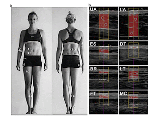
Fig. 1. B-Mode ultrasound measurement of uncompressed subcutaneous fat. (a) Sites for ultrasound measurement of subcutaneous adipose tissue (SAT) patterning: upper abdomen (1, UA), lower abdomen (2, LA), erector spinae (3, ES), distal triceps (4, DT), brachioradialis (5, BR), lateral thigh (6, LT), front thigh (7, FT) and medial calf (8, MC). Body height was used as the reference length for all distances. A detailed description of the standardized marking and of the ultrasound measurement technique can be found in M€uller et al. (2016). (b) Ultrasound images and evaluations of SAT thicknesses. Participant A: male, body mass index 5 25.5, body mass 5 80.7 kg, stature 5 1.78 m. (c) Survey plot of SAT patterning according to (b). The columns represent the mean values of the semi-automatic multiple thickness measurements for the eight ultrasound sites. The mean thickness value of the SAT thickness in a given ultrasound image (within the region of interest) is termed dINCL (gray) when fibrous structures are included and dEXCL (black) when fibrous structures are subtracted. Sum of the eight thicknesses DINCL 5 44.3 mm (DEXCL 5 39.8 mm). (d) Survey plot of a participant B with similar body mass index. Body mass index 5 25.4 kgm22 ; body mass 5 75.5 kg, stature 5 1.72 m. The SAT thickness sum was 86.9 mm (77.9 mm), 96% higher than in participant A.
(E-mail: wolfram.mueller@medunigraz.at) 2016 The Authors. Published by Elsevier Inc. on behalf of World Federation for Ultrasound in Medicine & Biology. This is an open access article under the CC BY-NC-ND license (http://creativecommons.org/licenses/by-nc-nd/4.0/).
Key Words: Body composition, Subcutaneous adipose tissue, Overweight, Obesity, Ultrasound measurement precision.
How can unnecessary ABUS recalls be avoided?
By Kate Madden Yee, AuntMinnie.com staff writer
April 16, 2018 -- Automated breast ultrasound (ABUS) can yield lower recall rates than handheld ultrasound, but unnecessary recalls still occur -- and it's important to find ways to reduce them, according to research presented at the recent American Institute of Ultrasound in Medicine (AIUM) annual meeting in New York City.
Why? Because having fewer recalls leads to fewer unnecessary biopsies and also less-stressed patients, lead researcher Dr. Beverly Hashimoto of Virginia Mason Medical Center in Seattle told AuntMinnie.com via email.

Dr. Beverly Hashimoto of Virginia Mason Medical Center.
"One of the weaknesses of screening breast ultrasound is that it results in a high number of women being called back for additional imaging," Hashimoto said. "When we started our automated breast ultrasound program, we wanted to determine if there were lessons we could learn from reviewing our ultrasound callbacks to reduce the number of patients recalled in the future."
Reducing recalls
ABUS offers benefits over handheld ultrasound for breast cancer screening, in part because it reduces the modality's dependency on operator skill. But the technology doesn't completely eliminate screening downsides such as unnecessary recalls, study presenter Elissa Picozzi from the University of Washington told session attendees.
She and her colleagues examined 18 months of their experience with screening ABUS. All patients included in the study were asymptomatic and had heterogeneous or extremely dense breast tissue. The researchers focused their attention on patients who were called back for additional imaging due to a category 0 ABUS screening assessment ("incomplete").

Elissa Picozzi from the University of Washington.
At Virginia Mason, patients called back for a possible ABUS abnormality undergo a diagnostic handheld ultrasound exam; if this exam is a category 4 or 5 (suspicious or highly suspicious of malignancy), then the woman has an ultrasound-guided core biopsy. The researchers reviewed ABUS images, handheld ultrasound images, and any pathology results from biopsy.
In that first year and a half of the medical center's screening ABUS practice, 867 exams were performed. Of these, 229 patients (26%) were called back for a possible ABUS lesion and had a diagnostic handheld ultrasound exam; 69 (8%) had biopsies.
Of the 229 patients called back, 222 were recalled for category 2 or 3 lesions. Results from the handheld ultrasound exams in these 222 patients were as follows:
- 68 patients (30.6%) with solid hypoechoic masses
- 71 patients (32%) with no abnormality -- the "lesions" on ABUS were actually artifact shadowing
- 35 patients (15.8%) with nonsimple cysts
- 20 patients (9%) with simple cysts
- 28 patients (12.6%) with miscellaneous lesions, including duct ectasia, scars, fat lobules, fat necrosis, or lymph nodes
The 69 biopsies revealed seven malignancies; of these, four were found only on ABUS. The cancer detection rate for ABUS was 4.6 per 1,000 cases.
Better evaluation?
Hashimoto and colleagues found that image artifacts masquerading as lesions were a frequent cause of unnecessary recalls. Many of these recalls could be avoided by using alternative reconstruction techniques and more careful consideration of different ABUS views, they noted.
"We were surprised to find that the second most common reason for calling a patient back was artifactual ultrasound shadowing," she told AuntMinnie.com. "As a result, we have developed techniques to better analyze shadowing on screening ultrasounds. We also discovered that many of the solid masses that were called back could have been resolved by changing our reviewing process of prior exams."
Thứ Bảy, 7 tháng 4, 2018
NAFLD and Ultrasound Elastography
Abstract
An increasingly common cause of chronic liver disease in
adults and children is nonalcoholic fatty liver disease (NAFLD). The diagnosis
of NAFLD was traditionally based on the histopathological changes of the liver,
evaluated by needle liver biopsy, an invasive method, with potential adverse
effects and great inter and intraobserver variability. The noninvasive methods
for the assessment of both fibrosis and steatosis in patients with NAFLD have
increasingly been studied lately. Of these noninvasive methods, in this
chapter, we will focus on the methods assessing the stiffness of liver
parenchyma, i.e. elastographic methods, of which, the most widely used are
ultrasound elastography techniques. We will discuss the principal elastographic
methods of some utility in NAFLD, i.e. shear wdave elastography (SWE)
(quantitative elastography), and especially transient elastography, point SWE
(acoustic radiation force impulse elastography, ARFI) and two-dimensional
real-time SWE (Supersonic). For each method usable in NAFLD cases, we will
review the method principle, examination technique and performance in NAFLD
evaluation.
Keywords: nonalcoholic fatty liver disease, fibrosis,
steatosis, noninvasive, elastography
SSI, Fibroscan® or ARFI?
After comparing the performance in the assessment of NAFLD
of the three elastographic methods discussed above, we can conclude, on the
preliminary results, that the diagnostic performance according to the AUROC
values for the diagnosis of significant fibrosis, severe fibrosis and cirrhosis
is good for SSI (0.86–0.89); good for Fibroscan® (0.82–0.87) and fair or good
for ARFI (0.77–0.84) . The AUROC values for diagnosing severe fibrosis or
cirrhosis are particularly good for SSI or Fibroscan® (0.86 and 0.89) [50]. The
prediction of steatosis, however, can at this moment only be made using the
controlled attenuation parameter measured with Fibroscan®. As for the causes of
measurement failure or unreliable results, we mention clinical factors related
to obesity (BMI > 30 kg/m2 , waist circumference ≥ 102 cm or increased wall
thickness), which are associated with liver stiffness measurement failures when
using SSI or Fibroscan® and with unreliable results when using ARFI [50]. In
conclusion, SSI, Fibroscan® and ARFI are valuable diagnostic tools for the
staging of liver fibrosis in NAFLD patients. However, the diagnostic accuracy
of SSI appears to be superior to that of ARFI for the diagnosis of F2 or above. Most of the cut-off values for SSI for the diagnosis of different stages
of liver disease are quite similar to those of Fibroscan®; this is an issue of
great importance for the applicability of this technique and its wide
dissemination among radiologists and hepatologists in their daily practice.
However, as for the M probe of Fibroscan®, the SSI technique remains limited by
a high failure rate in cases of obesity, whereas ARFI has a high rate of
unreliable results.
Thứ Tư, 4 tháng 4, 2018
POCUS helps handle out-of-hospital cardiac arrest
By Kate Madden Yee, AuntMinnie.com staff writer
April 2, 2018 -- Point-of-care ultrasound (POCUS) shows promise as a tool for emergency responders to help predict survival and guide interventions for cardiac arrests experienced outside of the hospital, according to research presented at the recent American Institute of Ultrasound in Medicine (AIUM) meeting in New York City.
POCUS is becoming more commonplace as a tool used for resuscitation of patients in cardiac arrest in the hospital, but there are not much data on its use outside of the hospital setting, presenter Dr. Katherine German of the University of Pittsburgh told session attendees.
"We know that out-of-hospital cardiac arrest survival is low, and we were curious whether point-of-care ultrasound could be a useful tool to predict survival and guide therapy or the decision to transport in the field," German said.
A helpful tool
German and colleagues explored the feasibility of POCUS when used by emergency staff in patients experiencing cardiac arrest outside of hospitals. A portable ultrasound unit (iViz, Fujifilm SonoSite) was used in vehicles as part of the Pittsburgh Emergency Medical Services (EMS) system; all participating physicians received POCUS education and watched an instructional video on how to use the device. Physicians provided radio and in-person consultation to Pittsburgh city paramedics.
The field staff used ultrasound to identify cardiac motion during pulse checks -- without interrupting resuscitation -- and to guide management of the situation at the physicians' discretion. The POCUS users recorded data from still images and six-second video clips, as well as through electronic charting within the patient care report using emsCharts, a tool for air and ground emergency medical personnel.
The study's primary purpose was to track the frequency of POCUS use during out-of-hospital cardiac arrest events; the secondary purpose was to have an expert faculty reviewer analyze images acquired during these events and to identify any barriers to the use of the technology.
German's group included 348 field responses that occurred between November 2016 and March 2017. Of these, 127 were out-of-hospital cardiac arrest. Field doctors used POCUS in 56 (44%) of the out-of-hospital cardiac arrest cases, with 48 (85%) still images and 34 (61%) video images recorded and reviewed.
Agreement between interpretations of emergency responder physicians and those of expert reviewers was found in 91% of video cases. Responders noted the following reasons for not using POCUS:
- Return of spontaneous circulation (ROSC) close to physician arrival (30%)
- Emergency personnel busy with other interventions (21%)
- Provider preference (23%)
- Mechanical issues (6%)
- Cessation of resuscitation per advanced directives (6%)
In the field
German did note some of the study's limitations, including the user dependency of ultrasound, as well as the fact that POCUS use was optional and that interpretations were recorded after the technology was used in the field. She also cited the small sample size of video images.
However, overall, POCUS showed promise as a tool for dealing with cardiac arrest in the field, she concluded.
"POCUS use by novice physician ultrasonographers to detect cardiac wall motion in out-of-hospital cardiac arrest is not only feasible, it also correlates with expert interpretation," German said. "Barriers to its use can be addressed through educational interventions and increased familiarity with the device."
Thứ Năm, 29 tháng 3, 2018
Về sử dụng trí tuệ nhân tạo trong phân loại xơ hóa gan của siêu âm
DOWNLOAD 2 bài sau theo link
DEEP LEARNING on LIVER FIBROSIS
https://static.livemedia.gr/livemedia/documents/al22186_us75_20180323092028_op25_icim2018.pdf
DEEP LEARNING on FATTY LIVER [NAFLD]
https://static.livemedia.gr/livemedia/documents/al22186_us75_20180323092030_op26_icim2018.pdf
Như tin bài trước từ AuntMinnie, nhóm nghiên cứu Hy lạp [Greece] dẫn đầu bởi Athanasios Angelakis, PhD, từ National Technical University of Athens, đã tạo một deep neural network có thể xác định bệnh nhân có đang trong giai doạn sớm gan hóa xơ không hoặc đã hóa xơ gan rõ. Qua kiểm chứng thuật toán này có mức hoàn thiện cao hơn thuật toán [machine learning] đã công bố trên y văn.
Thuật toán đã công bố chưa có AUC [area under the curve] quá 0.87.
Nhóm Athenes, Greece dùng deep learning để phân loại cho dữ liệu của 103 bệnh nhân viêm gan mạn có sinh thiết xác chẩn giai đoạn hóa xơ gan= 62 ca có Metavir scores F0 hay F1, còn lại 41 ca có scores ít nhất là F2. Mạng deep-learning network sử dụng thông số giới tính và 4 thông số hình thái= dài cắt dọc gan T, thùy đuôi, gan P cũng như lách. Thuật tóan này sẽ đánh giá phân loại hóa xơ gan nhỏ hơn F1 hoặc lớn hơn F 2 (≤ F1 or ≥ F2).
Thông tin này đặc biệt có lợi cho người sử dụng còn thiếu kinh nghiệm về siêu âm tổng quát và siêu âm đàn hồi shearwaves.
Sau khi đánh giá chéo 10 lần, thuật toán deep learning mới về phân loại hóa xơ gan bằng siêu âm này có độ chính xác cao hơn
- độ nhạy: 90.2%
- độ chuyên biệt: 91.9%
- giá trị tiên đoán dương: 88.1%
- giá trị tiên đoán âm: 93.4%
- AUC: 0.9126
Tất nhiên bệnh nhân cần được đánh giá thêm thuật toán này.
Trong tương lai:
Bước cuối cùng của thuật toán deep-learning là cần kết hợp thêm tuổi và giới tính trong phần dân số [demographic data] nghiên cứu, và trong phần morphologic, hemodynamic, biochemical, image analysis,[hình thái, huyết động học, hóa sinh, tạo hình] và dữ liệu siêu âm đàn hồi [elastographic data]. Với thuật toán này, nên có thêm nhiều lớp [layers], không chỉ phân loại hóa xơ gan mà còn về gan thấm mỡ, cao áp tĩnh mạch cửa, và hội chứng viêm.
Đã có thuật toán và mô hình cho vấn đề gan thấm mỡ .
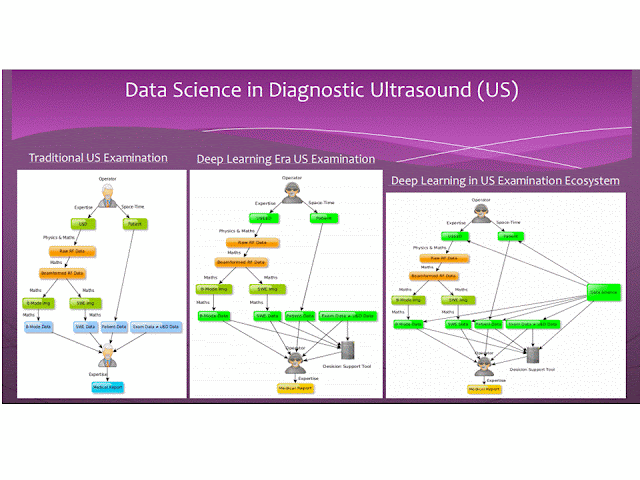
Deep learning và data science sẽ giữ vai trò quan trọng của siêu âm trong tương lai. Trong 10 năm tới hoặc ít hơn, data science sẽ được cài đặt trong máy siêu âm và sẽ sử dụng ngay trong tiến trình đánh giá siêu âm gan.
----------------
Thứ Tư, 28 tháng 3, 2018
AIUM: Can deep learning classify liver fibrosis on US?
By Erik L. Ridley, AuntMinnie staff writer
March 26, 2018 -- NEW YORK CITY - A deep-learning algorithm that analyzes B-mode morphological ultrasound data and patient demographic information can classify the severity of liver fibrosis in cases of chronic liver disease, according to research presented at the American Institute of Ultrasound in Medicine (AIUM) conference.
A team of researchers from Greece trained a deep neural network that could determine if patients were in the early stages of fibrosis or if they had advanced to significant fibrosis. In testing, the algorithm yielded a higher level of performance than existing methods previously reported in the literature, according to presenter Athanasios Angelakis, PhD, from the National Technical University of Athens.
A binary classification problem
Early-stage fibrosis can be reversed with treatment and prevented from reaching cirrhosis. It's also important to be able to diagnose significant fibrosis, classified as Metavir score F0 or F1, from more severe fibrosis (≥ F2).
"Given an individual's biomarkers, we would like to conclude if his or her fibrosis stage is either [Metavir score] ≤ F1 or ≥ F2," he said in a March 24 presentation "It's a significant binary classification problem."
Traditional methods used for this assessment include biopsy; biochemical tests; imaging, such as shear-wave elastography, strain elastography, and FibroScan transient elastography cutoff values; as well as image analysis or radiomic approaches. Recently, machine learning has also been applied in image analysis and radiomics. However, none of the biochemical, imaging, image analysis/radiomics, or machine-learning methods reported in the literature has yielded an area under the curve (AUC) from receiver operating characteristic analysis of more than 0.87, according to Angelakis.
The researchers sought to apply deep learning to this classification task. They used a dataset of 103 patients with chronic liver disease and biopsy-validated fibrosis stages; 62 patients had Metavir scores of F0 or F1, while 41 had scores of at least F2. The deep-learning network was given one demographic parameter (patient gender) and four morphologic parameters -- the longitudinal diameter of the left lobe, caudate lobe, and right lobe of the liver, as well as the spleen. The algorithm then provides its assessment of the patient's fibrosis class (≤ F1 or ≥ F2).
This information could be of particular benefit to inexperienced users, according to the researchers.
"It reduces the experience [needed] for the examiner [to perform these studies]," Angelakis said. "One of the goals of using deep learning in ultrasound in general and shear-wave elastography is to reduce this need for experience with the exam."
After performing tenfold cross-validation, the researchers found that the algorithm yielded a high level of accuracy for liver fibrosis classification:
- Sensitivity: 90.2%
- Specificity: 91.9%
- Positive predictive value: 88.1%
- Negative predictive value: 93.4%
- AUC: 0.9126
They acknowledged, though, that more patients are needed to further validate the algorithm.
The future
The ultimate holy grail for a deep-learning algorithm in the liver would be to incorporate patient demographic data such as gender and age, along with morphologic, hemodynamic, biochemical, image analysis, and elastographic data. Such an algorithm, which would have many layers, would not only classify fibrosis but also steatosis, portal hypertension, and inflammation, Angelakis said.
"We have already created algorithms and models that also attack the steatosis problem, because our datasets are bigger now," he said.
Deep learning and data science will likely take on a significant role in ultrasound in the future, according to Angelakis.
"The output from the [deep-learning] model will be one more input for the expert [to consider during interpretation]," he said. "That is how we reduce the expertise [requirement] for the reader. Looking ahead 10 years from now, maybe less, data science will also be used during the manufacturing of ultrasound devices and it will be in every [aspect of the ultrasound acquisition and interpretation process].
Thứ Ba, 27 tháng 3, 2018
NHÂN 03 CA DIFFUSE LARGE B CELL LYMPHOMA CÓ TRIỆU CHỨNG HÀM MẶT
Trong số 20 ca lymphom lan tỏa dòng tế bào B lớn [diffuse large B cell lymphoma] ở Trung tâm Y khoa Medic từ 2008, có 03 ca có triệu chứng đau răng và hàm mặt điều trị giảm đau, nhổ răng..không bớt, sau đó xuất hiện nhiều tổn thương khác ngoài hạch. Trong khi đó, tìm được một bài báo về đau xương hàm dưới trong lymphom ác. Sau đây là một hồi cứu về đề tài này.
and Appearences in Oral Cavity, and Maxillary Area
Nguyen Thien Hung,
Jasmine DCB Thanh Xuan, Le van Tai, Le
Thong Nhat, Le Thanh Liem, Phan Thanh Hai
Medic
Medical Center, HCMC, Vietnam
INTRODUCTION:
May there had been an initial symptom in oral cavity, maxillary
and mandibular area for diffuse infiltration of lymphoma? We will represent some cases of diffuse
lymphoma with appearences in oral
cavity, and maxillary areas.
MATERIALS and METHODS:
We retrospectively reviewed
20 cases of diffuse lymphoma that were histopathologically diagnosed in
our center from 2008 in which presented 03 cases with oral cavity, maxillary symptoms and bone
involvement [01 case in male 42 yo and 02 cases in female gender, 33 and 40 year-old].
RESULTS:
CASE 1= A 33 year-old female patient with pain in lower maxillary bone for one month and
tension in both 2 breats), hyperpigmented edema of areolar area both 2
sides without pregnancy. ABVS scanning
detected multiple nodules infiltrating in 2 breasts.
Biopsy of 2 breasts
reported microscopic with immunohistochemistry scanning, diffuse large B cell lymphoma.
MRI full body with gado
detected bone marrow changing, 2 breats hypercaptured
contrast, ascites and kidney infiltration. In pelvis 2 ovarian
tumors and big uterine cervix were detected. Blood tests showed lower
platelets, EGFR lower 46, beta2 microglobuline raised
3816, ferritin raised 911, LDH-l raised 1360.
CASE 2= A 40 year-old female patient with toothache but pain
remained after removing tooth, and in total body examination detected right
pleural effusion and some nodular tumors
in thyroid, left breast and subcutaneous area of right neck, left chest all
and lumbar region. Erosions of left
clavicle and right 1st rib. Biopsy of left breast tumor reported diffuse large B cell lymphoma.
CASE 3= Man 42 year-old, one month ago,
pain in oral sinus, difficult eating and 2 days after, pain appears in
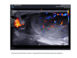
left testis. Ultrasound of left big and hot testis represented hypoechoic
infiltration, hypervascular of one
part of testis and elastoscan ultrasound value # 10.5 kPa).
DISCUSSION:
There are published reports of pain in mandibular area as initial symptom
but symptoms in oral cavity and maxillary bone as onset symptom seems to be not
represented in literature. But it exists a case of pain in oral cavity for
lymphoma infiltrating in testis and an another case of pain in maxillary area
for lymphoma in whole body in this report.Toothache and pain in maxillary region remained with management guided a survey in all body and detected extranodal
CONCLUSIONS:
Seveval reports of
lymphoma in mandible were published with pain as initial symptom, but in cases of diffuse infiltration of
lymphoma in whole body, the appearences in oral cavity, maxillary and mandibular
areas with pain symptom may be added criteria of diagnosis for lymphoma in
clinical examination.
REFERENCE
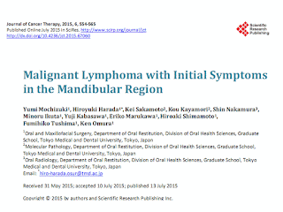
Mochizuki, Y., Harada, H., Sakamoto, K., Kayamori, K., Nakamura, S., Ikuta, M., Kabasawa, Y., Marukawa, E., Shimamoto, H., Tushima, F. and Omura, K. (2015) Malignant Lymphoma with Initial Symptoms in the Mandibular Region. Journal of Cancer Therapy, 6, 554-565
Abstract
Primary intraosseous lymphoma is rare and there are few case reports manifesting with a mass in the mandible. Thus, we retrospectively reviewed and analyzed the clinical characteristics, treatment, and outcome of extranodal non-Hodgkin’s lymphoma (NHL) with initial mandibular symptoms in our department. At initial treatment of dental clinics, dentists had diagnosed as dental or gingival diseases and had performed dental treatment. Neurological disorder to involvement of the inferior alveolar nerve was present in 80.0% of our cases. On dental or panoramic radiography a specific radiolucent lesion in the mandible was not detected, except for dental lesions. On CT, NHL of the mandible region has no widening and no clear destruction but a slit-like the cortex bone destruction pattern with keeping in shape of the mandibular body (62.5% of CT-examined cases), and extraosseous soft tissue mass are clearer on MRI (100.0% of MRI-examined cases).
Histopathologically, 80.0% of our cases were diagnosed as diffuse large B cell lymphoma (DLBCL).
One case as B-cell lymphoblastic lymphoma and one case as B-cell lymphoma unclassifiable with features intermediate between DLBCL and Burkitt lymphoma were Stage IV (Ann Arbor staging system) and had poor prognosis. The disease-specific survival rate was 77.8% at 5 years. If unexplained non-specific symptoms such as swelling of the jaw, pain, neurological disorder of the inferior alveolar nerve, tooth mobility are observed, oral surgeons and dentists should not perform
dental treatments. CT and MRI show disease specific appearance to be able to give a definitive diasnosis as NHL. PET/CT is useful for scaninng of whole body. A deep bone biopsy is preferred for
suspected malignant lymphoma.
Thứ Bảy, 24 tháng 3, 2018
Đăng ký:
Bài đăng
(
Atom
)

