Tổng số lượt xem trang

Thứ Sáu, 4 tháng 5, 2018

US features help define thyroid nodules in children


By AuntMinnie.com staff writers
May 3, 2018 -- Certain ultrasound characteristics can help clinicians identify malignant thyroid nodules in children, according to a study published online May 1 in Radiology
A team led by Dr. Danielle Richman of Brigham and Women's Hospital used data from 404 thyroid nodules in 314 patients (age range: 2-18 years) between January 2004 and July 2017. All nodules received ultrasound-guided fine-needle biopsy; the researchers recorded age, sex, background appearance of the thyroid gland for each patient, sonographic characteristics, and pathologic diagnosis for each nodule.
Of all the nodules, 77 (19.1%) were malignant, and most of these were papillary thyroid carcinoma (88.3%). The likelihood of malignancy did not differ by patient sex or puberty status, Richman and colleagues found. The cancer rate in children with a solitary nodule was higher than in those with multiple nodules (29.4% versus 14.2%, p = 0.003).
The researchers also identified a number of sonographic characteristics associated with malignant nodules, such as larger size, solid parenchyma, taller-than-wide shape, presence of speckled calcifications, lack of a smooth margin, and presence of abnormal lymph nodes.
"In children with thyroid nodules ... [these characteristics] raise concern for malignancy," they concluded.

Thứ Tư, 2 tháng 5, 2018

Automated 3D US effective for diagnosing hip dysplasia


By Kate Madden Yee, AuntMinnie.com staff writer
April 30, 2018 -- Automated 3D ultrasound is just as effective as 2D ultrasound for diagnosing developmental dysplasia of the hip (DDH) in infants: In fact, it reduces the number of studies characterized as borderline by more than two-thirds, according to research published online April 24 in Radiology.
The study's findings suggest that automated 3D ultrasound could serve as an even more effective tool for diagnosing this condition -- which, if untreated, can cause long-term damage, wrote the team led by Dornoosh Zonoobi, PhD, from the University of Alberta in Edmonton, Canada.
"Three-dimensional ultrasound has potential advantages in feasibility in a screening setting for hip dysplasia because the 3D indexes of dysplasia are calculated automatically from surface models generated with minimal user input, or potentially completely automatically calculated by using deep-learning tools," the researchers wrote.
Developmental hip dysplasia in infants is associated with premature osteoarthritis later in life, and it is the cause of 30% of hip arthroplasties in patients younger than 60. The condition is usually treated in infants with a harness, and a prompt and accurate diagnosis reduces its negative long-term effects. 2D ultrasound has long been used to identify DDH, but it has limitations, including operator variability and overdiagnosis.
3D ultrasound overcomes these limitations because it offers a more complete view of hip geometry than 2D ultrasound and also because it is automated, Zonoobi and colleagues wrote. The modality was first suggested for the diagnosis of DDH in the 1990s, but only recently has transducer technology evolved enough to make the use of 3D ultrasound for this application feasible.
For their study, Zonoobi and colleagues added 3D ultrasound to conventional 2D ultrasound exams of 1,728 infants (mean age, 67 days) to evaluate the children for DDH; the exams were performed between January 2013 and December 2016. Custom software automatically calculated measures such as 3D posterior and anterior alpha angle and osculating circle radius. Of the infants imaged, 1,347 were normal, 140 were borderline for the condition, and 241 were dysplastic.
The researchers found that 3D ultrasound helped correctly categorize 97.5% of the dysplastic and 99.4% of the normal hips, and no dysplastic hips were categorized as normal. 3D ultrasound provided a correct diagnosis in 69.3% of cases categorized as borderline at initial 2D ultrasound. The modality also reduced the number of borderline diagnoses to 39, compared with 140 with 2D ultrasound.
The study results justify generalized implementation of 3D ultrasound for DDH diagnosis in clinical settings, Dr. Diego Jaramillo of Nicklaus Children's Hospital in Miami wrote in an editorial that accompanied the study.

Thứ Sáu, 27 tháng 4, 2018

Spinal Pain in Space and US


By Kate Madden Yee, AuntMinnie.com staff writer
April 26, 2018 -- Astronauts armed with a compact ultrasound system successfully performed scans on each other while on the International Space Station. The scans were part of a study to assess spinal changes during long-term spaceflight that could lead to back pain, researchers wrote in the April issue of the Journal of Ultrasound in Medicine.

A group from Henry Ford Hospital worked with NASA to train astronauts on the International Space Station to use ultrasound for imaging the spines of their colleagues during flight. The researchers found that it was feasible to teach these novice users to use ultrasound effectively for this purpose. In addition, the data collected could help in the development of countermeasures to protect astronauts' spines during spaceflight, as well as the creation of protocols for treating injury once the astronauts have returned.
"Focused ultrasound monitoring of the spine for longitudinal changes during long-duration spaceflight may influence additional strategies or nutrition/drug therapies to reduce disk degeneration," lead author Kathleen Garcia and colleagues wrote. "[Our] study demonstrates a potential role for ultrasound in evaluating spinal integrity and alterations in the extreme environment of space."
Aches and pains
Starting with the Apollo program and continuing into the International Space Station era, moderate to severe back pain has been a common medical complaint among astronauts, corresponding author Dr. Scott Dulchavsky, PhD, told AuntMinnie.com.
"When there's no gravity, the spine loosens, making it less stable and putting stress on muscles and ligaments," he said. "The spine can actually elongate by as much as three inches, and that puts astronauts at higher risk of problems when they return."
MRI and CT are the clinical standards for spinal imaging, but they aren't available in space. Ultrasound can be carried on space vehicles thanks to its compact size, but a framework for imaging spinal structures in space hasn't been clearly formulated, Garcia's team wrote.
Dr. Scott Dulchavsky, PhD
Dr. Scott Dulchavsky, PhD, from Henry Ford Hospital.
To address this problem, the researchers developed an ultrasound protocol for spaceflight, and they investigated whether astronauts on the International Space Station could effectively perform ultrasound assessments of the lumbar and cervical regions of the spine. Seven astronauts participated in the study and served as both ultrasound operators and research subjects; two additional crew members were trained as backup operators. The exams were read remotely, and the researchers then compared these in-flight results with preflight and postflight MRI and ultrasound exams (J Ultrasound Med, April 2018, Vol. 37:4, pp. 987-999).
The astronauts were trained six months before their mission via an online program that included a review of spinal anatomy, procedure demonstrations, equipment setup orientation, and a software review, as well as a one-hour, hands-on session during which they alternated between patient and operator roles. The exams were conducted with GE Healthcare's Vivid q device, a laptop-sized ultrasound scanner. The astronauts were assisted remotely by experts at NASA's Lyndon B. Johnson Space Center in Houston.
When the astronauts underwent the exams, they were placed supine on a medical restraint system on board the space station. To evaluate the effects of a lack of gravity on the spine over time, each study participant had three in-flight ultrasounds: one at day 30, one at day 90, and one at day 150.
The astronauts easily obtained high-quality images of the lumbar and cervical vertebrae, the researchers found. Overall success rates for image acquisition were 95% in the lumbar spine and 90% in the cervical spine. In addition, there was "no appreciable difference in success rates for either image acquisition or image quality between expert operators and astronaut crew members in the lumbar and cervical regions," they wrote.
The study findings fill in a data gap, according to Garcia and colleagues.
"Given the previous void of in-flight spinal imaging capabilities in space, to our knowledge, this study represents the first attempt to monitor microgravity-associated acute changes to the spine while they are occurring," they wrote.
Greater purpose
One of the benefits of this kind of research is that the findings can influence healthcare on Earth, according to Dulchavsky.
"By putting smart people into constrained environments like space, we can find solutions to health problems that can be used beyond the space station," he said. "Our work here found not only that nonphysicians can be trained to effectively use imaging devices, but it also pointed to further research on exercise and dietary regimens that could help keep the spine healthy in patients on Earth."
As the U.S. sets its sights on sending astronauts on longer missions -- such as to Mars -- understanding how the human body is affected by space is crucial, Garcia and colleagues wrote.
"As the duration of space missions continues to increase, [ultrasound's utility] will only gain importance in monitoring crew health and diagnosing disorders," the group concluded. "Further investigations should be performed to corroborate this imaging technique and to create a larger database related to in-flight spinal disorders during long-duration spaceflights.

Chủ Nhật, 22 tháng 4, 2018

PROTOCOLS for SHOCK

LINK DOWNLOAD
PROTOCOLS FOR SHOCK


Shock is a major morbidity in emergency and critical care and is also one of the important prognostic factors affecting  in-hospital mortality [1]. Timely diagnosis and treatment of shock reduce the length of stay (LOS) and mortality rate at the emergency department (ED). The early use of the point of care ultrasound (POCUS) can reduce the diagnostic time as well as increase the accuracy of diagnosis [2]. The first protocol Undifferentiated hypotension protocol (UHP) was release on 2001, [3]; and more than 15 subsequent protocols were developed [4].

Future and discussion
Most currently available protocols are focused on diagnosis.Blanco et al. suggested that an ultrasound scan should also assess the efficacy of treatment [5]. For example, the use of velocity time integral (VTI) of LVOT to measure the responses to fluid and inotropic agent to help further management.
Point of care ultrasound is a powerful tool in emergency setting. The emergency ultrasound procedures for shock can reduce the LOS at the emergency department.
Therefore, POCUS is an essential skill for personnels in the emergency department and critical care units.

PROTOCOLS for SHOCK

Thứ Bảy, 21 tháng 4, 2018

CẮT TÚI MẬT NỘI SOI: TIÊU CHUẨN SIÊU ÂM TIÊN LƯỢNG KHÓ


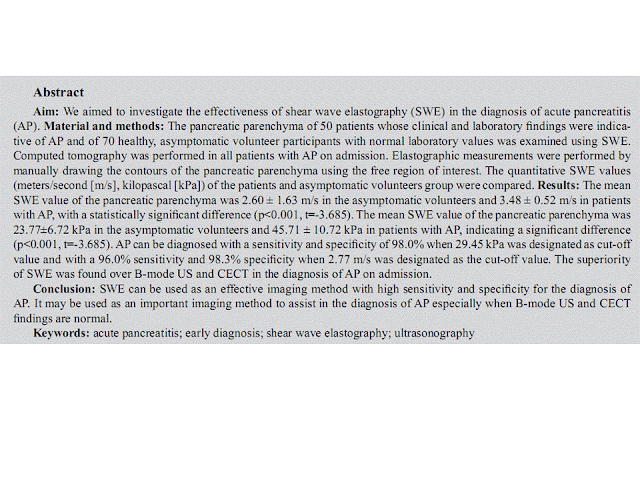
Abstract

Purpose: Laparoscopic cholecystectomy (LC) has become the treatment of choice for cholelithiasis. Still some patients required conversion to open cholecystectomy (OC). Our aim was to develop a standardized Ultrasound based scoring system for preoperative prediction of difficult LC.

 Methods and materials: Ultrasound findings of 300 patients who underwent LC were reviewed retrospectively. Four parameters (time taken, biliary leakage, duct or arterial injury, and conversion) were analyzed to classify LC as easy or difficult. The following ultrasound findings were analyzed: GB wall thickness, pericholecystic collection, distended GB, impacted stones, multiple stones, CBD diameter and liver size. Out of seven parameters, four were statistically significant in our study. A score of 2 was assigned for the presence of each significant finding and a score of 1 was assigned for the remaining parameters to a total score of 11. A cut-off value of 5 was taken to predict easy and difficult LC.
 Results: 66 out of 83 cases of difficult LC and 199 out of 217 cases of easy LC were correctly predicted on the basis of scoring system. A score of >5 had sensitivity 80.7% and specificity 91.7% for correctly identifying difficult LC. Prediction came true in 78.8% difficult and 92.6% easy cases. US findings of GB wall thickness, distended GB, impacted stones and dilated CBD were found statistically significant.
Conclusion: This indigenous scoring system is effective in predicting conversion risk of LC to OC. Patients having high risk may be informed and scheduled appropriately and decision to convert to OC in case of anticipated difficulty may be taken earlier.

ª 2017, Elsevier Taiwan LLC and the Chinese Taipei Society of Ultrasound in Medicine. This is an open access article under the CC BY-NC-ND license (http://creativecommons.org/licenses/ by-nc-nd/4.0/).

TI-RADS REDUCES BIOPSY

Thyroid Imaging Reporting and Data System Reduces Biopsies

Apr 18, 2018
Criteria from the American College of Radiology (ACR) Thyroid Imaging Reporting and Data System (TI-RADS) offers a meaningful reduction in the number of thyroid nodules recommended for biopsy, according to a study published in the journal Radiology
Researchers from several states performed a retrospective study to compare the biopsy rate and diagnostic accuracy before and after applying ACR TI-RADS criteria for thyroid nodule evaluation. Eight radiologists with three to 32 years of experience in thyroid ultrasonography were asked to review the ultrasound features of 100 thyroid nodules that were cytologically proven and/or pathologically proven. Nodules evaluated in five US categories and biopsy recommendations were provided based on the radiologists’ practice patterns without knowledge of ACR TI-RADS criteria. Three other expert radiologists were reference standard readers for the imaging findings. ACR TI-RADS criteria were retrospectively applied to the features assigned by the eight radiologists to produce biopsy recommendations. Comparison was made for biopsy rate, sensitivity, specificity, and accuracy.
The results showed 15 of the 100 nodules (15 percent) were malignant. The mean number of nodules recommended for biopsy by the eight radiologists was 80 ± 16 (standard deviation) based on their own practice patterns and 57 ± 11 with retrospective application of ACR TI-RADS criteria.
Without ACR TI-RADS criteria:
Sensitivity  95 percent
Specificity  20 percent
Accuracy  28 percent
With ACR TI-RADS criteria:
Sensitivity 92 percent
Specificity 44 percent
Accuracy 52 percent
Expert consensus:
Sensitivity 87 percent
Specificity 51 percent
Accuracy 56 percent

The researchers noted that although fewer malignancies were recommended for biopsy with ACR TI-RADS criteria, the majority met the criteria for follow-up US. Only three of 120 (2.5 percent) malignancy encounters required no follow-up or biopsy. Expert consensus recommended biopsy in 55 of 100 nodules with ACR TI-RADS criteria.
Not only did the ACR TI-RADS criteria offer a meaningful reduction in the number of thyroid nodules recommended for biopsy, the researchers wrote, they significantly improve the accuracy of recommendations for nodule management.