Tổng số lượt xem trang

Thứ Sáu, 4 tháng 6, 2021

SHORTER LUNG U S exams can help kidney disease patients


By Amerigo Allegretto, AuntMinnie.com staff writer

June 4, 2021 -- Lung ultrasound with an abbreviated scanning protocol can efficiently diagnose pulmonary congestion caused by fluid overload in hemodialysis patients, according to research published June 3 in the American Journal of Kidney Diseases.


team of researchers led by Dr. Nathaniel Reisinger from the University of Pennsylvania found that several abbreviated lung ultrasound protocols that focus on a limited number of lung zones (4, 6, or 8-zone) performed similarly to comprehensive 28-zone studies among patients with kidney failure on hemodialysis seeking care in an emergency department.

The researchers also did not find any differences in mortality between patients with no-to-mild and moderate-to-severe pulmonary congestion.

"Abbreviated lung ultrasound protocols take less time, are less bothersome to patients, and are nearly as accurate," Reisinger told AuntMinnie.com.

Pulmonary congestion from fluid overload is common among patients with kidney failure on hemodialysis and contributes to excess morbidity and mortality in patients. The team said that physical examination is an insensitive approach to detecting pulmonary congestion.

Lung ultrasound with a comprehensive 28-zone protocol has been shown to be sensitive for detecting pulmonary congestion, according to previous research. However, the 28-zone technique requires complete disrobing of the patient and longer scanning times for operators.

"In the constrained environment of the emergency department, this commitment is prohibitive to both patients and providers," the authors wrote.

Reisinger and his team wanted to see if abbreviated forms of lung ultrasound could produce similar results while saving time for operators and providing a more comfortable experience for patients.

The group started with research by Buessler and colleagues that studied the use of four-, six-, and eight-zone scanning for heart failure patients, as well as mapped the specific lung zones that would be most equivalent to a 28-zone study. The researchers noted that previous research has found that the regional distribution of pulmonary edema on imaging based on clinical condition has been well-demonstrated.

They then tested the protocol in 98 patients in the U.S. with kidney failure on hemodialysis participated, with a follow-up time of 30 days. Out of those, 84 were African American and 97 were non-Hispanic or Latino in ethnicity.

The team found high correlation and good agreement between 28-zone and abbreviated lung ultrasound studies. Each of the short-form studies was able to discriminate between no-to-mild pulmonary congestion versus moderate-to-severe pulmonary congestion when compared with the long-form 28-zone study, the authors wrote.

For short-form studies, the highest sensitivities were seen in 6C, 8C, and 8D zone ultrasound studies at 93.0%, 95.8%, and 98.6% respectively. Meanwhile, 6C and 8A studies had the highest specificities, at 96.3% and 100% respectively. Finally, 6C, 8C, and 8D studies had the highest area under the curve (AUC), at 0.95, 0.94, and 0.94 respectively.

Of the four-zone studies, lung zone 4C had the highest sensitivity at 90.1%, specificity at 92.6%, and AUC at 0.91.

"With AUC values of greater than 0.88, any of the 8-zone studies clearly seems sufficient for clinical use," the study authors wrote. "Similarly, the 4C pattern can easily be performed without removing a patient's shirt, while still achieving a point estimate of AUC greater than 0.90, demonstrating accuracy better than the current standard-of-care assessment of pulmonary congestion."

However, Reisinger said that despite the promise, abbreviated lung ultrasound protocols are "slightly less accurate" than more comprehensive tests and may miss focal pathology such as pneumonia.

He told AuntMinnie.com that the research team is looking at randomized controlled trials as the next step in investigating whether lung ultrasound-guided ultrafiltration therapy improves outcomes in patients with end-stage kidney disease chronically on hemodialysis.
Thứ Tư, 2 tháng 6, 2021

A I can help stratify COVID-19 risk on lung ultrasound

By Erik L. Ridley, AuntMinnie.com staff writer


June 1, 2021 -- Deep-learning algorithms can be used to automatically provide risk scores on lung ultrasound exams in COVID-19 patients, researchers from Italy reported in an article published online May 27 in the Journal of the Acoustical Society of America.


 A multi-institutional team of researchers trained deep-learning models to automatically score and provide semantic segmentations of lung ultrasound exams in COVID-19 patients. In testing, the deep-learning algorithms yielded a high level of agreement with lung ultrasound experts for stratifying patients as having either low- or high risk of clinical worsening, according to the group led by corresponding author Libertario Demi, PhD, of the University of Trento.

"These encouraging results demonstrate the potential of [deep-learning] models for the automatic scoring of [lung ultrasound] data, when applied to high-quality data acquired according to a standardized imaging protocol," the authors wrote.

Although lung ultrasound has been shown to be useful for evaluating COVID-19 patients, the modality is often limited to the visual interpretation of ultrasound data. As a result, there are concerns over reliability and reproducibility, according to the researchers.

Following up on prior work to develop standard lung ultrasound protocols and training deep-learning algorithms for COVID-19 patients, the researchers wanted to compare the performance of their models with that of expert clinicians. The first algorithm labels each video frame with a score, while the second provides semantic segmentation -- assigning one or more scores to each frame.

They used their algorithms to evaluate 314,879 ultrasound video frames from 1,488 lung ultrasound videos on 82 COVID-19 patients. The ultrasound exams were acquired using multiple ultrasound scanner types at the Agostino Gemelli University Hospital Foundation in Rome and the San Matteo Polyclinic Foundation in Pavia, Italy. None of the images were used in training the models.

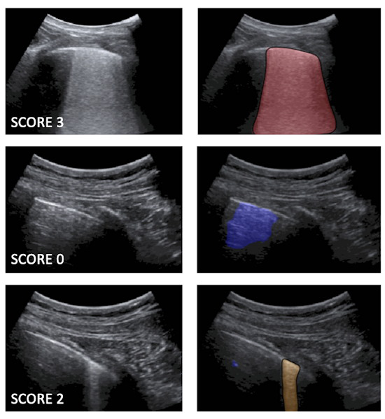
Examples of lung ultrasound images corresponding to score 3, score 0, and score 2
Left: Examples of lung ultrasound images corresponding to score 3 (top), score 0 (middle), and score 2 (bottom). Right: the corresponding semantic segmentations obtained with the described deep-learning algorithms. Color maps are informative of the score level (red for score 3, blue for score 0, orange for score 2), as well as indicate the relevant part of the image that determines the score. These maps explain the decision progress of the algorithms. Images and caption courtesy of Libertario Demi, PhD.

The algorithm's scores for each frame are then used to generate an aggregate score for each video. The researchers tested two methods for producing an aggregate score: one based only on the labeled video frames and another that combined the labeled frames with the segmented frames.

These video-level scores were then compared with the risk scores provided by expert clinicians.

AI algorithm performance for risk stratifying COVID-19 patients
 AI risk scores based on only labeled framesAI risk scores based on combination of labeled and segmented frames
Agreement with expert clinicians83.3%86%

Although the combined approach generally outperforms the first method, the improvement isn't significant. What's more, the first method seems more stable to threshold variations, according to the researchers.

"Therefore, the first approach can be considered as a self-standing strategy to classify [lung ultrasound] videos starting from a frame-based classification," the authors wrote. "Nevertheless, the semantic segmentation remains essential for clinicians, as it provides the explainability of the decision by highlighting the specific [lung ultrasound] patterns."

In future work, the researchers plan to expand their existing database and train deep-learning algorithms on video-labeled data instead of just frame-based labeled data. That approach would be more consistent with the process used by clinicians when evaluating lung ultrasound videos, they said.