Tổng số lượt xem trang

Thứ Tư, 29 tháng 9, 2021

Lung Ultrasound in Patients with Coronavirus COVID-19 Disease, Dirk-André Clevert, MD


Summary of Coronavirus COVID-19 examination and follow-up


In comparison between non-COVID-19 pneumonia and COVID-19 pneumonia, COVID-19 pneumonia is more likely to have a peripheral distribution [84] (Figure 23). In addition to HR-CT scan and X-ray of the lung, ultrasound can also be used for the diagnosis and follow-up of the disease [85]. By using a curved transducer (5–1 MHz), the morphology and changes of subpleural lesions are clearly displayed. Due to the option to use even the low-frequency of the transducer, changes of air and water contents in consolidated peri-pulmonary tissues and an air bronchogram sign can be depicted (Figure 24).


The blood supply and lesion progression in peripulmonary consolidation can be monitored by using  the color or power Doppler technique [86]. Currently, ultrasound of the lung is limited in the diagnosis and treatment of central lung diseases due  to the attenuation of sound waves by normal lung and bone tissues. The diagnosis of lung pathologies relies  on the artifacts of peri-pulmonary lesions [87–88]. The artifacts exist because of an abnormal ratio of air and water contents in alveoli and interstitial tissues. In order to improve the diagnostic ultrasound lung tool,  the use of an abdominal curved array probe (5–1 MHz) seems to be helpful. Typical for the COVID-19 disease  are the thickening of the pleural line with pleural  line irregularity. The pleural line could be unsmooth, discontinuous and interrupted [85, 89] (Figures 25–26).

The appearance of B-lines artifacts could vary from focal, to multifocal and confluent pattern. The consolidations could vary in different patterns, including multifocal small subpleural consolidations up to non-translobar and translobar with occasional air bronchograms [5]. Pleural effusions are uncommon in coronavirus COVID-19 disease.

An indirect sign for recovering is the appearance of A-lines during the recovery phase [85] (Figure 27).

In summary, in our experience, we consider that lung ultrasound will have a major utility for the management of COVID-19 pneumonia in the ICU due to its safety, repeatability, low cost and point of care use. HR-CT may be reserved in the follow-up if lung ultrasound is not  able to answer the clinical question. In our personal experience lung ultrasound could be used for rapid assessment of the severity of SARS-CoV-2 pneumonia,  to track the evolution of disease during follow-up and  to monitor lung recruitment maneuvers. Additional ultrasound can track the response to prone position and the management of extracorporeal membrane therapy [85]. With increased use of bedside ultrasound in the ICU, patients can be protected from unnecessary radiation and therapy delays. The transport of high-risk patients to X-ray examinations can be avoided.

From White Paper: Lung Ultrasound in Patient with Coronavirus-19 Disease, SIEMENS.
https://www.siemens-healthineers.com/ultrasound/lung-ultrasound-covid-19

Thứ Ba, 28 tháng 9, 2021

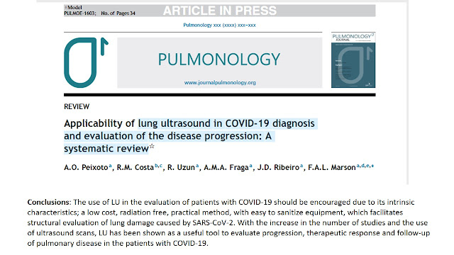
Applicability of lung ultrasound in COVID-19 diagnosis and evaluation of the disease progression: A systematic review


Abstract

Introduction

The COVID-19 pandemic originated in China and within about 4 months affected individuals all over the world. One of the limitations to the management of the COVID-19 is the diagnostic imaging to evaluate lung impairment and the patients’ clinical evolution, mainly, in more severe cases that require admission into the intensive care unit. Among image examinations, lung ultrasound (LU) might be a useful tool to employ in the treatment of such patients.

Methods

A survey was carried out on PubMed to locate studies using the descriptors: ((Lung ultrasound OR ultrasound OR lung ultrasonography OR lung US) AND (coronavirus disease-19 OR coronavirus disease OR corona virus OR COVID-19 OR COVID19 OR SARS-CoV-2)). The period covered by the search was November 2019 to October 2020 and the papers selected reported LU in COVID-19.

Results

Forty-three studies were selected to produce this systematic review. The main LU findings referred to the presence of focal, multifocal and/or confluent B lines and the presence of pleural irregularities.

Conclusions

The use of LU in the evaluation of patients with COVID-19 should be encouraged due to its intrinsic characteristics; a low cost, radiation free, practical method, with easy to sanitize equipment, which facilitates structural evaluation of lung damage caused by SARS-CoV-2. With the increase in the number of studies and the use of ultrasound scans, LU has been shown as a useful tool to evaluate progression, therapeutic response and follow-up of pulmonary disease in the patients with COVID-19.

Keywords

Image examination 
Lung disease 
Lung ultrasound 
SARS-CoV-2


Highlights

LU findings presented correlation with HRCT images.

LU can be used in respiratory system propaedeutics as an alternative to the “stethoscope use”. Special clothing and individual protection equipment are indispensable, since the manipulation of the stethoscope in pulmonary evaluation might create contamination risks for the health professionals and patients.

COVID-19 normally induces a bilateral and diffuse interstitial pneumonia with asymmetric lesions and uneven distribution, mainly involving the lung periphery, which makes it particularly suitable for investigation using LU.60

Studies have identified potential correlation between the LU patterns and the patients’ clinical outcome. One of the assays in this study reported that each pulmonary area could be in a different stage of the disease, therefore, the global evaluation of the lungs is fundamental.69

The POCUS allows for hemodynamic, cardiac and vascular evaluations (thromboembolic phenomena — deep venous thrombosis).

LU should be associated to the multisystem point-of-care exam, since the SARS-CoV-2 infection might be linked to myocarditis and a high incidence of thromboembolic events. Thus, multiorgan ultrasound evaluation in early treatment is useful to screening these complications at the bedside.

Perspectives

More studies on LU application in the pediatric population are necessary.

LU in COVID-19 score standardization.

Improvement of reading/automatic identification of B line software, as reported in this study is still needed.74

The advancement of the remote robotic ultrasound scanning technology assisted by the 5G network in real time by the use of big data, cloud storage and artificial intelligence must be improved.73