Tổng số lượt xem trang

Thứ Bảy, 16 tháng 7, 2022

Hepatorenal Index vs Fatty Liver Index in NAFLD and ALD.


Abstract

Objectives

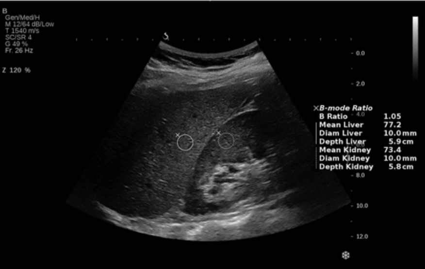
We aimed to evaluate the accuracy of the hepatorenal index by B-mode ratio to diagnose hepatic steatosis, compared to ultrasound steatosis score, controlled attenuation parameter, and the fatty liver index using histology as the gold standard.

Methods

We prospectively included participants with alcohol-related or nonalcoholic fatty liver disease for same-day noninvasive investigations and liver biopsy.

Results

We included 137 participants, 72% male, median age 60 years (53–65) and body mass index 32 kg/m2 (28–38). Eighty percent had steatosis (S0/S1/S2/S3 = 20/37/24/19%). B-mode ratio had moderate diagnostic accuracy for any steatosis (≥S1, area under the receiver operating characteristics curve [AUROC] = 0.79; 95% confidence interval 0.70–0.88), significant steatosis (≥S2, AUROC = 0.76; 0.66–0.85), and severe steatosis (=S3, AUROC = 0.74; 0.62–0.86), independent of disease etiology. The cutoff values to rule-out and rule-in any steatosis were 1.09 and 1.45. While B-mode ratio and controlled attenuation parameter correlated poorly, their diagnostic accuracies were comparable to each other and to ultrasound steatosis scoring. Fatty liver index did not differ from B-mode ratio in detecting any steatosis but had poor accuracy to detect higher steatosis grades. B-mode ratio measurements failed in 12% of patients, compared to 1% for ultrasound steatosis scoring and 2% for controlled attenuation parameter.


Conclusion

The hepatorenal index by B-mode ratio diagnose steatosis with moderate accuracy in patients with alcohol-related or nonalcoholic fatty liver disease, comparable to B-mode ultrasound steatosis scoring and controlled attenuation parameter. However, its clinical use is limited by a high failure rate.

Chủ Nhật, 3 tháng 7, 2022

SWE vs FIBROSCAN in Chronic Hepatitis


Key points

  • Chronic liver disease is one of the commonest chronic diseases worldwide.
  • The degree of fibrosis is important to determine the treatment strategy.
  • SWE and fibroscan are non-invasive tools for liver fibrosis grading.
  • SWE offers almost similar diagnostic accuracy as fibroscan with overestimation tendency.


Abstract

Background

The assessment of liver stiffness and the degree of fibrosis are important factors affecting the management strategy. Multiple non-invasive tools are now available to offer an adequate alternative to biopsy. In this study, we tried to compare the performance of 2D shear wave elastography (SWE) to the transient elastography/fibroscan as a non-invasive tool in the prediction of liver stiffness. This is a prospective study of 215 patients confirmed by serology to have positive virus C or B infection. 2D SWE was done followed by vibration-controlled transient elastography (VCTE) known as fibroscan at the same session. Biopsy results were collected.

Results

The mean age was 51.07 years ± 6.07 SD. Five cases were excluded due to insufficient data. Fibroscan failed in 30 cases out of 210 cases (failure rate of 14.3%) compared with only 12 patients (6.7% failure rate) while using SWE. Only 180 patients completed the study to the result analysis. SWE results showed significant agreement to the fibroscan results with 86.7% agreement with a tendency for overestimation of the degree of fibrosis (11.7%). The efficacy of SWE was the highest during the assessment of patients with F0 (98.9%), F1 (97.8%), and F4 (93.3%) respectively and relatively low in F2 (92.8%) and F3 (90.6%).


Conclusion

2D SWE is a relatively recent non-invasive tool in the assessment of liver fibrosis grading which can be used as an alternative to the fibroscan with almost similar diagnostic performance especially when fibroscan is not capable to obtain adequate results such as in obesity and ascites.