https://www.e-ultrasonography.org/journal/view.php?number=214
Abstract
Central nervous system (CNS) malformations play a role in all fetal malformations. Ultrasonography (US) is the best screening method for identifying fetal CNS malformations. A good echographic study depends on several factors, such as positioning, fetal mobility and growth, the volume of amniotic fluid, the position of the placenta, the maternal wall, the quality of the apparatus, and the sonographer’s experience. Although US is the modality of choice for routine prenatal follow-up because of its low cost, wide availability, safety, good sensitivity, and real-time capability, magnetic resonance imaging (MRI) is promising for the morphological evaluation of fetuses that otherwise would not be appropriately evaluated using US. The aim of this article is to present correlations of fetal MRI findings with US findings for the major CNS malformations.
Introduction
Fetal imaging evaluation has improved over the years. Faced with more complex diagnoses, magnetic resonance imaging (MRI) has become widely used as an important complement to prenatal ultrasonography (US). Due to its higher contrast resolution than US, fetal MRI allows normal versus abnormal tissue to be better differentiated, providing detailed imaging information on fetal structures, particularly the brain. To date, fetal MRI has been shown to play an important role in the evaluation of structural brain development and in the assessment of abnormalities suspected on US [1,2]. Moreover, the use of fetal MRI has been shown to help in counseling parents during pregnancy and in discussions about treatment [1].
As the indications for fetal brain MRI are mainly based on abnormal US findings, fetal MRI is usually performed during the second half of gestation, from 18 to 20 weeks onward. After that point, the utility of prenatal US is limited due to decreased amniotic fluid volume, fetal positioning, and acoustic shadowing from the ossifying calvaria. For these reasons, MRI represents an important modality for morphologic evaluation of the fetal brain in the second half of gestation [1,3].
The most common indications for imaging the fetal brain are briefly discussed below, and include anencephaly, ventriculomegaly, corpus callosum agenesis, holoprosencephaly, hydranencephaly, schizencephaly, porencephaly, microcephaly, Chiari malformation, iniencephaly, the Dandy-Walker malformation, vein of Galen malformations, tuberous sclerosis, and encephalocele.
anencephaly= vô não
ventriculomegaly= phì đại não thất
corpus callosum agenesis = teo thể chai
holoprosencephaly= tiền não hoàn toàn do không có phân cách vỏ não, (gồm alobar, semilobar và lobar forms).
hydranencephaly = não úng thủy toàn bộ
schizencephaly= não chẻ (nứt)
porencephaly= rỗ não, thông não thất bên ra bề mặt não
microcephaly= não nhỏ
Chiari malformation
iniencephaly = dị dạng não củ hành
dị dạng Dandy-Walker
dị dạng tĩnh mạch Galen
xơ cứng củ
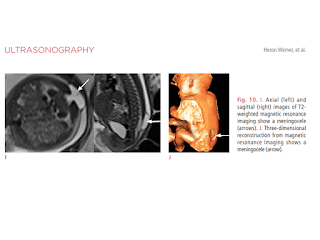
encephalocele= thoát vị não


Không có nhận xét nào :
Đăng nhận xét