Background:
Ultrasound is used to observe the imaging manifestations of COVID-19 in order to provide reference for real-time bedside evaluation.
Purpose: To explore the ultrasonic manifestations of peripulmonary lesions of non-critical COVID-19, so as to provide reference for clinical diagnosis and efficacy evaluation.
Materials and Methods: The clinical and ultrasonic data of 20 patients with clinically diagnosed non-critical COVID-19 treated in Xi'an Chest Hospital during January and February 2020 were retrospectively analyzed. Conventional two-dimensional ultrasound and color Doppler ultrasound were used to observe the characteristics of lesions.
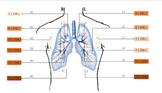
Results: All 20 patients (40 lungs and 240 lung areas) had a history of travel, residence or close contact in/with Wuhan, and 5 of them caught COVID-19 after family gatherings. Lesions tended to occur in both
lungs. Lesions in the lung areas: 14 in L1+R1 area (14/40), 17 in L2+R2 area (17/40), 17 in L3+R3 area (17/40), 17 in L4+R4 area (17/40), 20 in L5+R5 area (20/40), and 28 in L6+R6 area (28/40). Lesion types: rough and discontinuous pleural line (36/240), subpleural consolidation (53/240), air bronchogram sign or air bronchiologram sign in subpleural peripleural consolidation (37/240), visible B lines (91/240), localized pleural thickening (19/240), localized pleural effusion (24/240), poor blood flow in the consolidation detected by color Doppler ultrasound (50/53).
Conclusion: The non-critical COVID-19 has characteristic ultrasonic manifestations, which are visible in the posterior and inferior areas of the lung. The lesions are mainly characterized by a large number of
B lines, subpleural pulmonary consolidation and poor blood flow. Lung ultrasound can provide reference for the clinical diagnosis and efficacy evaluation.
Key Words: lung ultrasound; ultrasonic manifestations; novel coronavirus pneumonia (COVID-19)


Không có nhận xét nào :
Đăng nhận xét