B-lines
The “comet-tail” ultrasonographic sign was first described by Ziskin and colleagues in 1982 when an intrahepatic shotgun pellet was observed to create an artifact like what is seen in lung comets.
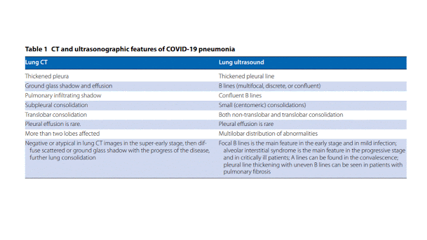
B-lines are not to be confused with normal comet-tail artifacts that originate at the pleura but fade before reaching the edge of the screen (Figure 8). The B-lines are vertical, highly dynamic, hyperechoic artifacts originating from the pleura or consolidation areas. These lines indicate accumulation of fluid in the pulmonary interstitial space (“lung rockets”) or alveoli (“ground glass”).
Multiple B-lines are associated with pulmonary edema of cardiogenic and noncardiogenic or mixed origin. They occur when sound waves pass through the superficial soft tissues and cross the pleural line encountering a mixture of air and water. One or two B-lines are not too concerning but when they increase in number or spread out in one zone, they are an indication of lung interstitial syndrome (Figures 9–10).
How are you using ultrasound to assess COVID-19 patients in your practice?
Prof. Chao: The answer depends on the different condition. Once a patient develops symptoms such as a cough, fatigue or fever, the most important thing is to focus on what is happening in the lungs. Most patients will have a favorable prognosis, but about 10 percent will become worse. To help determine prognosis in this phase, lung ultrasound is easier (than X-ray, CT scan or a laboratory test) under conditions of high patient volume and low resources. Ultrasound is also helpful to follow the patient’s condition, recording the lung images for the patient in the machine and comparing from one exam to the next.
Prof. Wang: I also want to point out that looking at the other organs – the kidneys, the liver and the heart – is very important. COVID-19, because of low oxygen, will affect those organs. Echocardiography is an important part of critical care ultrasound because the heart is the center of oxygen transport. So, timely and routine echocardiography examination for the patient is very important. For the kidney, we look at it with imaging, with color and with Doppler. Even in traditional sepsis, the secondary injury is also important, and we need to pay more attention to it.
Dr. Jalil: When I’m putting a probe on a patient, it’s usually after I’ve talked to them or am trying to figure out what’s going on. I’m trying to add ultrasound to my physical exam. On the ventilator you can look at the compliance of the lungs, you can look at trends. Another way is looking at bilateral B-lines. It can tell you which direction the lungs are going and if the treatment is helping or not.
Dr. Villén: I look at three particular things: B-lines, subpleural consolidations and the thickness of the pleural line. In my opinion, the most sensitive places to put the probe and find something is the posterior below the arm and the axillar point. If you find just subpleural consolidation posterior, probably these are related to mild disease. Keep in mind this is a quite new virus for us. We are learning every day.
How are you using ultrasound to scan the lungs of COVID19 patients?
Prof. Wang: Lung ultrasound is very important in monitoring the lung deterioration in COVID-19 patients, especially for critical patients. ICU doctors have learned that critical care ultrasonography can be used to manage and monitor the lungs of critical COVID patients. We have two methods of monitoring these patients. ICU doctors are using lung ultrasound at the very beginning [when patients are admitted] and when patients are critical, like respiratory and circulatory issues.
Dr. Villén: The findings in lung ultrasound of patients with COVID-19 or any other viral pneumonia is based on three findings: 1) The subpleural consolidations, which is an area of small pneumonia in the border of the lung. Generally, it's triangular with a base with the pleura and the vortex pointed towards the lung; 2) The B-lines--so, an appearance which indicates no edema--in this case there is no fluid. It’s not a matter of fluid, but a matter of initial inflammation which cells fiber; 3) A thickened pleura, which is kind of the same of the subpleural consolidation, but it's more related to an area not a small spot.
In my opinion, the most sensitive places to put the probe and find something is in the posterial, below the arm and the axillar point – they are the most sensitive and not common when performing a lung examination. Most subpleural consolidations are posterior or between lateral and posterior and these are not normal points we normally use for lung examination. And the anterior chest is only affected in severe patients in my experience. If you move only into the anterior lateral, you will not find anything if the patient has coronavirus. So, you will go posterior for more superior and apical more axillar and this are not as tender of points of examinations.
How does point-of-care ultrasound compare with other imaging options in the context of COVID-19?
Prof. Wang: More and more doctors are recognizing the role of ultrasound for monitoring of COVID-19 patients. Point of care ultrasound is portable and can be right at the beside making it convenient and repeatable.
Dr. Jalil: I don’t think this replaces any modalities that I use today. The whole point of being able to do point of care ultrasound it to extend your physical exam. It gives quick information. If I walk into a patient’s room, I can very quickly get images and figure out if this is a pneumothorax, the heart or something different. In the emergent situation, ultrasound is easier to squeeze into a room than an X-ray machine. Handheld ultrasound takes it one step further – just having it in my pocket is convenient.
Dr. Villén: Right now, we cannot afford to send the patient to a CT scan and wait two hours until it is clean again. We need something quick, fast, and reliable that can be made at the bedside, if possible, so I am using ultrasound. With ultrasound, first you must control the environment, cover the machine or at least the probes, dress yourself in protective gear. After performing the examination, you have to go out of the room, take your clothes off and clean the machine for every patient. This process takes 10 to 15 minutes.
What advice would you share with other healthcare providers using ultrasound to help treat and monitor COVID-19 patients?
Prof. Wang: The key principle is do everything early. Detection, monitoring, testing, treatment, isolation, and IPC - infection prevention and control. The earlier you diagnose, the better the outcome.
Dr. Jalil: We can minimize daily x-rays and prevent some of the spread by thinking of trends, for example, what the lungs look like. A lot of times in the ICU you’re trying to keep their volume to the lowest. One or two way to achieve this – on the ventilator you can look at the compliance of the lungs or you can look at the trends. Another way is looking at bilateral B-lines. It can also tell you which direction the lungs are going and if the treatment is helping or not.
Dr. Villén: Go more posterior than you think. Don’t rely on the basic views of lung ultrasound.
Go posterior.
Go axilar.
Check the bases of the lungs
and if the patient is severe, look for big subpleural consolidations with a lot of B-lines colliding.
And, sliding is another thing, because the more affected areas in my opinion and experience are less ventilated, so the amount of sliding is important. A normal lung will slide a lot with wide extortions, but the infection with subpleural consolidated white lungs are less ventilated, so the perception of sliding is much less than a normal thing.
This was already described for acute respiratory distress syndrome (ARDS) which is what COVID is. We are seeing ARDS in early stages at least at the emergency department and in different states of the progression of the disease. That’s why it’s quite new for us, because we know how ARDS behaves in the ICU, but not in the emergency department. It’s a matter of time evolving of the disease.


Không có nhận xét nào :
Đăng nhận xét