Tổng số lượt xem trang

Thứ Năm, 29 tháng 3, 2018

Về sử dụng trí tuệ nhân tạo trong phân loại xơ hóa gan của siêu âm

DOWNLOAD 2 bài sau theo link

DEEP LEARNING on LIVER FIBROSIS

https://static.livemedia.gr/livemedia/documents/al22186_us75_20180323092028_op25_icim2018.pdf


DEEP LEARNING on FATTY LIVER [NAFLD]

https://static.livemedia.gr/livemedia/documents/al22186_us75_20180323092030_op26_icim2018.pdf


Như tin bài trước từ AuntMinnie, nhóm nghiên cứu Hy lạp [Greece] dẫn đầu bởi Athanasios Angelakis, PhD, từ National Technical University of Athens, đã tạo một deep neural network có thể  xác định bệnh nhân có đang trong giai doạn sớm gan hóa xơ không hoặc đã hóa xơ gan rõ. Qua kiểm chứng thuật toán này có mức hoàn thiện cao hơn thuật toán [machine learning] đã công bố  trên y văn.
Thuật toán đã công bố chưa có AUC [area under the curvequá 0.87.
Nhóm Athenes, Greece dùng deep learning để phân loại cho dữ liệu của 103 bệnh nhân viêm gan mạn có sinh thiết xác chẩn giai đoạn hóa xơ gan= 62  ca có Metavir scores  F0 hay F1, còn lại 41 ca có scores ít nhất là F2. Mạng deep-learning network sử dụng thông số giới tính và 4 thông số hình thái= dài cắt dọc gan T, thùy đuôi, gan P cũng như lách. Thuật tóan này sẽ đánh giá phân loại hóa xơ gan nhỏ hơn F1 hoặc lớn hơn F 2 (≤ F1 or ≥ F2).

Thông tin này đặc biệt có lợi cho người sử dụng còn thiếu kinh nghiệm về siêu âm tổng quát và siêu âm đàn hồi shearwaves.


Sau khi đánh giá chéo 10 lần, thuật toán deep learning mới về phân loại hóa xơ gan bằng siêu âm này có độ chính xác cao hơn
  • độ nhạy: 90.2%
  • độ chuyên biệt: 91.9%
  • giá trị tiên đoán dương: 88.1%
  • giá trị tiên đoán âm: 93.4%
  • AUC: 0.9126
Tất nhiên bệnh nhân cần được đánh giá thêm thuật toán này. 

Trong tương lai:

Bước cuối cùng của thuật toán deep-learning là cần kết hợp thêm tuổi và giới tính trong phần dân số [demographic data] nghiên cứu, và trong phần morphologic, hemodynamic, biochemical, image analysis,[hình thái, huyết động học, hóa sinh, tạo hình] và dữ liệu siêu âm đàn hồi [elastographic data]. Với thuật toán này, nên có thêm nhiều lớp [layers], không chỉ phân loại hóa xơ gan mà còn về gan thấm mỡ, cao áp tĩnh mạch cửa, và hội chứng viêm.
 Đã có thuật toán và mô hình cho vấn đề gan thấm mỡ .
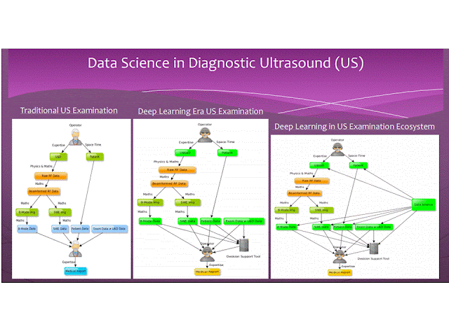
Deep learning và data science sẽ giữ vai trò quan trọng của siêu âm trong tương lai. Trong 10 năm tới hoặc ít hơn, data science sẽ được cài đặt trong máy siêu âm và sẽ sử dụng ngay trong tiến trình đánh giá siêu âm gan. 
----------------
ĐỌC THÊM=


https://tech.fpt.com.vn/ai-machine-learning-va-deep-learning-dinh-nghia-va-cach-phan-biet-chung/


  1. Trí tuệ nhân tạo (artificial intelligence - AI) giờ xuất hiện ở khắp mọi nơi. Nó là thứ được sử dụng để trả lời email tự động trên Gmail, học cách lái xe cho chúng ta ngồi chơi, sắp xếp lại ảnh của những chuyến đi chơi thành từng album riêng biệt, thậm chí còn giúp quản lý ngôi nhà hay đi mua sắm nữa. Nhưng bạn có biết là trí tuệ nhân tạo không chỉ đơn giản là một thực thể mà nó còn được chia thành nhiều loại nhỏ hơn? Những hạn chế hiện tại của sản phẩm trí tuệ nhân tạo là gì? Và vì sao chúng ta không cần (hay chưa cần) phải lo lắng về việc trí tuệ nhân tạo bùng lên chiếm lấy thế giới?

    Neural networkmachine learningdeep learning là gì?

    Đây là những cụm từ mà bạn rất thường hay thấy trong những thông tin liên quan đến trí tuệ nhân tạo. Về cơ bản, bạn có thể nghĩ về những thứ này như các lớp cấu thành trí tuệ nhân tạo.

    Neural network, tạm gọi là mạng lưới thần kinh nhân tạo, sẽ nằm dưới cùng. Đây là hệ thống các máy tính và thiết bị hệ thống điện toán nói chung được kết nối theo một cách nào đó để mô phỏng lại một phần cách hoạt động của các nơ-ron thần kinh trong não người. Những chiếc máy tính trong neural network có thể nằm gần nhau trong cùng một phòng hay cách xa nhau cả nghìn cây số, mỗi một chiếc máy tính trong đó có thể được xem như một đơn vị thần kinh, gọi là node.

    Cập nhật: Neural network không nhất thiết phải dựa trên phần cứng, nó vẫn có thể là phần mềm và các giải thuật.

    Khái niệm về neural network đã có từ những năm 1950 với sự ra đời của ngành nghiên cứu về trí tuệ nhân tạo. Người ta nói rằng khi nằm riêng biệt, những node máy tính này chỉ chạy những gì được lập trình sẵn và chỉ có thể trả lời những câu hỏi đơn giản, hay nói cách là nó "không thông minh". Cũng giống như trong cơ thể người, một nơ-ron không chưa làm nên chuyện, nhưng khi kết nối chúng thành một mạng lưới dày đặt thì mọi chuyện sẽ khác đi rất nhiều. Khi các hệ thống máy tính được nối lại với nhau, chúng có thể giải quyết những vấn đề khó khăn hơn. Và quan trọng nhất, khi áp dụng đúng thuật toán, người ta có thể "dạy học" cho máy tính.

    Lớp kế tiếp là machine learning. Từ này mình không biết nên dịch sang tiếng Việt như thế nào cho chính xác nên tạm thời cứ để nguyên tiếng Anh. Đây là một chương trình chạy trên neural network, nó sẽ làm nhiệm vụ huấn luyện máy tính để "học hỏi" một vấn đề gì đó, ví dụ như học hỏi từ hàng nhìn mẫu chữ viết tay của người dùng để đoán xem đó là kí tự gì, hoặc học từ hàng nghìn bức ảnh chụp bãi biển để tìm ra điểm chung và sau đó nhìn phát là biết ngay tấm này chụp biển chứ không phải chụp núi.

    Deep learning nằm ở trên cùng, cũng không biết dịch ra tiếng Việt như thế nào. Đây là một nhánh đặc biệt của ngành khoa học machine learning. Deep learning trở nên phổ biến trong thập kỉ gần đây nhờ vào sự gia tăng nhanh chóng của lượng dữ liệu số mà loài người tạo ra, ngoài còn nhờ sức mạnh xử lý của máy tính gia tăng trong khi giá thành giảm xuống. Sẽ nói kĩ hơn về deep learning ở bên dưới.

    Trí tuệ nhân tạo hoạt động ra sao?

    Giả sử như bạn muốn một cái máy tính biết cách băng qua đường. Theo cách truyền thống, chúng ta sẽ lập trình cho nó cách nhìn trái, nhìn phải, cách đợi xe chạy qua hết, cách đi đúng vạch băng đường theo luật và nhiều thứ khác, sau đó để cho máy tự đi.

    Còn với kĩ thuật trí tuệ nhân tạo, mà cụ thể là một chương trình machine learning, bạn sẽ cho máy tính xem 10.000 đoạn video về cách băng đường an toàn. Kế tiếp bạn lại cho nó xem thêm 10.000 đoạn video nữa nhưng lần này chiếu cảnh người ta bị xe tông khi băng đường ẩu. Lúc này bạn mới thả cho nó tự băng đường.

    Đang tải google_self_driving_car.0.gif… ​

    Phần khó nhất đó là bạn phải làm sao cho máy tính hiểu và tiếp thu được thông tin từ các video này, cũng giống như phần khó nhất khi đi dạy học là phải làm cho học sinh hiểu được bạn nói gì và ghi nhớ điều đó. Trong nhiều thập kỉ qua, người ta đã thủ nhiều phương pháp khác nhau để dạy cho máy tính học. Một trong những cách đã được xài đó là "reinforcement learning", tức là bạn sẽ "thưởng" cho máy tính khi nó làm đúng thứ bạn muốn rồi từ từ tối ưu để đạt kết quả tốt nhất. Người ta vẫn thường hay huấn luyện thú vật theo cách này. Một cách khác nữa là "chọn lọc tự nhiên", tức là nhiều cách giải quyết cùng một vấn đề sẽ được áp dụng cho chạy song song, cái này giải được nhanh và chính xác nhất sẽ thắng những cái còn lại.

    Còn trong thời đại ngày nay, người ta xài một phương pháp gọi là deep learning. Deep learning sử dụng nhiều lớp trong một neural network để phân tích dữ liệu theo nhiều khía cạnh khác nhau. Ví dụ, nếu bạn đưa cho máy tính học một tấm ảnh theo kĩ thuật deep learning, mỗi lớp trong mạng lưới thần kinh nhân tạo này sẽ nhìn nhận vấn đề theo một cách riêng. Lớp dưới cùng sẽ chỉ đơn giản là vẽ ra một cái lưới 5x5 lên tám ảnh và đánh dấu "có" hoặc "không" khi có một đối tượng xuất hiện trong ô. Nếu "có", lớp bên trên sẽ bắt đầu nhìn vào từng ô này một cách kĩ càng hơn, nó phân tích xem đây có phải là điểm đầu của một đường thẳng không, hay đây là một cái góc nghiêng? Thật nhiều lớp như thế sẽ giúp phần mềm hiểu được những vấn đề phức tạp, tất cả đều dựa trên các bẻ nhỏ nó ra rồi "điều tra" từ từ. Cũng chính vì lý do này mà người ta gọi đây là "deep", tức là sâu và có nhiều lớp.

    Đang tải DeepFace.jpg…
    Kĩ thuật được Facebook áp dụng để nhận diện gương mặt, trong đó chia bức ảnh thành lớp khác nhau để học hỏi

    Yann LeCun, trưởng bộ phận trí tuệ nhân tạo của Facebook, cho biết: "Khi bạn đi lên các lớp cao hơn thì những thứ được phát hiện sẽ càng lúc càng rộng hơn. Càng lúc càng có nhiều khía cạnh hơn được phân tích. Và khi bạn leo lên đến lớp cao nhất, bạn sẽ có những công cụ để cho bạn biết bức hình đó đang chụp người hay một chút chó hay một chiếc máy bay".

    Nãy giờ chúng ta chỉ mới nói đến chuyện nhận biết, giờ thì đến lúc dạy cho máy tính biết thứ mà nó vừa nhận ra là gì. Một hệ thống neural network lại được sử dụng, nhưng lần này nó sẽ xem xét nhiều đặc điểm của một con mèo. Rất nhiều bức hình chụp mèo cũng sẽ được đưa cho hệ thống xem kèm theo lời dặn: đây là ảnh con mèo đó nha. Rồi người ta lại cho máy xem thêm một loạt ảnh khác nữa chụp chó, heo, gấu, vịt và dặn: đây không phải là con mèo. Thông qua hàng loạt dữ liệu như vậy, phần mềm sẽ biết được những con mèo thường có điểm chung gì, móng, lông, tay chân, đầu, đuôi của chúng ra sao thì mới được gọi là mèo...

    Theo thời gian, máy sẽ ghi nhớ những dữ liệu này và sắp xếp theo thứ tự quan trọng. Ví dụ, móng vuốt không chỉ mèo mới có, nhưng nếu móng đi chung với bàn chân to và ria mép thì đây đích thị là con mèo. Các mối liên hệ như thế này cũng sẽ được cung cấp theo thời gian trong quá trình phần mềm machine learning học hỏi các ảnh. Quy trình này diễn ra trong thời gian dài và lặp đi lặp lại nhiều lần. Cứ lần sau nó sẽ tốt hơn lần trước vì được góp ý từ con người hoặc thậm chí là từ những hệ thống trí tuệ nhân tạo khác.

    Bạn có thể thấy rằng chỉ để nhận biết được một con mèo thôi đã phức tạp quá chừng, trong khi các hệ thống machine learning của Facebook, Google hay Microsoft phải nhận biết nhiều thứ khác nữa trong đời sống. Thế nên, việc Microsoft tự hào khi phát hành một ứng dụng có khả năng nhận biết các giống chó khó nhanh nghe có vẻ đơn giản nhưng phía sau nó là cả một mạng lưới thần kinh nhân tạo phức tạp và đã bắt đầu chạy học hỏi trong thời gian rất dài rồi.

    Đang tải dogknight_3346130k.0.jpg…
    Hình ảnh được tạo ra từ dự án trí tuệ nhân tạo Google Drea

    Đây có phải là thứ mà Google, Facebook và nhiều công ty khác đang sử dụng?

    Câu trả lời chung sẽ là "Đúng rồi".

    Deep learning hiện tại đang được xài cho nhiều tác vụ khác nhau. Các công ty công nghệ lớn thường lập riêng một bộ phận trí tuệ nhân tạo. Google, Facebook còn mở những hệ thống phần mềm machine learning của mình ra cho mọi người sử dụng. Google hồi tháng trước cũng mới mở một khóa học kéo dài 3 tháng vè machine learning và deep learning.

    Một vài ví dụ về machine learning đó là công cụ Google Photos. Nó có khả năng nhận biết và phân loại hình ảnh mà bạn chụp theo từng chủ đề khác nhau, thậm chí là theo từng khuôn mặt khác nhau một cách tự động. Hay Facebook M, một trợ lý ảo cá nhân nửa người nửa máy có thể giúp bạn đặt một số món hàng mà bạn muốn. Microsoft với Cortana, Google với Google Now và Apple với Siri đều là những ví dụ rất thực của trí tuệ nhân tạo.

    Đây cũng là lý do vì sao mà trong thời gian gần đây chúng ta bắt đầu nghe nói nhiều về machine learning, deep learning và trí tuệ nhân tạo. Đó là vì các hãng tiêu dùng lớn đã bắt đầu nhảy vào cuộc chơi và đưa ra những sản phẩm thực tế mà người ta có thể cầm nắm và trải nghiệm. Trước đó, deep learning và machine learning chỉ nằm trong các phòng thí nghiệm ở những viện nghiên cứu và trường đại học mà thôi.

    Hạn chế của trí tuệ nhân tạo hiện nay

    Deep learning đang được sử dụng cho những thứ như nhận dạng giọng nói và nhận dạng hình ảnh, những thứ có nhiều tiềm năng thương mại hóa. Nhưng song song đó, nó cũng còn nhiều hạn chế.

    Đầu tiên, deep learning cần một lượng dữ liệu đầu vào khổng lồ để máy tính có thể học hỏi. Quy trình này mất nhiều thời gian, nhiều sức mạnh xử lý mà chỉ có các server cỡ lớn mới làm được. Nếu không có đủ dữ liệu đầu vào, hay có đủ dữ liệu nhưng không đủ sức mạnh để xử lý, thì mọi thứ không thể diễn ra đúng như ý định, kết quả do máy học sẽ không chính xác.

    Thứ hai, deep learning vẫn chưa thể nhận biết được những thứ phức tạp, ví dụ như các mối liên hệ thông thường. Chúng cũng sẽ gặp khó khăn khi cần nhận biết những thứ tương tự nhau. Lý do là vì hiện chưa có kĩ thuật nào đủ tốt để trí tuệ nhân tạo có thể rút ra những kết luận đó một cách logic. Bên cạnh đó, vẫn còn nhiều thách thức trong việc tích hợp kiến thức trừu tượng vào các hệ thống machine learning, ví dụ như thông tin về vật đó là gì, nó dùng để làm gì, người ta hay xài nó làm sao... Nói cách khác, machine learning chưa có được những kiến thức thông thường như con người.

    Một ví dụ rất cụ thể cho các bạn dễ hiểu: trong một dự án của Google, một mạng lưới thần kinh được sử dụng để tạo ra bức ảnh của một quả tạ mà người ta hay cầm tập trong phòng gym. Kết quả đưa ra khá ấn tượng: hai vòng tròn màu xám được kết nối bằng một cái ống ngang. Nhưng ở giữa cái ống này còn có cánh tay của con người, và đây không phải là thứ nằm trong "đề bài". Lý do thì khá là dễ đoán: hệ thống được dạy về quả tạ bằng những tấm hình chụp người ta đang cử tạ, vậy nên dính tay người vào là chuyện hiển nhiên. Hệ thống có thể biết được quả tạ ra sao, nhưng nó không biết là quả tạo sẽ không bao giờ có cánh tay cả.

    Đang tải dumbbells.0.png… ​

    Với một số bức hình đơn giản hơn nhưng mahcine vẫn nhầm lẫn. Thử nghiệm của một nhóm nhà nghiên cứu cho thấy rằng khi họ đưa máy tính xem một loạt ảnh chỉ có các pixel ngẫu nhiên nhưng máy lại chắc chắn đây 95% là một cái... xe tải, hay là một con sao biển...

    Đó chưa phải là tất cả. Theo lời của nhà khoa học máy tính Hector Levesque, những công cụ trí tuệ nhân tạo hiện nay sử dụng nhiều "mánh" để xóa đi những lỗ hổng thật sự trong kiến thức của chúng. Các trợ lý ảo như Siri hay Cortana thường làm cho bạn có cảm giác bạn đang nói chuyện với người thật vì chúng sử dụng các câu nói đùa, câu trích dẫn, các biểu hiện cảm xúc và nhiều thứ khác, cốt chỉ để bạn phân tâm.

    Bạn hãy thử hỏi những thứ cần đến suy nghĩ thông thường, ví dụ như "con cá xấu có lái xe được không" hay "một cầu thủ đá banh có được phép gắn cánh để bay hay không". Các câu hỏi dạng này quá phức tạp với những hệ thống trí tuệ nhân tạo hiện nay nên thường sẽ không có kết quả nào được trả về cho bạn, nếu có cũng là những thứ không liên quan hay chỉ đơn giản là search câu nói của bạn trên Internet.

    Mùa đông AI

    Ngành khoa học trí tuệ nhân tạo là một ngành dễ lên nhưng cũng dễ xuống. Năm 1958, tờ New York Times từng nói về một chiếc máy có khả năng phân biệt bên trái và bên phải như là một dạng mầm sống của robot thông minh. Nhưng đến nay, chúng ta vẫn chưa thể tạo ra được một con robot nào có trí thông minh đến vậy. Và khi những lời hứa hẹn đó không được thực hiện, người ta dùng từ "mùa đông AI". Đó là giai đoạn mà lượng tiền đầu tư cho AI giảm mạnh, ít còn ai nhắc về nó, và người ta cũng tỏ ra hoài nghi về những kết quả có thể đạt được. Tính đến nay đã có khoảng 6 "mùa đông AI" nhỏ và 2 mùa lớn từng xuất hiện, vào cuối những năm 70 và đầu các năm 90.

    Trí tuệ nhân tạo có tri giác?

    Nhiều người đang làm việc trong ngành trí tuệ nhân tạo cho rằng chúng ta sẽ rất khó để tạo ra một trí tuệ nhân tạo có tri giác. "Có rất ít bằng chứng ở thời điểm hiện tại cho thấy hi vọng về việc tạo ra một trí tuệ nhân tạo có tính linh hoạt cao và làm được những thứ mà chúng không được tạo ra để thực hiện", theo lời giáo sư Andrei Barbu từ đại học MIT. Ông nhấn mạnh việc nghiên cứu trí tuệ nhân tạo hiện nay chỉ tạo ra được những hệ thống được tối ưu để giải quyết một vấn đề cụ thể mà thôi.

    Cũng đã có một số công trình nghiên cứu về việc cho máy tính học nhưng không cần sự giám sát, tức là cứ đưa dữ liệu cho máy học mà không dán nhãn đúng sai hay giải thích gì cả. Tuy nhiên, Andrei Barbu nhận xét những dự án như thế này vẫn chưa có tiến triển nào và vẫn còn rất xa xời để đến được ngày có kết quả. Một ví dụ đã từng xuất hiện đó là một hệ thống neural network của Google đã từng lấy ngẫu nhiên ảnh thumbnail của 10 triệu video trên YouTube để tự dạy mình con mèo trông giống như thế nào. Thế nhưng, Google nói đây chỉ là một thử nghiệm và không nói gì về độ chính xác của nó cả.

    Nói cách khác, chúng ta vẫn chưa biết cách làm cho máy tính tự học mà không cần giám sát. Đó là rào cản lớn nhất. Tức là vẫn còn rất xa mới tới cái ngày mà robot có tri giác và đánh trả lại con người :D

    Như Elon Musk đã nói, công ty ông có tạo ra một trí tuệ nhân tạo cho tính năng tự lái xe trên những chiếc Tesla. Nhưng ông chưa bao giờ nói rằng nó sẽ biết hết mọi thứ. Đây chỉ đơn giản là một mạng lưới giúp các xe học hỏi lẫn nhau. Khi một chiếc này học được điều gì thì những chiếc khác cũng biết điều tương tự. Kết quả cuối cùng không phải là những chiếc xe có thể làm mọi thứ trên đời, nó chỉ để giải quyết một vấn đề rất cụ thể mà thôi.

Thứ Tư, 28 tháng 3, 2018

AIUM: Can deep learning classify liver fibrosis on US?


By Erik L. Ridley, AuntMinnie staff writer
March 26, 2018 -- NEW YORK CITY - A deep-learning algorithm that analyzes B-mode morphological ultrasound data and patient demographic information can classify the severity of liver fibrosis in cases of chronic liver disease, according to research presented at the American Institute of Ultrasound in Medicine (AIUM) conference.
A team of researchers from Greece trained a deep neural network that could determine if patients were in the early stages of fibrosis or if they had advanced to significant fibrosis. In testing, the algorithm yielded a higher level of performance than existing methods previously reported in the literature, according to presenter Athanasios Angelakis, PhD, from the National Technical University of Athens.
A binary classification problem
Early-stage fibrosis can be reversed with treatment and prevented from reaching cirrhosis. It's also important to be able to diagnose significant fibrosis, classified as Metavir score F0 or F1, from more severe fibrosis (≥ F2).
"Given an individual's biomarkers, we would like to conclude if his or her fibrosis stage is either [Metavir score] ≤ F1 or ≥ F2," he said in a March 24 presentation "It's a significant binary classification problem."
Traditional methods used for this assessment include biopsy; biochemical tests; imaging, such as shear-wave elastography, strain elastography, and FibroScan transient elastography cutoff values; as well as image analysis or radiomic approaches. Recently, machine learning has also been applied in image analysis and radiomics. However, none of the biochemical, imaging, image analysis/radiomics, or machine-learning methods reported in the literature has yielded an area under the curve (AUC) from receiver operating characteristic analysis of more than 0.87, according to Angelakis.
The researchers sought to apply deep learning to this classification task. They used a dataset of 103 patients with chronic liver disease and biopsy-validated fibrosis stages; 62 patients had Metavir scores of F0 or F1, while 41 had scores of at least F2. The deep-learning network was given one demographic parameter (patient gender) and four morphologic parameters -- the longitudinal diameter of the left lobe, caudate lobe, and right lobe of the liver, as well as the spleen. The algorithm then provides its assessment of the patient's fibrosis class (≤ F1 or ≥ F2).
This information could be of particular benefit to inexperienced users, according to the researchers.
"It reduces the experience [needed] for the examiner [to perform these studies]," Angelakis said. "One of the goals of using deep learning in ultrasound in general and shear-wave elastography is to reduce this need for experience with the exam."
After performing tenfold cross-validation, the researchers found that the algorithm yielded a high level of accuracy for liver fibrosis classification:
  • Sensitivity: 90.2%
  • Specificity: 91.9%
  • Positive predictive value: 88.1%
  • Negative predictive value: 93.4%
  • AUC: 0.9126
They acknowledged, though, that more patients are needed to further validate the algorithm.
The future
The ultimate holy grail for a deep-learning algorithm in the liver would be to incorporate patient demographic data such as gender and age, along with morphologic, hemodynamic, biochemical, image analysis, and elastographic data. Such an algorithm, which would have many layers, would not only classify fibrosis but also steatosis, portal hypertension, and inflammation, Angelakis said.
"We have already created algorithms and models that also attack the steatosis problem, because our datasets are bigger now," he said.
Deep learning and data science will likely take on a significant role in ultrasound in the future, according to Angelakis.

"The output from the [deep-learning] model will be one more input for the expert [to consider during interpretation]," he said. "That is how we reduce the expertise [requirement] for the reader. Looking ahead 10 years from now, maybe less, data science will also be used during the manufacturing of ultrasound devices and it will be in every [aspect of the ultrasound acquisition and interpretation process].

Thứ Ba, 27 tháng 3, 2018

NHÂN 03 CA DIFFUSE LARGE B CELL LYMPHOMA CÓ TRIỆU CHỨNG HÀM MẶT

Trong số 20 ca lymphom lan tỏa dòng tế bào B lớn [diffuse large B cell lymphoma] ở Trung tâm Y khoa Medic từ 2008,  có 03 ca có triệu chứng đau răng và hàm mặt điều trị giảm đau, nhổ răng..không bớt, sau đó xuất hiện nhiều tổn thương khác ngoài hạch. Trong khi đó, tìm được một bài báo về đau xương hàm dưới trong lymphom ác. Sau đây là một hồi cứu về đề tài này.

             Diffuse Large B Cell Lymphoma
and Appearences  in Oral Cavity, and Maxillary Area
Nguyen Thien Hung, Jasmine DCB Thanh Xuan, Le van Tai, Le Thong Nhat, Le Thanh Liem, Phan Thanh Hai
Medic Medical Center, HCMC, Vietnam

INTRODUCTION:
May there had been an initial symptom in oral cavity, maxillary and mandibular area for diffuse infiltration of lymphoma?  We will represent some cases of diffuse lymphoma with appearences  in oral cavity,  and maxillary areas.

MATERIALS and METHODS:
We retrospectively reviewed  20 cases of diffuse lymphoma that were histopathologically diagnosed in our center from 2008 in which presented 03 cases  with oral cavity,  maxillary symptoms and  bone involvement [01 case in male 42 yo and 02 cases in female gender, 33 and 40 year-old].

RESULTS:

CASE 1= A 33 year-old female patient with pain in lower maxillary bone for one month and tension in both  2 breats), hyperpigmented edema of areolar area both 2 sides without pregnancy.  ABVS scanning  detected  multiple nodules infiltrating in 2 breasts.
Biopsy of 2 breasts  reported  microscopic with immunohistochemistry scanning, diffuse large B cell lymphoma.
MRI  full body with gado detected  bone marrow changing, 2 breats  hypercaptured contrast,  ascites and kidney infiltration. In pelvis  2 ovarian tumors and big uterine cervix were detected. Blood tests showed   lower platelets,  EGFR  lower  46, beta2 microglobuline raised  3816,  ferritin raised  911, LDH-l  raised  1360.


CASE 2= A 40 year-old female patient with toothache but pain remained  after removing tooth, and in total body examination detected right pleural effusion  and some nodular tumors in thyroid, left breast and subcutaneous area of right neck, left chest all and  lumbar region. Erosions of left clavicle and right 1st rib. Biopsy of left breast tumor reported diffuse large B cell lymphoma.


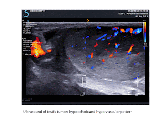
CASE 3= Man 42 year-old, one month ago, pain in oral sinus, difficult eating and 2 days after,  pain appears in left testis. Ultrasound of left big and  hot testis represented  hypoechoic  infiltration,  hypervascular of one part of testis and elastoscan ultrasound value #  10.5 kPa).
 FNAC of this mass of left testis  having abnormal cells. Biopsy of  tumor in oral area represented diffuse large  B cell lymphoma.


DISCUSSION:
There are published reports of pain in mandibular area as initial symptom but symptoms in oral cavity and maxillary bone as onset symptom seems to be not represented in literature. But it exists a case of pain in oral cavity for lymphoma infiltrating in testis and an another case of pain in maxillary area for lymphoma in whole body in this report.Toothache and pain in maxillary region remained with management guided a survey in all body and detected extranodal lesions: bone erosions, perirenal edema, ovarian, uterine cervix, testicular edema, subcutaneous nodules. But in literature, only mandibular lesion with tooth pain were published which not included.

CONCLUSIONS:
Seveval  reports of lymphoma in mandible were published with pain as initial symptom,  but in cases of diffuse infiltration of lymphoma in whole body, the appearences in oral cavity, maxillary and mandibular areas with pain symptom may be added criteria of diagnosis for lymphoma in clinical examination.

REFERENCE

 Mochizuki, Y.,  Harada, H.,  Sakamoto, K.,  Kayamori,  K.,  Nakamura,  S.,  Ikuta, M.,  Kabasawa, Y., Marukawa, E., Shimamoto, H., Tushima, F. and Omura, K. (2015) Malignant Lymphoma with Initial Symptoms in the Mandibular Region. Journal of Cancer Therapy, 6, 554-565


Abstract
Primary intraosseous lymphoma is rare and there are few case reports manifesting with a mass in the mandible. Thus, we retrospectively reviewed and analyzed the clinical characteristics,  treatment, and outcome of extranodal non-Hodgkin’s lymphoma (NHL) with initial mandibular symptoms in our department. At initial treatment of dental clinics, dentists had diagnosed as dental or gingival diseases and had performed dental treatment. Neurological disorder to involvement of the inferior alveolar nerve was present in 80.0% of our cases. On dental or panoramic radiography a specific radiolucent lesion in the mandible was not detected, except for dental lesions. On CT, NHL of the mandible region has no widening and no clear destruction but a slit-like the cortex bone destruction pattern with keeping in shape of the mandibular body  (62.5% of CT-examined cases), and extraosseous soft tissue mass are clearer on MRI  (100.0% of MRI-examined cases).
Histopathologically, 80.0% of our cases were diagnosed as diffuse large B cell lymphoma (DLBCL).
One case as B-cell lymphoblastic lymphoma and one case as B-cell lymphoma unclassifiable with features intermediate between DLBCL and Burkitt lymphoma were Stage IV  (Ann Arbor staging system) and had poor prognosis. The disease-specific survival rate was 77.8% at 5 years. If unexplained non-specific symptoms such as swelling of the jaw, pain, neurological disorder of the inferior alveolar nerve, tooth mobility are observed, oral surgeons and dentists should not perform
dental treatments. CT and MRI show disease specific appearance to be able to give a definitive diasnosis as NHL. PET/CT is useful for scaninng of whole body. A deep bone biopsy is preferred for
suspected malignant lymphoma.

Chủ Nhật, 18 tháng 3, 2018

Acute and Chronic DVT and Ultrasound (US)

SOURCE: Advanced imaging in acute and chronic deep vein thrombosis, Gita Yashwantrao Karande , Sandeep S. Hedgire , Yadiel Sanchez , Vinit Baliyan , Vishala Mishra , Suvranu Ganguli , Anand M. Prabhakar © Cardiovascular Diagnosis and Therapy. Cardiovasc Diagn Ther 2016;6(6):493-507

DVT and Ultrasound (US)[extracted]

US is used in evaluation of both symptomatic and asymptomatic DVT (patients at high risk of DVT). It is useful not only in assessing DVT but can also identify other conditions causing signs and symptoms indistinguishable from DVT.

Compression US
Compression US has been procedure of choice for investigation of suspected upper and lower extremity DVT for decades (14). Other modification to this technique like two-point compression US (15), extended compression US (16) and complete compression US (17) are used in different combinations at different institutions.


Venous duplex US
 Lower extremity venous duplex US combines 2 components to assess for DVT: B-mode or gray-scale imaging with transducer compression maneuvers and Doppler evaluation consisting of color-flow Doppler imaging and spectral Doppler waveform analysis. Respiratory phasicity and cessation of flow with the valsalva maneuver offer indirect evidence of patent abdominal and pelvic veins (18). The primary diagnostic US criteria for acute DVT remains non-compressibility of the vein with secondary diagnostic criteria being echogenic thrombus within the vein lumen, venous distention, complete absence of spectral or color Doppler signal within the vein lumen, loss of flow phasicity, and loss of response to valsalva or augmentation (18). US can also be used to differentiate acute from chronic thrombus. In acute thrombosis, vein is distended by hypoechoic thrombus and shows partial or no compressibility without collaterals (Figure 1). In chronic thrombosis, the vein is incompressible, narrow and irregular and shows echogenic thrombus attached to the venous walls with development of collaterals (Figure 2). According to American College of Radiology (ACR) guidelines and technical standards, lower extremity US should include compression, color and spectral Doppler sonography with assessment of phasicity and venous flow augmentation (19,20). Advantages of lower extremity venous Duplex US are that it is readily available, quick, cost effective, noninvasive, devoid of ionizing radiation, lacks need for intravenous contrast and can be portable for critically ill patients prone for developing DVT.


Limitations include that it is difficult and less sensitive in patients with obesity, edema, tenderness, recent hip or knee arthroplasty, cast, overlying bandages and immobilization devices. It also has limitations in patients who had previous DVT and have new symptoms shortly after the treatment. False-positive results include extrinsic compression of a vein by a pelvic mass or other perivascular pathology (21) and thrombosis in the distal popliteal vein. False-negative studies may occur in the presence of calf DVT, proximal DVT in asymptomatic (even high-risk) patients or in the presence of a thrombosed duplicated venous segment. In a systematic review of accuracy of US in diagnosis of DVT in asymptomatic patients, Kassai et al. suggested that US was accurate in proximal veins for diagnosis of DVT in patients hospitalized for orthopaedic surgery (11) with lower sensitivity in other settings.


Sonographic elasticity imaging (SEI)
As described previously venous duplex US is considered primary noninvasive imaging for DVT. However, this method cannot assess the age and maturity of the thrombus i.e., it cannot distinguish pure post thrombotic syndrome (PTS) (which develops in 20–50% of patients after DVT) from new development of acute DVT with or without PTS (22). It is important to distinguish acute on chronic DVT from PTS as the latter doesn’t require anticoagulant therapy with highly potent fast acting anticoagulants associated with high risk of bleeding (23). Also chronic clots are treated with oral warfarin sodium, which has better safety profile than heparin. SEI is the latest promising technique for estimating age of the thrombus. It uses tissue deformation to assess the tissue hardness and hence clot maturity (24,25). It has shown promising results in animals and in a few studies in humans. As SEI requires tissue to be deformed during imaging, it is consistent with venous duplex US which also requires compression. The degree of compression required for standard strain measurements on SEI is lesser than compression US that is beneficial for patients with swollen painful legs (26-28). Thus, even if evaluation of clot age does not work, just the use of SEI to detect thrombi may be an improvement over the present compression US technique. SEI can hence be incorporated into standard duplex US so that thrombus can be diagnosed and presumably aged simultaneously. SEI is in preliminary stages of investigations and there is limited data on its ability to determine the age of DVT in human subjects (26,29,30). It is highly operator dependent. Another limitation is that comparison between the two clots requires an internal standard with the same hardness in both images, as it is difficult to know the force that was used to deform the tissue in each case. Rubin et al. used the wall of the vein as standard but the hardness difference estimate was conservative due to lack of another standard reference (31). Another limitation is that it is difficult to distinguish between subacute and chronic thrombi that are closer in age (29).
….

Conclusions


The incidence of DVT is increasing, not just in the lower extremity but also in upper extremity, where malignancy and central venous catheters are the major precipitating factors. While US still has advantages, it has various limitations and in such cases advanced imaging techniques such as MRI, should be considered. CTV, while sensitive, can be incorporated into CTPA in suspected PE but has the disadvantage of radiating sensitive pelvic organs especially in young patients. MRI will almost likely feature more commonly in DVT evaluation in the near future with new “blood pool” contrast agents allowing a comprehensive examination for PE and DVT in the same scan. Although advanced imaging techniques in nanotechnology/ biotechnology, molecular imaging and PET are also being investigated, these may not replace the established first line modalities in diagnosis, but may be useful as adjuncts in patients who are not good candidates for structural imaging like renal disease patients with contraindication for intravenous contrast. Most of these agents are under trial and will hopefully come into routine use in diagnosing this potentially fatal disease.

Thứ Tư, 14 tháng 3, 2018

PoCUS MEDIC:Foreign Body [Thorn # 02mm]

Thôn Dakrong.
Bé trai 10 tuổi . Mủ dưới da bàn chân. Siêu âm thấy có dị vật d# 2mm.
Xử trí: rạch da lấy dị vật tại chỗ, gai.

PoCUS MEDIC detected a 2mm foreign body in an abscess of left subcutaneous forefoot of a 10 year-old young boy.
Management: Removed right away a thorn in place.


Why does it need an abscess evaluation by ultrasound?
- to differentiate abscess from cellulitis.
- to find out a foreign body in an abscess.
- to use appropriate antibiotics in case of cellulitis.

Thứ Ba, 6 tháng 3, 2018

RENAL FIBROSIS in Ultrasound Elastography

Extracted from
 2017 Mar 7;7(5):1303-1329. doi: 10.7150/thno.18650. eCollection 2017. Ultrasound Elastography: Review of Techniques and Clinical Applications

Renal Fibrosis


Chronic kidney disease (CKD) in native kidneys and interstitial fibrosis in allograft kidneys are the two major kidney fibrotic pathologies where USE may be clinically useful. Both these conditions can lead to extensive morbidity, mortality, and high health care costs. CKD is a prevalent pathology affecting approximately 14% of the population [98] and it can progress to end-stage renal disease requiring dialysis or renal transplant. Allograft renal interstitial disease can lead to renal transplant failure. Currently, biopsy is the standard method for renal fibrosis staging. Ultrasound elastography methods of strain imaging and SWI can potentially be useful to noninvasively detect, stage and monitor renal fibrosis, reducing the need for renal biopsy [99]. The superficial location of allograft kidneys allows assessment by strain imaging. Orlacchio et al. evaluated 50 patients with allograft kidneys by SE (Philips) and compared USE results with the degree of histopathologic fibrosis (F1=mild, F2=moderate, F3=severe). SE was shown to be useful for predicting fibrosis in renal transplant patients, mainly for F2-F3 cases, with overall accuracy of 95%. Sensitivity, specificity, PPV and NPV were 85.7%, 95.5%, 96% and 84% respectively (using a tissue mean elasticity cut-off value of 46 a.u. – arbitrary units) to diagnose F2-F3 [100]. Strain imaging has also been used to assess native kidneys, although the difficulty of applying external compression to the native kidney in the retroperitoneal location can limit the accuracy of strain elastograms [99]. Menzilcioglu et al. used SE to compare native kidneys in patients with and without CKD. They found the mean strain index value of renal parenchyma in CKD patients (1.81±0.88) was significantly higher than in healthy individuals (0.42±0.30) (p under 0.001). However, SE was not able to distinguish between different stages of CKD (Table 4, [101]). SWI is advantageous to strain imaging in evaluating kidney fibrosis in both allograft and native kidneys since it does not depend on external compression [99]. The majority of studies using SWI to evaluate CKD (Table 4, [102-105]) have shown that the shear wave velocity of the renal parenchyma of CKD patients was significantly lower than in normal patients. Furthermore, studies have shown significant correlations between shear wave velocity and biochemical parameters of CKD. For example, Guo et al used VTQ/ARFI to show that shear wave velocity correlated significantly with estimated glomerular filtration rate, urea nitrogen and serum creatinine (Table 4,[105]), and Hu et al concluded that shear wave velocity correlated significantly with serum creatinine and glomerular filtration rate (Table 4, [103]). In contrast to the above promising results, Wang et al. used VTQ/ARFI to assess 45 patients with CKD referred for renal biopsy and concluded that shear wave velocity measurements did not correlate with any histologic indicators of fibrosis (glomerular sclerosis index, tubular atrophy, interstitial fibrosis) and could not distinguish between CKD stages (Table 4, [106]). Interestingly in SWI of kidney fibrosis, a negative correlation has been reported between shear wave velocity and the progression of CKD. For example, Bob et al. showed that the shear wave velocity decreased with increasing CKD stage (Table 4, [104]). This is supported by other studies that found significantly decreased shear wave velocity in CKD compared to normal kidneys (Table 4 [103, 105]). These findings are opposite to SWI findings in liver fibrosis, where increasing liver fibrosis corresponds to increasing shear wave velocity. The reason for this difference remains unclear. Asano et al. hypothesized that the decreased renal blood flow in patients with CKD reduces kidney stiffness, resulting in decreased shear wave velocities [107].

Limitations of Renal Ultrasound Elastography

The kidney has many unique properties that limit use of USE:
- The retroperitoneal position of native kidneys impairs the application of external compression.
- The kidney’s complex architecture and high tissue anisotropy can influence shear wave velocity. Anatomically, the renal cortex is not organized in linear structures since glomeruli are spherical and proximal/distal tubes have convoluted shape. The medulla consists of the loops of Henle, the vasa recta and the collecting ducts, which have a parallel orientation spanning from the renal capsule to the papilla. When the ARFI pulses are sent parallel to these anatomic structures, shear waves propagate perpendicular to them, creating various tubular and vascular interfaces, decreasing the shear wave velocity, thus, lowering elasticity values. Conversely, when the ARFI pulse is sent perpendicular to these structures, shear waves propagate parallel to them, without any interfaces, increasing their velocities and resulting in higher elasticity values [114]. As shown by Ries et al in a MR imaging-diffusion experiment, anisotropy is 40% in the medulla and 22% in the cortex [115], since the cortex anatomy is not organized in linear structures. Another study used
in vivo SWI and also demonstrated higher anisotropy in the medulla than in the inner and outer cortex: 31.8%,29.7% and 10.5% respectively [116]. This suggests SWS measurements are more reliable in the cortex than the medulla.
- The kidney has an outer fibrous covering,  similar to the liver’s Glisson’s capsule, making any stiffness measurements sensitive to blood and urinary pressure [99]. The effects of blood flow on kidney stiffness was demonstrated by an
in vivo study where ligation of the renal artery produced a decrease in renal elasticity, conversely, the ligation of the renal vein increased renal elasticity [116]. In this same study, urinary pressure was found to have a strong effect on elasticity measurements in the kidneys.


In summary, USE in both allograft and native kidneys has shown encouraging results in the detection of fibrosis, potentially providing a low-cost, non-invasive imaging alternative to renal biopsy. However, USE has not been reliable in differentiating between stages of CKD [101, 105] or grading fibrosis in transplanted kidneys [117]. Also, the reported negative correlation between shear wave velocity and the progression of CKD is poorly understood. Further work is needed with larger numbers of patients to evaluate renal fibrosis staging and to understand the relationship between the progression of fibrosis and kidney shear wave velocity. Also, only a few studies have used USE to characterize focal renal masses (primarily AML vs RCC) with controversial results so far using existing technology.