BÀI SOẠN VỀ SIÊU ÂM CHẨN ĐOÁN

BS NGUYỄN THIỆN HÙNG

Tổng số lượt xem trang

Sparkline

Thứ Năm, 12 tháng 3, 2020

SIÊU ÂM TÔN THƯƠNG PHÔI COVID-19


 Abstract
Background:
Ultrasound is used to observe the imaging manifestations of COVID-19 in order to provide reference for real-time bedside evaluation.
Purpose: To explore the ultrasonic manifestations of peripulmonary lesions of non-critical COVID-19, so as to provide reference for clinical diagnosis and efficacy evaluation.
Materials and Methods: The clinical and ultrasonic data of 20 patients with clinically diagnosed non-critical COVID-19 treated in Xi'an Chest Hospital during January and February 2020 were retrospectively analyzed. Conventional two-dimensional ultrasound and color Doppler ultrasound were used to observe the characteristics of lesions.
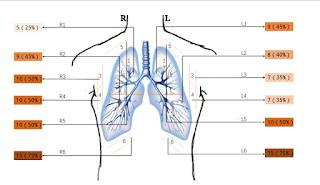
Results: All 20 patients (40 lungs and 240 lung areas) had a history of travel, residence or close contact in/with Wuhan, and 5 of them caught COVID-19 after family gatherings. Lesions tended to occur in both
lungs. Lesions in the lung areas: 14 in L1+R1 area (14/40), 17 in L2+R2 area (17/40), 17 in L3+R3 area (17/40), 17 in L4+R4 area (17/40), 20 in L5+R5 area (20/40), and 28 in L6+R6 area (28/40). Lesion types: rough and discontinuous pleural line (36/240), subpleural consolidation (53/240), air bronchogram sign or air bronchiologram sign in subpleural peripleural consolidation (37/240), visible B lines (91/240), localized pleural thickening (19/240), localized pleural effusion (24/240), poor blood flow in the consolidation detected by color Doppler ultrasound (50/53).
Conclusion: The non-critical COVID-19 has characteristic ultrasonic manifestations, which are visible in the posterior and inferior areas of the lung. The lesions are mainly characterized by a large number of
B lines, subpleural pulmonary consolidation and poor blood flow. Lung ultrasound can provide reference for the clinical diagnosis and efficacy evaluation.

Key Words: lung ultrasound; ultrasonic manifestations; novel coronavirus pneumonia (COVID-19)


Người đăng: VIETNAMESE MEDIC ULTRASOUND DIAGNOSIS vào lúc 19:48 Không có nhận xét nào :

Chủ Nhật, 8 tháng 3, 2020

U S guides bone metastases pain treatment.

By AuntMinnie.com staff writers

March 5, 2020 -- Using ultrasound to guide focused ultrasound treatment for bone metastases pain is feasible and effective, according to a study published online March 4 in Ultrasound Medicine & Biology.

The study findings offer patients and clinicians an alternative to MRI, which has been the modality most often used to perform the procedure but which has its limitations, wrote a team led by Leah Drost of the University of Toronto in Ontario.
"Previous generations of focused ultrasound devices have featured MR image guidance," the group wrote. "These devices have been found ... to be tolerable, effective, and noninvasive in the palliation of metastatic bone pain. ... However, potential drawbacks of such devices include the cost attached to MR equipment, the immobility of the unit, the complex positioning required of the patient, and the long treatment duration."
Drost and colleagues sought to evaluate whether a standalone, portable focused ultrasound device could be guided by diagnostic ultrasound rather than MRI for this application. The study included nine patients treated with focused ultrasound guided by ultrasound. The team assessed the procedure's safety and efficacy 10 days after it was performed.
The researchers found the following:
  • The procedure was safe and tolerable. Four patients reported minor skin irritations.
  • Average pain score decreased from 6.9 at baseline to 3.2 at day 10. P atients' use of analgesics also decreased from baseline to day 10.
  • Six patients reported "durable" pain relief over the assessment period.
"Our study provides evidence that ultrasound-guided focused ultrasound is a safe, tolerable and versatile procedure," the researchers concluded. "It appears to be effective in achieving durable pain response in patients with painful bone metastases. Further research is required to refine the technology and optimize its efficacy."
Người đăng: VIETNAMESE MEDIC ULTRASOUND DIAGNOSIS vào lúc 04:32 Không có nhận xét nào :

Thứ Năm, 5 tháng 3, 2020

Patient and U S features linked to breast cancer


  • By Theresa Pablos, AuntMinnie staff writer


March 4, 2020 -- Nine unique patient and ultrasound features may enable breast imagers to distinguish benign from malignant complex cystic or solid breast lesions, according to research published on February 24 in Ultrasound in Medicine and Biology.
Lesion type, size, orientation, and vascularity were just a few of the features that were found through handheld ultrasound scans and linked to breast cancer. The combination of all nine factors resulted in a sensitivity of 77% and specificity of 82%, the authors reported.
"In addition to identifying whether a lesion is cystic or solid, [handheld ultrasound] is widely applied to tumor diagnosis, biopsy guidance, and even further treatment," wrote the authors, led by Dr. Huiling Xiang from the department of ultrasound at the Sun Yat-sen University Cancer Center in Guangzhou, China. "However, studies related to the diagnostic yield of [handheld ultrasound] in distinguishing benign from malignant complex cystic and solid breast lesions are limited."
The authors defined complex cystic lesions as those that are oval or round in shape with circumscribed margins but also have debris with low-level echoes. These lesions are common in middle-aged women and are often benign, but they do carry a nontrivial risk of malignancy. The researchers therefore wondered whether features on handheld ultrasound might be better able to differentiate complex cystic lesions that needed biopsy and ones that were likely benign.
To find out, the authors used data from 453 patients with 472 complex cystic and solid breast lesions who visited a university cancer center between 2000 and 2018. Radiologists with at least three years of breast imaging experience examined the lesions using handheld ultrasound with a high-frequency linear array transducer.
They also subdivided the lesions into four categories:
  • Type I: Thick wall and/or septations, > 0.5 mm
  • Type II: One or more mural or papillary nodules
  • Type III: Mixed lesion that is more than 50% cystic
  • Type IV: Mixed lesion that is more than 50% solid
The study included a variety of lesion types. About 13% of the lesions were classified as type I, 10% were type II, 21% were type III, and 55% were type IV.
Following biopsy, a little more than half of the lesions were confirmed as benign, while the remaining 45% were deemed malignant. However, the results varied based on lesion type.
The positive predictive value was significantly higher for type III or IV lesions than type I or II lesions. Other factors independently associated with malignancy included the following:
  • Lesion diameter of more than 18 mm
  • Lesion with irregular shape
  • No parallel orientation
  • Uncircumscribed margin
  • Calcification
  • Vascularity in the tumor
  • Abnormal axillary lymph nodes
  • Patient age of 52 or older
A lesion type of III or IV and a lesion diameter of more than 18 mm had the highest sensitivity (87%) and negative predictive value (82%) of any of the factors significantly associated with malignancy. Abnormal findings in axillary lymph nodes had the highest specificity (94%) and positive predictive value (83%).
It is important to note that even type I and type II lesions, which were not linked to malignancy in the study, still met the 2% malignancy threshold to qualify for a BI-RADS 4 categorization and biopsy. While the authors couldn't rule out any lesions as benign, they were able to better categorize them.
"Type I, II, and IV lesions can be assessed as BI-RADS 4B, while type III lesions can directly go into BI-RADS 4C, which allows a more accurate subclassification of complex cystic and solid breast lesions in clinical practice," the authors wrote.
In addition, the researchers excluded complicated cystic lesions without solid components and only included patients who had already undergone handheld ultrasound. As a result, the study may have selection bias.
Nevertheless, the findings reaffirm the power of handheld ultrasound as a powerful, secondary screening tool, the authors noted.
"Therefore, we may safely conclude that the [handheld ultrasound] is a useful modality in differential diagnosis of benign and malignant complex cystic and solid breast lesions and a tool complementing [mammography] in lesion detection," they wrote.
Người đăng: VIETNAMESE MEDIC ULTRASOUND DIAGNOSIS vào lúc 17:34 Không có nhận xét nào :

Thứ Hai, 2 tháng 3, 2020

Older athletes have risk of AA.

By Theresa Pablos, AuntMinnie staff writer

February 28, 2020 -- Older endurance athletes may be prone to heart conditions with a relatively high risk of death. Echocardiographic imaging helped identify an enlarged aorta in 25% of older athletes in a new study, published on February 26 in JAMA Cardiology.
Dilation of the aorta is a risk factor for a group of related conditions tied to an in-hospital mortality rate of more than 25%, with age, male sex, and hypertension established as some risk factors for aortic dilation.
The new research suggests long-term endurance exercise may be a risk factor, too.
"Findings from this study fill an important gap in our understanding of how long-term participation in endurance sport affects the cardiovascular system," wrote the authors, led by Dr. Timothy Churchill from the cardiovascular performance program at Massachusetts General Hospital in Boston.
Previous research has found that intense exercise can contribute to aortic dilation, but no studies had looked at the impact of this phenomenon on older endurance athletes. For their study, the authors recruited 442 male and female runners and rowers who participated in competitive athletic activities, including the Boston Marathon and U.S. national-level rowing competitions.
All athletes were between the ages of 50 and 75 and had at least 10 years of endurance training after the age of 40. The authors screened out participants with a personal or family history of relevant heart conditions, including aortopathy and connective tissue disorders.
Transthoracic echocardiographic imaging was performed on the athletes to measure their myocardial structure and function. The authors took aortic measurements in triplicate over three cardiac cycles.
Out of the male athletes, almost one-third had at least one aortic dimension of 40 mm or larger; however, the findings varied by sport. Male rowers accounted for 61% of athletes with an enlarged aorta. The rowers also had a significantly larger raw aortic size and ascending aorta than male runners.
The difference in aortic dilation between male rowers and runners could be explained by the cardiovascular activity requirements of each sport, the authors noted. For instance, running requires sustained, even cardiovascular activity, whereas rowing requires different levels of strain during the stroke motion.
"While speculative, these findings suggest that the pressure stress uniquely present in rowing may represent an important hemodynamic driver of aortic dilation, particularly at the level of the aortic sinuses," the authors wrote.
For female athletes, the findings were much less pronounced. Only 6% of women had aorta measurements of 40 mm or larger, and there was no significant difference between runners and rowers. However, 38% of female athletes would meet the criteria for enlarged aorta if the threshold was lowered to 34 mm, another standard measurement.
Finally, aortic dilation was also linked to elite athlete status, defined as rowers who participated in the Olympics or world championships and marathon runners who completed races in under two hours and 45 minutes. Elite competitors and those with more cumulative years of athletic training had significantly larger aortic dimensions, the authors found.
"To our knowledge, this study presents the first detailed characterization of the prevalence of clinically relevant ascending aortic dilation among aging competitive athletes," the authors wrote.
What these findings mean for clinical outcomes has yet to be ascertained, the authors noted. They theorized the changes in aorta size could be a previously unrecognized but benign adaption to endurance sports. However, the changes could also mean that long-term training could result in overuse pathology with potentially deadly complications.
"Future studies aimed at defining the natural history of aortic dilation in this population with an emphasis on clinical outcomes ... will be required to resolve this fundamental uncertainty," the authors wrote. "In the absence of such data, clinical implications of our findings remain uncertain and will require individualized assessment."
Người đăng: VIETNAMESE MEDIC ULTRASOUND DIAGNOSIS vào lúc 13:34 Không có nhận xét nào :

Thứ Bảy, 29 tháng 2, 2020

ARFI predicts Prognosis of HCC post RF.


Người đăng: VIETNAMESE MEDIC ULTRASOUND DIAGNOSIS vào lúc 14:23 Không có nhận xét nào :

Thứ Sáu, 28 tháng 2, 2020

U S performance mixed in breast cancer survivors.

By Kate Madden Yee, AuntMinnie.com staff writer.

February 27, 2020 -- Supplemental screening ultrasound shows a mixed performance in women with a personal history of breast cancer, with lower sensitivity and a higher interval cancer rate. But the modality also had higher specificity, according to a study published online February 25 in Radiology
The findings suggest that the quest for effective ways to follow breast cancer survivors continues, wrote a team led by Dr. Soo-Yeon Kim, PhD, of Seoul National University Hospital in South Korea.
"Further research is needed to more accurately identify women who will benefit from supplemental ultrasound screening," the group wrote.
Breast cancer survivors are more vulnerable to developing second cancers, and they are tracked carefully with regular imaging. But mammography has shown lower sensitivity and higher interval cancer rates in women with a history of the disease compared with those without, Kim and colleagues noted.
Breast MRI is recommended for screening women at high risk of breast cancer, but it has its downsides, including high cost and the need for a contrast agent. Because screening breast ultrasound is readily available, doesn't require contrast, and is cost-effective, interest in using it to track breast cancer survivors has increased.
Kim and colleagues compared the performance of screening breast ultrasound between women with and without personal history of breast cancer. Their study included 3,226 breast cancer survivors and 3,226 women without history of the disease, all of whom underwent supplemental breast ultrasound and mammography between January and December 2013. The women were matched by age and breast density.
Fourteen cancers were identified among the breast cancer survivors (six on screening, eight interval) and 13 among the women with no personal history of the disease (12 on screening, one interval). Supplemental ultrasound's performance among the two groups of women varied.
Performance of supplemental screening ultrasound
Performance measureWomen with no personal history of breast cancerBreast cancer survivorsp-value
Abnormal interpretation rate11%7.3%< 0.001
Biopsy rate1.8%1.2%0.04
Interval cancer rate per 1,000 women0.32.50.02
Sensitivity92%43%0.03
Specificity89.3%92.8%< 0.001
"The low sensitivity and high interval cancer rate in women with a personal history of breast cancer imply that limited sensitivity of mammography in these women might have not been fully overcome by supplemental ultrasound," Kim and colleagues wrote. But, "regarding false-positive findings, supplemental ultrasound in women with a personal history of breast cancer showed lower abnormal interpretation rates, lower biopsy rates, and higher specificity than in women without a personal history of breast cancer."
It's clear that more research is necessary to determine how best to track breast cancer survivors, wrote Drs. Christoph Lee and Janie Lee, both of the University of Washington in Seattle, in an accompanying editorial.
"Additional studies are needed in larger and more diverse settings to better define the benefits and harms of supplemental screening among women with a previous history of breast cancer," they wrote.
Người đăng: VIETNAMESE MEDIC ULTRASOUND DIAGNOSIS vào lúc 04:41 Không có nhận xét nào :

Thứ Hai, 17 tháng 2, 2020

Which US method is best for DVT.

By  Theresa Pablos, AuntMinnie Staff writer

February 17, 2020 -- Three different compression ultrasound methods all performed well for imaging deep vein thrombosis (DVT) in a study published online on February 11 by PLOS One. The failure rates for single limited, serial limited, and whole-leg ultrasound ranged from 1% to 2%.


While the three methods are regularly used in clinical practice to help identify DVT, there is no consensus about which one works best. The authors reviewed dozens of studies with thousands of patients and found that no one ultrasound method stood above the rest.
"In the present meta-analysis, more recent studies using clinical follow-up as the reference method showed very comparable failure rates of the three strategies," wrote the authors, led by Dr. Noémie Kraaijpoel from the department of vascular medicine at the University of Amsterdam. "This could imply that there may not be a preferred strategy when taking only safety into account."
The authors searched the scientific literature for compression ultrasound studies from on January 1, 1989, the year the first high-quality research on the modality was published, through July 2019. They included studies using three methods of imaging adults with suspected DVT:
  1. Single limited compression ultrasound -- a single examination of the proximal deep veins
  2. Serial limited compression ultrasound -- two examinations about five to 10 days apart of proximal deep veins
  3. Whole-leg compression ultrasound -- a single examination of both distal and proximal deep veins
Six studies with 2,079 patients assessed single limited compression ultrasound. The analysis showed the modality had a 1.4% failure rate and 6.4% proportion of positive results, although the findings varied among the included individual studies.
For serial limited compression ultrasound, the authors included 11 studies with 3,360 patients. The modality had a 1.9% failure rate and 25% proportion of positive results.
Finally, seven studies with 3,159 evaluated whole-leg compression ultrasound. The findings showed a failure rate of 1% and 25% proportion of positive results.
While all included studies had comparable failure rates (p = 0.36), the proportion of confirmed DVT was significantly higher for serial limited and whole-leg compression ultrasound than for single limited compression ultrasound. The authors noted the difference in prevalence could be the result of patient selection.
Notably, single limited compression ultrasound had a lower DVT prevalence -- likely due to the selection of patients with a lower DVT risk. As a result, the modality could theoretically have a higher failure rate if more DVTs had been found in the group.
Nevertheless, the compression ultrasound methods produced similar results and may be considered equivalent for clinical practice.

"The failure rates of single limited, serial limited, and whole-leg [compression ultrasound] for DVT were found to be quite comparable in patients selected as per the individual study diagnostic algorithms," the authors wrote. "Preference for one of the strategies should be based on probability assessment, feasibility, and expertise."
Người đăng: VIETNAMESE MEDIC ULTRASOUND DIAGNOSIS vào lúc 20:15 Không có nhận xét nào :

Thứ Hai, 10 tháng 2, 2020

Penile Ultrasound Integral to Diagnosing Erectile Dysfunction Cause.


Penile Ultrasound Integral to Diagnosing Erectile Dysfunction Cause

Ultrasound of the penis can play a vital role in determining the underlying cause of erectile dysfunction in men who don’t respond to medication.
Although prescription drugs have significantly – and successfully – changed how providers treat erectile dysfunction (ED), for those men who don’t see improvement, relying on ultrasound can be the key to identifying next steps and possible treatments, according to an article published in the American Journal of Roentgenology.


“Radiologists must be familiar with the imaging protocol, the limitations of the technique, and the interpretations of its findings, to warrant an accurate diagnosis and appropriate patient management,” said lead study author Cristian Gómez Varela, from the radiology department in Complejo Hospitalario Universitario de Pontevedra in Spain. “It is essential to differentiating between the vascular and nonvascular causes of the ED and, therefore, determining appropriate management of the patient.”
The Conditions
When used correctly – with a high-frequency linear array (7.5-12 MHz) and full-length images of the penis in both flaccid and erect states – ultrasound can contribute to successfully diagnosing three vascular-related causes of ED, Varela said.


Peyronie’s Disease (PD): This penile deformity caused by scar tissue that develops after repeated injury results in painful erections. In most cases, MRI is the preferred modality to assess PD. But, gray-scale ultrasound can be very helpful in diagnosis and follow-up, assessing size and location of plaques, detecting small non-palpable lesions or the involvement of the penile septum, or evaluating disease progression. In addition, sonoelastography can estimate tissue stiffness, as well as identify plaques that go undetected on gray-scale ultrasound.
Penile Fracture: Caused by trauma to the penis, usually during sexual intercourse, fracture can cause a rupture in the membrane responsible for trapping blood in the penis to sustain an erection. Ultrasound can make pinpointing any ruptures easier, as fractures are seen as dark breaches in the membrane.
Priapism: This condition can occur in two forms – both high-flow and low-flow. Low-flow priapism is considered a medical emergency as it can cause tissue death and, if left untreated, permanent ED. Ultrasound can identify tissue thickening and scarring in the arteries of the penis, and Doppler ultrasound can also pinpoint inadequate blood retention.
Given its effectiveness is identifying these vascular-related conditions behind ED, according to Varela and colleagues, ultrasound is the preferred method for initially evaluating the penile anatomy and blood flow. Not only is it readily available, but it is minimally invasive and can be well tolerated by patients.

Người đăng: VIETNAMESE MEDIC ULTRASOUND DIAGNOSIS vào lúc 07:38 Không có nhận xét nào :

Thứ Tư, 5 tháng 2, 2020

AI helps characterize breast masses on ultrasound


By Erik L. Ridley, AuntMinnie staff writer
February 4, 2020 -- A breast ultrasound artificial intelligence (AI) algorithm was able to differentiate breast masses at a high level of accuracy by combining analysis of B-mode and color Doppler images, according to research published online January 31 in European Radiology. It even yielded comparable performance to experienced radiologists.
A research team led by Xuejun Qian, PhD, of the University of Southern California and Bo Zhang, PhD, of Central South University in Hunan, China, found that their deep-learning algorithm had substantial agreement with radiologists for providing BI-RADS categorization. It also yielded high sensitivity and specificity.
"The decisions determined by the model and quantitative measurements of each descriptive category can potentially help radiologists to optimize clinical decision-making," the authors wrote.
Breast ultrasound interpretation has been characterized by variable inter- and intrareader reproducibility, with higher false positives than other imaging tests, according to the researchers. Seeking to develop an automated breast classification system that could improve consistency and performance, they gathered a training set of 103,212 breast masses and a validation set of 2,748 independent breast masses at two Chinese hospitals between August 2014 and March 2017. They also assembled a test set of biopsy-proven 605 breast masses classified as BI-RADS 2 to 5 from March 2017 to September 2017.
Next, the researchers trained two convolutional neural networks: one based just on B-mode images and the other based on both B-mode and color Doppler images. On the validation cases, the model based on both B-mode and color Doppler image analysis had a higher level of agreement (kappa = 0.73) with the original interpreting radiologists for BI-RADS categorization than the network based only on B-mode images (kappa = 0.58). The difference was statistically significant (p < 0.001).
They then evaluated the performance of both models on the test set of 605 masses.
Performance of neural networks for classifying breast masses on test set
 Model based on B-mode imagesModel based on both B-mode and color Doppler images
Sensitivity96.8%97.1%
Specificity75.5%88.7%
Accuracy85.3%92.6%
Area under the curve (AUC)0.9560.982
The researchers noted that the addition of color Doppler information improved the algorithm's specificity and accuracy on a statistically significant basis (p < 0.001). The small increase in sensitivity was not statistically significant, however.
"Overall, Doppler information should be incorporated into breast [ultrasound] examination protocols for breast masses, and the use of such a dual-modal system may improve cancer diagnostics," the authors wrote.
The researchers also had 10 radiologists with three to 20 years of breast imaging experience assess the 605 masses in the test set. Nine of the 10 radiologists had a sensitivity that ranged from 87.8% to 94.6%, while the last radiologist had 64.7% sensitivity. That same radiologist had the highest specificity (98.5%), with the remaining participants producing sensitivity ranging from 84.1% to 91.7%. Overall, the 10 readers produced an AUC of 0.948.
"These results indicate that the performance of model 2 reached the levels of the human experts," the authors concluded.


Người đăng: VIETNAMESE MEDIC ULTRASOUND DIAGNOSIS vào lúc 04:44 Không có nhận xét nào :

Thứ Bảy, 1 tháng 2, 2020

ARFI TRONG ĐÁNH GIÁ HẠCH NGOẠI BIÊN.

ARFI TRONG ĐÁNH GIÁ HẠCH NGOẠI BIÊN @ MEDIC HOÀ HẢO
Tao-hinh-xung-luc-buc-xa am-arfi-trong-danh-gia-hach-ngoai-bien


Người đăng: VIETNAMESE MEDIC ULTRASOUND DIAGNOSIS vào lúc 22:02 Không có nhận xét nào :

Thứ Sáu, 31 tháng 1, 2020

ULTRASOUND, CT, MRI Offer Options for Diagnosing Appendicitis.

By AuntMinnie.com staff writers

June 22, 2018 -- Ultrasound, CT, and MRI are all viable supplemental imaging modalities for assessing acute appendicitis after initial ultrasound, according to a study published online June 19 in Radiology.
The team led by Dr. Kevin Eng conducted a literature search on Medline and Embase, identifying studies that used surgery or histopathologic exam information alone or in combination with clinical follow-up or chart review to assess the diagnostic accuracy of supplemental imaging modalities for appendicitis.
Studies included the following:
  • For children -- ultrasound: six studies and 548 patients; CT: nine studies and 1,498 patients; MRI: five studies and 287 patients
  • For adults -- ultrasound: three studies and 169 patients; CT: 11 studies and 1,027 patients; MRI: six studies and 427 patients
All three modalities had comparable, high accuracy for diagnosing the disease, both in children and adults, the researchers found.
Pooled sensitivities and specificities of second-line imaging for diagnosing appendicitis
MeasureUltrasoundCTMRI
Sensitivity: Adults83.1%89.9%97.4%
Sensitivity: Children91.3%96.2%89.9%
Specificity: Adults90.9%93.6%97.1%
Specificity: Children95.2%94.6%93.6%
"All three modalities may be valid as second-line imaging in a clinical imaging pathway for diagnosis and management of appendicitis," they concluded.
Người đăng: VIETNAMESE MEDIC ULTRASOUND DIAGNOSIS vào lúc 05:24 Không có nhận xét nào :
Bài đăng mới hơn Bài đăng cũ hơn Trang chủ
Đăng ký: Bài đăng ( Atom )

Giới thiệu về tôi

VIETNAMESE MEDIC ULTRASOUND DIAGNOSIS
Xem hồ sơ hoàn chỉnh của tôi
Được tạo bởi Blogger.