Tổng số lượt xem trang

Thứ Tư, 5 tháng 6, 2019

Lung Comet US Reveal Subclinical Congestion in HF Patients


ATHENS, Greece — A small but groundbreaking randomized trial has strengthened the case for lung ultrasound (LUS) examinations, which can show likely subclinical pulmonary congestion, in outpatients with heart failure (HF).
The blinking appearance of "B lines" on LUS images, an artifact caused by echo differences between tissue and accumulated fluid, is a confirmed diagnostic and prognostic indicator of congestion. More B lines, also called ultrasound lung comets for the way they streak across the scan from the pleural line, mean more fluid.
The current study suggests the lines could potentially serve as a target for managing volume-depletion therapy, in that adding diuretics in response to them might improve clinical outcomes.
There was a marginally significant 48% decline in 6-month risk for a clinical composite primary endpoint, driven by a more highly significant 75% drop in urgent clinic visits for worsening HF in recently discharged patients whose outpatient diuretic therapy was guided by B lines on LUS.
Scans were obtained using highly portable, pocket-sized systems in all patients, and clinicians who used their findings to adjust diuretics in those assigned to guided therapy didn't follow a defined treatment protocol.
Because of that, the patient population numbering only about 120 from one center, the marginal primary outcome, and other reasons, the study dubbed LUS-HF is more food for thought than an endorsement of LUS-guided HF therapy.
"We propose lung ultrasound as a tool to complement clinical examination and to detect subclinical congestion," said Mercedes Rivas-Lasarte, MD, Hospital de la Santa Creu i Sant Pau, Barcelona, Spain, during the presentation of LUS-HF here at European Society of Cardiology Heart Failure (ESC-HF) 2019.
"The lung-ultrasound guided strategy was safe and reduced the number of decompensations," Lasarte said. "We think that lung ultrasound is a rapid, easy, inexpensive, and broadly available tool that may be recommended in heart failure follow-up to improve outcomes."
However, regarding the use of B lines on LUS to guide diuretic therapy, Lasarte added, "We have to take our study as a proof of concept, and we think that multicenter studies are needed to confirm our results and to test harder endpoints."
Even though there was no treatment protocol in the study, how clinicians managed diuretics for the patients was a good reflection of real-world practice, said Peter S. Pang, MD, Indiana University, Indianapolis, an emergency physician and early adopter of LUS in patients with HF.
The trial's primary endpoint, which included mortality and urgent clinic visit or rehospitalization for worsening HF, may have been significantly reduced in the LUS-guided group, "but I think we need to be careful how we interpret the positive trial because it was driven only by urgent heart-failure visits," Pang, who was not involved in LUS-HF, told theheart.org | Medscape Cardiology.
Still, that can be important. "I think it's fair to say that many patients don't want to come back to the doctor to say they feel worse. So perhaps by using lung ultrasound as a measure of congestion, we can make patients feel better."
Lung ultrasound is safe and it sharpens diagnosis and prognostic evaluations, "so adding it to the bedside examination is strongly encouraged," Pang said.
As for whether resolution or improvement of B lines on serial lung scans after diuretic intensification predicts improved clinical outcomes, "the jury is still out."
The reported number needed to treat with LUS-guided therapy to avoid one primary endpoint was a mere five patients, Pang had pointed out earlier as the invited discussant following Lasarte's presentation.
That indicates an absolute risk reduction of 20%, "an impressive finding, in fact so impressive that we should be cautious. It is unlikely such an effect size would be observed in other populations or in larger studies," he said.
"The good thing about this technology is that it's very easy to do. It's noninvasive, and once you have the ultrasound in your hand, there's no additional cost to it," Mandeep R. Mehra, MD, Brigham and Woman's Hospital, Boston, who is not connected to LUS-HF, observed for theheart.org | Medscape Cardiology.
Although he is cautious about the magnitude of its significance, "this study is at least a step in the right direction. But it's small study, and its confounding by detection of a problem is not to be ignored," he said. That is, because all the patients received LUS, clinicians treating those in the control group could potentially have become aware of and been influenced by the ultrasound findings.
"I always look at these kinds of data with some degree of skepticism."
The LUS-HF design specified that only clinicians who treated patients in the guided-therapy group would have access to the ultrasound results. Treating physicians could take their lead from the scans on any treatment adjustments.
Indeed, they "were strongly directed to change treatment in relation to number of B lines," Lasarte said when presenting LUS-HF.
The trial included 124 patients recently discharged from hospitalization with a primary diagnosis of acute HF. They were required to have had dyspnea and X-ray evidence of pulmonary congestion, high age-adjusted natriuretic peptide levels, but no severe lung diseases.
They were randomized single-blind prior to discharge to receive standard care with guidance from LUS in 61 patients and without LUS guidance in 63 patients. The groups were similar at baseline with respect to mean left-ventricular ejection fraction, natriuretic peptide levels, cardiovascular and pulmonary comorbidities, 6-minute walk distance, and number of B lines on LUS.
Natriuretic peptides were measured and LUS performed thereafter at 2 weeks, 1 month, 3 months, and 6 months.
Six-month rates for death or urgent clinic visits or rehospitalization for worsening HF were 23% in the LUS-guided group and 40% in the control group, for a hazard ratio (HR) of 0.52 (95% CI, 0.27 - 0.99; P = .046).
There were no significant differences in natriuretic peptide levels, measures of quality of life, or the individual components of the primary endpoint except for urgent visits for worsening HF, a prespecified secondary endpoint.
Six-Month Secondary Endpoint Outcomes, LUS-HF
EndpointsLUS Guidance,
n = 61
Non-LUS Guidance, n = 63P
Urgent visits for worsening heart failure, %521.008
Change in 6-minute walk distance, m+60+37.023
Proportion receiving loop diuretics, %9175.023

Other potential applications for LUS using hand-held ultrasound systems in the chronic HF setting, Pang said, include use in a broader population to monitor for signs of impending decompensation, in the hope that early therapy can avoid hospitalization. "The promise for that is great," he said.
"It's not going to replace things like history or physical exam, but maybe it's another thing to add that helps us better decide how to treat patients. That's what I think it adds more than anything else."
Lasarte has reported no relevant financial relationships. Pang has previously disclosed consulting for Baxter, Bristol-Myers Squibb, and Novartis; and receiving support from Bristol-Myers Squibb, Roche, Novartis, Ortho Diagnostics, and Abbott. Mehra has previously disclosed being a consultant for Abbott, Portola, Bayer, and Xogenex; a trial steering committee member for Medtronic and Janssen; a scientific advisory board member for NuPulseCV and FineHeart; and a data safety monitoring board member for Mesoblast; and receiving travel support from Abbott.
ESC-HF 2019. Presented May 25, 2019. Late breaking trial I, Abstract 25.
Follow Steve Stiles on Twitter: @SteveStiles2. For more from theheart.org | Medscape Cardiology, follow us on Twitterand Facebook.

Thứ Hai, 3 tháng 6, 2019

SWE and plantar fasciitis diagnosis.


By Kate Madden Yee, AuntMinnie.com staff writer
June 3, 2019 -- Shear-wave elastography (SWE) boosts diagnostic accuracy in patients with plantar fasciitis, according to a new study published online May 30 in Academic Radiology.
And its diagnostic power is even greater when it's combined with B-mode ultrasound, yielding 100% sensitivity, according to a team led by Dr. Matthias Gatz of University Hospital RWTH Aachen in Germany.
"[Our] study showed for the first time that SWE has an additive diagnostic value for diagnosing plantar fasciitis, with a sensitivity of 100% for the combined usage of SWE and B-mode ultrasound," the group wrote. "Additionally, the diagnostic accuracy increased from 79% using B-mode ultrasound to 84% using SWE."
The current standard for diagnosing plantar fasciitis is B-mode ultrasound to identify hypoechoic areas, border irregularities of the fascia, and calcifications. In particular, plantar fascia thickness of more than 4 mm is the key diagnostic sign for diagnosing fasciitis with B-mode ultrasound. But since fascia thickness can actually decrease over time, even in patients with fasciitis, B-mode ultrasound can miss fasciitis cases.
Gatz and colleagues hypothesized that SWE would be more effective in diagnosing fasciitis because it measures tissue stiffness and can therefore identify the condition even if the fascia isn't thickened, according to the group. The technique may also be more effective than B-mode ultrasound for monitoring the effects of treatment, according to the team.
To investigate, the group conducted a study that included 82 patients with plantar fasciitis. These patients were divided into three groups: symptomatic (39), asymptomatic (23) and bilateral asymptomatic (20). The reference standard for the study was B-mode ultrasound findings of a plantar fascia thickness greater than 4 mm. The researchers measured shear-wave tissue elasticity at the calcaneus, at 1 cm away from the calcaneus, and at the central part of the calcaneus. The group then calculated sensitivity, specificity, and diagnostic accuracy of SWE compared with B-mode ultrasound.
Gatz's team found that SWE was more sensitive and more diagnostically accurate than B-mode ultrasound alone, although it was less specific.
SWE compared with B-mode ultrasound for diagnosis of plantar fasciitis
Performance measureB-modeSWE
Sensitivity61%85%
Specificity95%83%
Diagnostic accuracy79%84%
Sensitivity and diagnostic accuracy were highest when the two techniques were combined, at 100% and 90%, respectively.
The study results show that SWE can boost B-mode ultrasound's performance -- good news for patients with a condition like plantar fasciitis, which doesn't necessarily present in a way ultrasound can identify, according to Gatz's team.
"SWE provides a quantitative assessment of plantar fasciitis integrity and can distinguish between symptomatic and asymptomatic patients better than B-mode ultrasound," the group concluded.

Thứ Bảy, 1 tháng 6, 2019

Ultrasound for evaluating liver steatosis.


By Kate Madden Yee, AuntMinnie.com staff writer
May 31, 2019 -- Ultrasound is a reliable alternative to MRI for assessing liver steatosis in the clinical setting, according to a study published in the July issue of Clinical Radiology. The findings offer clinicians a cost-effective, accessible option for evaluating steatosis.
While MRI has become an accepted modality for detecting liver steatosis, it comes with a high cost and is generally less available than other modalities, according to a team led by Dr. Marie-Luise Kromrey of University Medicine Greifswald in Germany. "Ultrasonography is commonly used to detect liver steatosis and has the advantage of being cost-effective, simple, and widely available," the team wrote.
Fat storage in the liver can be an indicator of metabolic syndrome, a condition characterized by insulin resistance and a precursor to type 2 diabetes. Since liver steatosis is also a risk factor for a variety of other diseases, having an effective way to assess it is important, Kromrey and colleagues noted.
Yet although ultrasound is regularly used to detect liver steatosis, its diagnostic accuracy and reliability for assessing the severity of fatty liver have been unclear. So Kromrey and colleagues conducted a study to compare the modality's performance to that of MRI.
The study included 2,783 patients who underwent 1.5-tesla MRI scans of the liver; from these MRI exams, the group calculated proton-density fat fraction and transverse relaxation rate to estimate liver steatosis and iron overload. Patients also underwent B-mode ultrasound. Kromrey's team then assessed the sensitivity and specificity of ultrasound to identify different degrees of steatosis and amounts of liver iron (Clin Radiol, July 2019, Vol. 74:7, pp. 539-546).
MRI showed liver steatosis in 40% of participants (mild, 68.9%; moderate, 26.7%; severe, 4.4%), while ultrasound found liver steatosis in 37.8%, which corresponded to a sensitivity of 74.5% and a specificity of 86.6%.
The group also found that ultrasound sensitivity increased with the amount of liver fat present (65.1% for low fat content, 95% for moderate fat content, and 96% for high fat content). Liver iron did not affect ultrasound's ability to detect liver steatosis, Kromrey and colleagues noted.
"The present results show excellent sensitivity and specificity of ultrasound for the estimation of fatty liver disease in patients with moderate and high liver fat content," the group wrote.
However, since ultrasound didn't perform as well in patients with low liver fat content, additional evaluation methods may still need to be used, according to the team.
"The weakness of ultrasound in assessing small amounts of liver fat should be considered and compensated by additional liver enzyme quantification or MRI," the authors concluded.

Thứ Tư, 29 tháng 5, 2019

Top 4 Priorities for AI Research in Medical Imaging

By Erik L. Ridley, AuntMinnie staff writer
May 29, 2019 -- Bringing radiology artificial intelligence (AI) technology to routine clinical practice will require four major priorities: structured use cases, data sharing methods, validation and monitoring tools, and new standards and data elements, according to a report published online May 28 in the Journal of the American College of Radiology.
"An active AI ecosystem in which radiologists, their professional societies, researchers, developers, and government regulatory bodies can collaborate, contribute, and promote AI in clinical practice will be key to translating foundational AI research to clinical practice," wrote a team of authors led by Dr. Bibb Allen Jr. of the American College of Radiology (ACR) Data Science Institute.
Following up on an initial medical imaging artificial intelligence roadmap published April 16 in Radiology, which covered the challenges, opportunities, and priorities for foundational research in AI for medical imaging, Allen and colleagues turned their attention to the key priorities for translational research. Both articles were produced as a summary of last year's U.S. National Institute of Biomedical Imaging and Bioengineering (NIBIB) workshop on medical imaging, which was co-sponsored by the RSNA, the ACR, and the Academy for Radiology & Biomedical Imaging Research.
In their latest report, the authors highlighted four key translational research priorities:
  • Create structured use cases to define and highlight the clinical challenges that AI could potentially solve.
  • Create methods to encourage data sharing to support the training and testing of AI algorithms. This would promote generalizability of these algorithms to widespread clinical practice and mitigate unintended bias.
  • Establish tools for validating and monitoring the performance of AI algorithms in clinical practice, to facilitate regulatory approval.
  • Develop standards and common data elements to facilitate seamless integration of AI tools into existing clinical workflows.
In defining and prioritizing AI use cases, the medical imaging community should describe exactly what's important to radiology and what data scientists -- including researchers and developers -- can do to improve patient care, according to the authors.
"Those descriptions should go beyond narratives and flowcharts," they wrote. "Human language should be converted to machine-readable language using standardized data elements with specific instructions for standard inputs, relevant clinical guidelines that should be applied, and standard outputs so that inferences can be ingested by downstream HIT resources."
Standardized inputs would enable algorithms to run on the modality, on a local server, or in the cloud. Meanwhile, application programming interfaces (APIs) could be developed based on these standardized outputs to integrate AI into any system or electronic resource, according to the researchers.
Furthermore, structured use cases should include specifications for data that should be collected to inform the developer how the algorithm performs in actual clinical use, according to the researchers.

"Understanding performance variances that occur in different patient populations, across different equipment manufacturers, or using different acquisition protocols can then be used to refine the algorithm, modify the use case specifications, or inform regulatory agencies," they wrote.

Chủ Nhật, 26 tháng 5, 2019

SWE and POLYCYSTIC OVARIAN SYNDROME


US and BREAST TUMOR SIZE


Abstract
 Aims: To determine the factors influencing ultrasound breast tumor size assessment accuracy. Material and methods: Five factors (tumor type, molecular subtype, histological size, histological grade, and breast density) were used to assess the measurement accuracy of breast ultrasound in tumor size. Size underestimation was defined as ultrasound index lesion diameter < histological size by at least 5 mm.
 Results: Breast ultrasound underestimated tumor size significantly, especially in cases with intraductal components (p=0.002). There was a tendency for higher size underestimation in breast cancer tumors with high–histological grade (p=0.03), human epidermal growth factor receptor type 2 (HER2)-overexpressing breast cancer tumors (p=0.02) and hormone receptor (HR)−/HER2+ breast cancer tumors (p=0.008). Furthermore, core biopsy revealed higher probability of size underestimation with intraductal components (p=0.002). Size underestimation was more frequent with larger histological size (p <0 .001="" breasts.="" breasts="" compared="" dense="" in="" masses="" nbsp="" non-dense="" p="" significantly="" to="" underestimated="" were="">Conclusions: The size underestimation was influenced by pathological type, molecular subtype, and histological size. The pathological results of core biopsy were conducive for predicting tumor size pre-surgery in precise breast cancer diagnosis.
Keywords: breast cancer; ultrasound; tumor size; molecular subtype; core biopsy


Thứ Bảy, 25 tháng 5, 2019

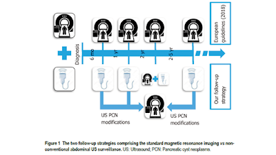
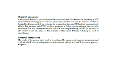
ULTRASOUND vs MRI in SURVEILLANCE FOLLOW-UP PANCREATIC NEOPLASM


EVALUATION of HYPERFERRITINEMIA in DIABETIC PATIENTS

https://www.slideshare.net/hungnguyenthien/evaluation-of-hyperferritinemia-in-diabetic-patients


Hyperferritinemia with normal transferrin saturation, with or without iron overload is 

often found in patients with hepatic steatosis and/or hepatitis. The metabolic 

hyperferritinaemia (disorder of iron and glucose and/or lipid metabolism) may occur with the i
ncidence up to 49% in type 2 diabetes mellitus patients.


Thứ Bảy, 18 tháng 5, 2019

Shear-wave elastography helps diagnose Graves' disease


By Kate Madden Yee, AuntMinnie.com staff writer
April 24, 2019 -- Shear-wave elastography (SWE) is an effective additional tool to complement conventional ultrasound for diagnosing Graves' disease, since it can characterize the tissue stiffness of the thyroid gland, according to a new study published in the May issue of the American Journal of Roentgenology.
The findings support the use of SWE to gather more information about thyroid tissue stiffness than may be possible with conventional ultrasound alone, wrote a team led by Dr. Shimei Li of Sun Yat-Sen University in Guangzhou, China.
"Conventional ultrasound can provide a variety of information, such as size, morphologic features, borders, internal echo characteristics, presence or absence of nodules, and blood flow conditions," the group wrote. "Recently, SWE has been proposed as a complementary technology to conventional ultrasound that can help provide tissue stiffness information. It not only provides a new reference index for the diagnosis of Graves' disease but also helps to monitor the changes in thyroid stiffness in different stages of disease progression."
Graves' disease tends to present in younger people and prompts overactivity of the thyroid gland, the team wrote. Ultrasound has long been used to diagnose the condition, revealing disease features such as an enlarged thyroid gland, diffuse limited hypoechogenicity, and intralesional vascularization on color Doppler (AJR, May 2019, Vol. 212:5, pp. 950-957).
But ultrasound elastography offers additional information about the thyroid gland's tissue stiffness, making SWE more objective than palpation, which is the typical manner of characterizing the tissue, the authors noted.
Li's group included 207 patients in the study. Of these, 162 had Graves' disease and 45 were healthy. All patients underwent an SWE exam that recorded three elasticity values in kilopascals (kPa) for each thyroid gland: SWE mean, SWE minimum, and SWE maximum.
The researchers found that the SWE elasticity values were higher in patients with Graves' disease than in healthy subjects.
SWE elasticity values in patients with Graves' disease
MeasureHealthy subjectsSubjects with Graves' disease
SWE mean14.3 ± 2.7 kPa17.6 ± 6.4 kPa
SWE minimum8.4 ± 2.4 kPa10.7 ± 6.4 kPa
SWE maximum22.1 ± 5.4 kPa25.6 ± 10.6 kPa
All results were statistically significant.
The team also found that the duration of disease, thyroid size, and isthmus thickness, as well as levels of thyroid peroxidase, thyroglobulin, and thyrotropin receptor antibodies correlated with SWE mean in patients with the disease.
It's possible that the results could help clinicians forecast how patients will respond to treatment, according to the group.
"Perhaps [assessing] the thyroid stiffness before treatment could predict the outcome of [radioactive iodine] treatment in patients with Graves' disease," the authors concluded.

Shear-wave elastography useful for assessing rectal tumor depth


By Kate Madden Yee, AuntMinnie.com staff writer
May 17, 2019 -- Shear-wave elastography (SWE) is a useful way to evaluate the depth of invasion of rectal tumors, offering clinicians another tool for preoperative surgery staging, according to a study published online May 10 in Ultrasound in Medicine and Biology.
Accurate staging of rectal cancer before surgery provides the crucial information needed to select appropriate treatment, and SWE can be used as an adjunct to traditional staging modalities such as endorectal ultrasonography (ERUS), wrote a team led by Dr. Zhihui Fan and colleagues of Peking University Cancer Hospital and Institute in Beijing.
"Shear-wave elastography can provide quantitative indicators of the depth of invasion of rectal tumors," the researchers wrote. "This method is tolerable, noninvasive, and easy to perform."
Increasing incidence
Rectal cancer is a common gastrointestinal disease, and its incidence continues to increase, according to Fan's group. The depth of the invasion of rectal cancer is a key prognostic factor and affects clinical treatment options, making preoperative staging crucial. Traditionally, this staging has been performed using ERUS or MRI, each of which has its limitations, according to the authors.
"MRI visualizes the rectal wall involved in rectal cancer adequately, but the examination time is long and the cost is high," the group wrote. "ERUS can visualize the structure of the different layers of the rectal wall clearly and determine the depth of tumor invasion, and the cost is low. ... Although the accuracy rate of ERUS is relatively high, there are still too many cases of overstaging or understaging. In addition, cancer staging by ERUS is heavily dependent on the operator's experience."
Use of SWE has increased, primarily for liver, breast, thyroid, and cervical lesions, but also for assessing prostate lesions. It's beneficial in characterizing tissue stiffness, and cancer tissue tends to be stiffer than healthy tissue. Yet SWE's utility for evaluating preoperative rectal cancer staging has not been studied. So Fan's team investigated the technique's value for this application compared with ERUS and MRI.
The study included 55 patients with rectal cancer who underwent ERUS, SWE, and MRI exams between September 2016 and April 2018. The average distance of each tumor from the anal verge was 6.9 cm. The average length was 3.1 cm and thickness 1.4 cm. All patients had undergone surgery, so the researchers used pathologic results as the gold standard for each modality/technique's performance.
Fan and colleagues found that SWE's overall concordance rate with pathologic stage was better than ERUS or MRI -- although the differences were not statistically significant, which the team attributed to the study's small sample size.
Performance of ERUS, MRI, and SWE for assessing rectal cancer lesion depth
ERUSMRISWE
Rate of concordance78.2%74.6%85.5%
What was statistically significant, however, was SWE's characterization of lesion stiffness by cancer stage: tumor stiffness increased as T-stage increased (p < 0.001).
Finally, the study found that among 27 cases of T1 rectal cancer, four ERUS cases and 10 MRI cases were overstaged; among 18 cases of T3 and T4 rectal cancer, eight ERUS cases and three MRI cases were understaged, according to the authors.
"At present, overstaging and understaging occur with both ERUS and MRI, which may result in unnecessary therapy or loss of opportunity for neoadjuvant therapy," they wrote.
Promising adjunct
Shear-wave elastography offers clinicians important rectal cancer treatment staging information, especially in cases with uncertain diagnosis, Fan and colleagues noted. But SWE should serve as a supplement to ERUS, not a replacement for it.
"A combined evaluation strategy, giving play to the strengths of the two methods, may be more sensible," they wrote.
More research is needed, according to the team.
"SWE is noninvasive, real-time, and inexpensive compared with MRI. It can provide quantitative diagnostic indicators and may be used as a supplement to conventional ultrasound," the group wrote. "To validate our findings, prospective studies with large samples are needed."

Thứ Hai, 6 tháng 5, 2019

Ultrasound-on-Chip Transforms Field Diagnostics.


By Medimaging International staff writers
Posted on 01 May 2019
A handheld, single-probe whole body system is making ultrasound technology universally accessible and affordable.

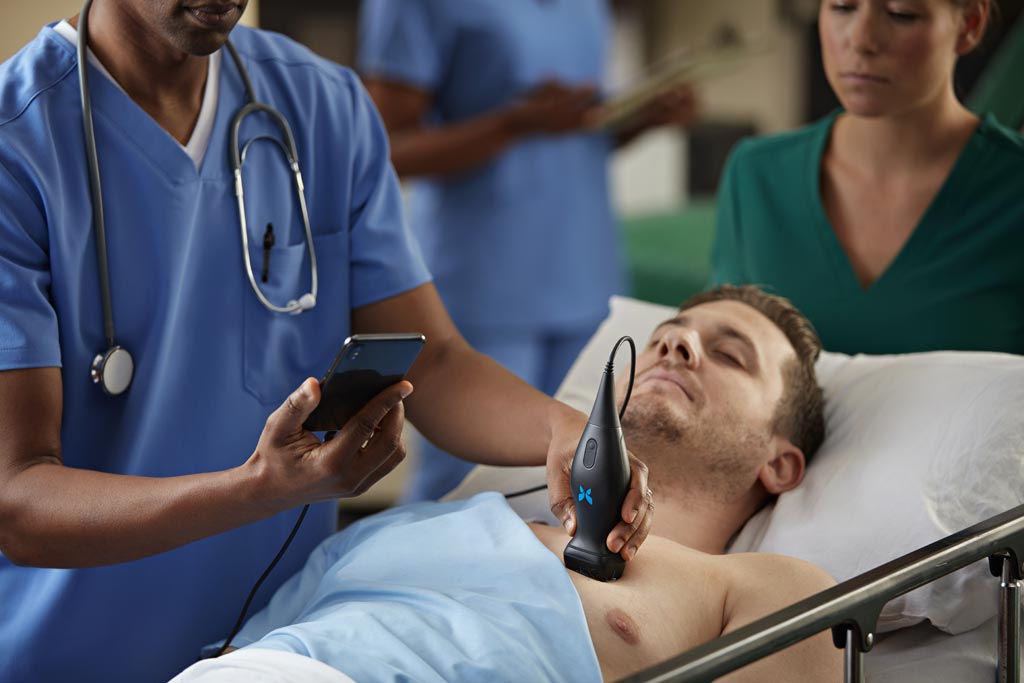
The Butterfly Network (New York, NY, USA) Butterfly iQ is a multi-mode (M-mode, B-mode, and Color Doppler) ultrasound device less than 15 centimeters long and weighing just 313 grams, allowing it to easily fit into a pocket. A rugged anodized aluminum body encases and protects the device, which connects via a USB or lightening cable to a standard handheld Apple iPhone or iPad mobile device. All data is stored and managed on the Butterfly iQ app and in the Butterfly Cloud with 256-bit encryption for tight monitoring and security.

Image: Affordable ultrasound at the point-of care is now possible (Photo courtesy of Butterfly Network).

Butterfly IQ is programmed with 18 preset scanning programs, which enable diagnostic ultrasound imaging of peripheral vessels, including for carotid and arterial studies and procedural guidance; and cardiac, abdominal, urology, gynecological; fetal/obstetric, and musculoskeletal use. In addition, Butterfly Network has developed deep learning-based artificial intelligence (AI) applications that are tightly coupled to the hardware and assist clinicians with both image acquisition and interpretation, which will ultimately enable less skilled users to reliably extract life-saving insights from ultrasound.

The Butterfly iQ is powered by capacitive micromachined ultrasonic transducer (CMUT) technology, which replaces the traditional piezoelectric transducer with a single silicon chip that incorporates an array of 9,000 programmable microelectromechanical systems (MEMS) sensors directly overlaid onto an integrated circuit encompassing the electronics backbone of a high performance ultrasound system, allowing it to emulate any type of transducer - linear, curved, or phased. An integrated 400 mAh Lithium Ion battery provides up to two hours of continuous use.

“Just as putting a camera on a semiconductor chip made photography accessible to anyone with a smart phone and putting a computer on a chip enabled the revolution in personal computing before that,” said Jonathan Rothberg, founder and chairman of Butterfly Network. “Two thirds of the world's population has no access to medical imaging; that's not ok. Butterfly's Ultrasound-on-a-Chip technology enables a low-cost window into the human body, making high-quality diagnostic imaging accessible to anyone.”

Thứ Bảy, 27 tháng 4, 2019

SIÊU ÂM TỤY

Abstract


An ultrasound (US) study is often the first imaging approach in patients with abdominal symptoms or signs related to abdominal diseases, and it is often part of the routine workup. The pancreatic gland, despite its retroperitoneal site, can be efficiently examined with US thanks to advances in US technologies. Nowadays, a pancreatic US study could be considered complete if multiparametric, including the use of Doppler imaging, US elastography, and contrast‐enhanced imaging for the study of a pancreatic mass. A complete US examination could contribute to a faster diagnosis, especially if the pancreatic lesion is incidentally detected, addressing second‐step imaging modalities correctly.

Thứ Hai, 22 tháng 4, 2019

Ultrasound bests x-ray for identifying pulmonary edema.


April 22, 2019 -- Bedside ultrasound is more sensitive than chest x-ray for identifying pulmonary edema in patients presenting with dyspnea, according to a study published in the April issue of the Journal of Ultrasound in Medicine.
And the modality's greater sensitivity isn't its only benefit, wrote a team led by Dr. William Wooten of Mount Carmel Health System in Columbus, OH.
"Bedside ultrasound appears to offer several advantages over chest radiography in the workup of patients with dyspnea beyond its superior sensitivity in the diagnosis of pulmonary edema," the team wrote. "First, bedside ultrasound exposes the patient to less radiation. Second, imaging costs to payers may be reduced."
Chest x-ray has been the commonly used imaging modality to assess patients for pulmonary edema, but its interpretation can be variable, affected by clinicians' level of expertise, Wooten and colleagues wrote. That's why lung ultrasound may be a better tool.
"Lung ultrasound may produce more objective findings through evaluation of vertical comet tail artifacts known as B-lines, which are created by a decrease in the ratio of alveolar air to fluid pulmonary content," the group explained.
The study included 99 patients who presented in the emergency room with dyspnea between August 2016 and March 2017. Of these, 32.3% had congestive heart failure, and 40.4% had chronic obstructive pulmonary disease. Each patient underwent an ultrasound exam within an hour of having a chest x-ray. The researchers used the final diagnosis from the patient's discharge summary as the reference standard (J Ultrasound Med, April 2019, Vol. 38:4, pp. 967-973).
The team found that bedside ultrasound had higher sensitivity compared with chest x-ray, at 96% versus 65%. Specificity was comparable between the two modalities. Of 18 patients with negative x-ray findings and a discharge diagnosis of pulmonary edema, 16 (89%) had positive findings on ultrasound.
Chest x-ray vs. ultrasound for pulmonary edema
MeasureChest x-rayUltrasoundp-value
Sensitivity65%96%< 0.001
Specificity96%90%0.26
Additionally, patients may be more comfortable with bedside ultrasound, Wooten and colleagues noted.
"Anecdotally, we observed that patients appeared to prefer the bedside ultrasound, as it kept the provider at the bedside longer, and the patient did not have to leave the room or family to undergo the chest radiography," the group wrote.
Bedside ultrasound shows promise for patient care when it comes to diagnosing pulmonary edema, but it could also benefit hospitals, according to the authors.

"This study has the potential to affect the care of patients with congestive heart failure, which is the most common cause of hospitalization in the Medicare population and is growing substantially as people are living longer," the team concluded. "As hospitals are being penalized for 30-day readmissions for congestive heart failure, it is crucial that these patients have an accurate diagnosis and are properly treated."

Thứ Tư, 17 tháng 4, 2019

Ultrasound in late pregnancy could reduce C-section rate.


By Kate Madden Yee, AuntMinnie.com staff writer
April 17, 2019 -- Performing ultrasound scans late in pregnancy helps women avoid undiagnosed breech presentation of their babies, translating to improved clinical outcomes, lower rates of emergency cesarean sections (C-sections), and perhaps even lower healthcare costs, according to a study published April 16 in PLOS Medicine.
The findings are good news not only for women and their babies but also for the healthcare system, wrote a team led by David Wästlund of the University of Cambridge in the U.K.
"According to our estimates, universal late pregnancy ultrasound in nulliparous women would virtually eliminate undiagnosed breech presentation, would be expected to reduce fetal mortality in breech presentation, and would be cost-effective if fetal presentation could be assessed for less than 19.80 pounds [$25.95 U.S.] per woman," the group wrote.
Fetal breech presentation increases the risk of complications for the baby and the mother, Wästlund and colleagues noted. Typically, a baby's position is assessed by palpating the woman's abdomen, but this technique's sensitivity varies by practitioner.
"Despite the relative ease with which breech presentation can be identified through ultrasound screening, the assessment of fetal presentation at term is often based on clinical examination only," the researchers wrote. "Due to limitations in this approach, many women present in labor with an undiagnosed breech presentation."
The investigators performed screening ultrasound at 36 weeks gestation in 3,879 English women having first pregnancies between January 2008 and July 2012. Of these, 179 (4.6%) were diagnosed with breech presentation. In more than half of those (54%), breech presentation had not been suspected prior to labor.
Women with babies in the breech position were offered a procedure called an external cephalic version (ECV) to try to turn the baby; for those who did not want this procedure or for whom it did not work, a C-section was scheduled.
The investigators also estimated the cost of universal late pregnancy ultrasound scans using data from the English National Health Service (NHS) to compare birth outcomes of breech pregnancies screened with and without ultrasound.
The ECV procedure was attempted in 84 (46.9%) of the women with breech babies and was successful in 12 (14.3%). Of the 179 women with breech babies, the researchers found the following:
  • 10.6% delivered vaginally (following either a planned or spontaneous version).
  • 61.5% delivered via elective C-section.
  • 27.9% delivered via emergency C-section (due to labor starting before the scheduled cesarean date).
"No woman in the cohort had a vaginal breech delivery or experienced an intrapartum cesarean for undiagnosed breech," the researchers noted.
Wästlund and colleagues estimated that routine late pregnancy ultrasound could prevent nearly 15,000 undiagnosed breech presentations, more than 4,000 emergency C-sections, and seven to eight baby deaths per year. But the effect of the intervention on healthcare costs needs more research, the group wrote: "If universal ultrasound could be provided for less than 12.90 pounds [$16.91] per scan, the policy would also be cost saving." But it's unclear if this is possible.
"If this procedure could be implemented into routine care, for example, by midwives conducting a routine [scan at 36 weeks gestation] and using a portable ultrasound system, it is likely to be cost-effective," the group concluded. "Such a program would be expected to reduce the consequences to the child of undiagnosed breech presentation, including morbidity and mortality."