BÀI SOẠN VỀ SIÊU ÂM CHẨN ĐOÁN

BS NGUYỄN THIỆN HÙNG

Tổng số lượt xem trang

Sparkline

Thứ Bảy, 13 tháng 6, 2020

Ultrasound for Primary Breast Screening

By Theresa Pablos, AuntMinnie staff writer


June 11, 2020 -- Could ultrasound be used as a primary screening method for breast cancer? A long-term systematic review published on June 1 in BMC Cancer found ultrasound scans performed well as both a primary and secondary cancer screening modality.
The review, conducted by researchers in China, pooled data from almost two dozen studies evaluating the effectiveness of ultrasound for breast cancer screening. Ultrasound screening performed comparably to mammography in most categories and may work particularly well as a screening method for women with dense breasts, the authors noted.
"Up to now, there have been no consistent conclusions concerning whether [ultrasound] screening should be recommended as the primary screening method for women in the screening guidelines for breast cancer," wrote lead authors Lei Yang and Shengfeng Wang from Peking University in Beijing.
Cancer detection, recall, and biopsy rates
The review included 23 English-language studies published between January 2003 and May 2018. Twelve of the studies evaluated the use of ultrasound as a secondary screening method after a negative mammography result, while the remaining 11 studies compared the effectiveness of ultrasound and mammography as primary screening modalities.
Mammography vs. ultrasound for breast cancer screening
 Mammography, primary screeningUltrasound, primary screeningUltrasound, secondary screening
Cancer detection rate per 1,000 examinations4.64.63.0
Recall rate4.6%5.9%8.8%
Biopsy rate1.5%2.3%3.9%
Percentage of all cancers detected65%68%N/A
Percentage of invasive cancers detected65%87%74%
The review found no statistically significant differences in sensitivity and specificity between ultrasound and mammography as primary breast cancer screening methods. Mammography detected 65% of cancers, comparable to the 68% of cancers detected through ultrasound scans. Similarly, mammography identified 97% of women without cancer, while ultrasound detected 98% of women without cancer.
Furthermore, both mammography and ultrasound identified 4.6 cancers per 1,000 examinations. Ultrasound scans detected significantly more invasive cancers than mammography, but the modality also resulted in a higher percentage of recalled patents. The researchers found no statistically significant differences between mammography and ultrasound for the percentage of biopsies or for detection of node-negative invasive cancers
When used as a secondary screening method after a negative mammogram result, ultrasound identified 96% of occult breast cancers and 93% of healthy patients. The modality also identified 3 cancers per 1,000 scans, with an 8.8% recall rate and a 3.9% biopsy rate. As a secondary screening method, ultrasound found 74% of invasive cancers and 71% of node-negative invasive cancers.
"Our study is the first systematic review and meta-analysis to investigate the performance of [primary ultrasound] screening for breast cancer, and that is also an important up-to-date systematic review and meta-analysis investigating the performance of [secondary ultrasound] screening," the authors wrote.
Benefits and limitations of ultrasound screening
Ultrasound has some benefits as a breast screening method, including that it is radiation-free and may be more accessible in low-resource countries and areas. However, it is also not without its limitations. For instance, ultrasound cannot image the whole breast at once or show microcalcifications, and it requires a skilled operator.
"However, as shown in our study, these concerns seemed not to cause significant differences in the sensitivity and specificity, or even in cancer detection rates and cancer characteristics between [primary ultrasound] screening and [primary mammography] screening," the authors wrote.
All of the studies in the review were graded as high or fair quality by the authors, but the review itself had some noteworthy limitations. Importantly, some of the ultrasound studies in the review included repeated screenings using the same group of individuals. For the analysis, the authors counted each screening as an individual person, which could have overestimated the benefit of ultrasound screening.
Despite its limitations, the authors noted the findings highlight the need for future studies to investigate the long-term benefits and risks of using ultrasound as a primary breast cancer screening method, particularly for use in women with dense breasts and those who live in rural or resource-poor areas.

"We hope that [ultrasound] screening for breast cancer should deserve more attention in the future, not only because [ultrasound] is comparable to [mammography] in women with dense breasts ... but also because ultrasound is not a radiation modality and is easier to access in low-resource areas, such as Chinese rural areas," the authors concluded.
Người đăng: VIETNAMESE MEDIC ULTRASOUND DIAGNOSIS vào lúc 20:30 Không có nhận xét nào :

LIVER 2D-SWE ARTIFACTS

 Two-dimensional shear wave elastography artifacts, although seen very frequently in a clinical setting, are poorly recognized. Our review emphasizes that interpretation of 2D-SWE images must incorporate knowledge of US artifacts.


Người đăng: VIETNAMESE MEDIC ULTRASOUND DIAGNOSIS vào lúc 20:21 Không có nhận xét nào :

Thứ Năm, 11 tháng 6, 2020

ULTRASOUND and SARCOPENIA


ABSTRACT

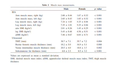
Our aim of this study is to compare the thigh muscle thickness measurements obtained using ultrasound and bioelectrical impedance analysis (BIA) methods, and to investigate the validity and cutoff value of the ultrasonography.
We analyzed a total of 201 participants (99 male and 102 female participants, mean age, 66.2 years) participated in the annual health checkup in the Yakumo Study, 2014. Thigh muscle thickness (TMT, sum of the rectus femoris and vastus intermedius muscle thickness) was measured using ultrasound at mid-thigh in the sitting position. Appendicular skeletal muscle mass (aSMI) was measured using BIA. Cutoff value of TMT was determined through the receiver operating characteristic analysis. We defined sarcopenia with the diagnostic algorithm of Asian Working Group for Sarcopenia.
TMT was significantly reduced in subject with sarcopenia than in those without sarcopenia in both gender. Muscle measurements obtained using the BIA methods (aSMI) and ultrasound methods (TMT) showed a significant correlation, with a correlation coefficient of 0.38 (P < 0.001). Cutoff value, sensitivity, and specificity of TMT in diagnosis of muscle loss were 36 mm, 72.0%, and 73.9%, respectively, for the male participants, and 34 mm, 72.2%, and 72.4%, respectively, for the female participants.
In conclusion, the ultrasonography for thigh muscle might be a simple diagnostic method for sarcopenia.

Keywords: ultrasonography, thigh muscle thickness, sarcopenia, community-dwelling people, cut-off value


Người đăng: VIETNAMESE MEDIC ULTRASOUND DIAGNOSIS vào lúc 14:50 Không có nhận xét nào :

Thứ Tư, 10 tháng 6, 2020

C B F in Neonatal Sepsis

 Abstract
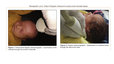
Background: Neonatal sepsis is an important cause of morbidity and mortality among newborns. As there is paucity of literature regarding early alteration of the cerebral blood flow (CBF) in neonatal sepsis our study aims to evaluate the changes in the CBF velocities and Doppler indices in neonates with early‑onset neonatal sepsis (EONS) and to evaluate the predictive accuracy of cerebral blood flow velocities (CBFV) by using ultrasound Doppler as a diagnostic marker of EONS.
Methods: This cross‑sectional analytical study was conducted over a period of 2 years with 123 neonates enrolled in the study. The neonates were divided into two groups: Group I (with 54 neonates) ‑ neonates with EONS and group II (with 69 neonates) ‑ age‑matched neonates without any signs of sepsis. Ultrasound Doppler examination was performed and the cerebral hemodynamics assessed in neonates during the first seventy two hours of life. Doppler indices and CBFV were measured in the internal carotid artery (ICA), middle cerebral artery (MCA), and vertebral artery (VA) of either side. Data were analyzed using the statistical program SPSS version 23.0 (SPSS Inc., Chicago, IL, USA). Sensitivity, specificity, positive predictive value (PPV), negative predictive value (NPV), and diagnostic accuracy were calculated at different selected cutoff values for CBFV parameters.
Results: Lower resistance and higher peak systolic velocity and end diastolic velocity have been documented in neonates with EONS.
Conclusion: Our study shows that the cerebral hemodynamics in neonates with EONS is altered which can be assessed bedside by noninvasive ultrasound Doppler examination.


Người đăng: VIETNAMESE MEDIC ULTRASOUND DIAGNOSIS vào lúc 21:55 Không có nhận xét nào :

Thứ Sáu, 5 tháng 6, 2020

PoCUS for Syncope

Point-of-care ultrasound to complete physical exam and to reach the diagnosis in a young man with syncope

  • Fatemeh Rasooli, 
  • Mehrnoosh Aligholi Zahraie & 
  • Maryam Bahreini 
The Ultrasound Journal volume 12

Abstract

Background

Cardiac syncope can result from an atrial myxoma due to outflow obstruction. Myxoma is the most common primary cardiac tumor that may cause sudden death and the nonspecific symptoms may make early diagnosis difficult.

Case presentation

A 27-year-old man presented to our emergency department after two episodes of syncope and severe fatigue. He had no complaint of fever, weight loss, sweating, chest pain or dyspnea. Vital signs were within normal limits. A loud heart S1 was detected and normal neck veins. Other systemic examinations including neurological assessment were normal. Electrocardiography showed normal sinus rhythm. An obvious variability in heart rate was noticed on cardiac monitor changing by the patient’s position. Point-of-care ultrasonography (PoCUS) showed a large hyperechoic lesion with a well-defined stalk originating from the left atrium (LA). Thus, the patient was transferred to a cardiac surgery center for surgical intervention. Histopathology reported an LA mass compatible with myxoma.

Conclusions

Emergency physicians should be familiar with the vague presentations of cardiac tumors to improve patient outcomes. It is beneficial to take advantage of bedside ultrasound for prompt diagnosis and subsequent treatment.
Người đăng: VIETNAMESE MEDIC ULTRASOUND DIAGNOSIS vào lúc 12:03 Không có nhận xét nào :

Feasibility of renal resistive index

Feasibility of renal resistive index measurements performed by an intermediate and novice sonographer in a volunteer population

  • Mårten Renberg, 
  • Naima Kilhamn, 
  • […]
  • Max Bell 
The Ultrasound Journal volume 12

Abstract

Background

The Doppler-derived renal resistive index (RRI) is emerging as a promising bedside tool for assessing renal perfusion and risk of developing acute kidney injury in critically ill patients. It is not known what level of ultrasonography competence is needed to obtain reliable RRI values.

Objective

The aim of this study was to evaluate the feasibility of RRI measurements by an intermediate and novice sonographer in a volunteer population.

Methods

After a focused teaching session, an intermediate (resident), novice (medical student) and expert sonographer performed RRI measurements in 23 volunteers consecutively and blinded to the results of one another. Intraclass correlation coefficients and Bland–Altman plots were used to evaluate interobserver reliability, bias and precision.

Results

Both non-experts were able to obtain RRI values in all volunteers. Median RRI in the population measured by the expert was 0.58 (interquartile range 0.52–0.62). The intraclass correlation coefficient was 0.96 (95% confidence interval 0.90–0.98) for the intermediate and expert, and 0.85 (95% confidence interval 0.69–0.94) for the novice and expert. In relation to the measurements of the expert, both non-experts showed negligible bias (mean difference 0.002 [95% confidence interval − 0.005 to 0.009, p = 0.597] between intermediate and expert, mean difference 0.002 [95% confidence interval − 0.011 to 0.015, p = 0.752] between novice and expert) and clinically acceptable precision (95% limits of agreement − 0.031 to 0.035 for the intermediate, 95% limits of agreement − 0.056 to 0.060 for the novice).

Conclusions

RRI measurements by both an intermediate and novice sonographer in a volunteer population were reliable, accurate and precise after a brief course. RRI is easy to learn and feasible within the scope of point-of-care ultrasound. 
Người đăng: VIETNAMESE MEDIC ULTRASOUND DIAGNOSIS vào lúc 11:57 Không có nhận xét nào :

AA in Patient < 50years of age


Abstract


Introduction

Practitioners of US routinely include a survey of the abdominal aorta during abdominal US in accordance with international guidelines. Such practice is of uncertain value in younger patients.


Results

The most common indications for US were abdominal pain (1337, 44%), deranged liver function tests (453, 15%), nausea and/or vomiting (229, 8%), elevated inflammatory markers (146, 5%), pancreatitis (134, 4%) and pyrexia (127, 4%). Fewer than half (977, 49%) of the reports contained a comment regarding the aorta. Aortic pathology was reported in 2 (0.1%) cases. Both were reported as aortic ectasia and both represented a false‐positive diagnosis. One male patient had a known abdominal aortic aneurysm with endovascular aortic repair. No new aortic aneurysms were found. All cases of atherosclerotic disease were ignored, and none were reported. Periaortic pathology was encountered on 1 patient, but this was known. No case of new periaortic pathology was detected.
Người đăng: VIETNAMESE MEDIC ULTRASOUND DIAGNOSIS vào lúc 04:50 Không có nhận xét nào :

Thứ Tư, 3 tháng 6, 2020

US autopsies help doctors understand COVID-19

  By Theresa Pablos, AuntMinnie staff writer

 June 3, 2020 -- A hospital in São Paulo used a portable ultrasound machine to perform autopsies on 10 patients with fatal cases of COVID-19. The authors described the modality's benefits for studying the effects of the disease caused by the novel coronavirus in a paper published on May 22 in Histopathology.
Using a technique called ultrasound-based minimally invasive autopsies (MIA-US), an ultrasound examiner and technologist took tissue samples from the most affected parts of each patient's organs. The findings confirmed that COVID-19 affects multiple organs and tissues, including the kidneys, spleen, lymph nodes, brain, testicles, and skin.
The ultrasound team also wore head-to-toe personal protective equipment, including two aprons, rubber boots, plastic sleeves, three glove layers, a rubber cap, an N95 mask, a surgical mask, and eye protection.
During the autopsies, the examiner and technologist used a portable ultrasound machine with multifrequency broadband transduces and standard image quality. They scanned patients' organs to identify the most-affected locations, then cut 10-cm openings at the appropriate locations to take ultrasound-guided tissue samples from the lung, liver, kidneys, spleen, and heart. They also took unguided samples from the quadriceps, skin, and brain.
The samples revealed significant lung findings, including exudative and/or proliferative diffuse alveolar damage. The authors also found severe alveolar epithelial changes, which they described as more intense and prevalent than findings for other respiratory viruses.
Many but not all findings outside of the lungs could be attributed to the comorbidities of the patients or to septic shock, the authors noted. For instance, eight patients had fibrinous thrombi in alveolar arterioles and a high density of alveolar megakaryocytes, which the authors believed could be evidence of a hypercoagulative state in severely ill patients. The researchers also found cases with superficial perivascular dermatitis, myositis, orchitis, and myocarditis.

Người đăng: VIETNAMESE MEDIC ULTRASOUND DIAGNOSIS vào lúc 22:40 Không có nhận xét nào :

Thứ Bảy, 30 tháng 5, 2020

Abdominal imaging reveals bowel injuries in COVID-19 patients


Matt O'Connor | May 12, 2020 | Advanced Visualization
https://www.healthimaging.com/topics/advanced-visualization/imaging-bowel-injuries-covid-19?utm_source=newsletter&utm_medium=hi_monthly

A number of studies have shown how COVID-19 can impact a patient’s lungs, but new research suggests individuals may experience bowel abnormalities as well.
Boston-area physicians retrospectively looked at more than 400 patients admitted to a single center to reach their conclusions, published May 11 in Radiology. They found that abnormalities were most commonly seen in sicker patients with the coronavirus who were also admitted to the intensive care unit.

... "abdominal manifestations” in those who are infected, said study author Rajesh Bhayana, MD, abdominal imaging fellow at Massachusetts General Hospital’s Department of Radiology.
“Some findings were typical of bowel ischemia, or dying bowel, and in those who had surgery we saw small vessel clots beside areas of dead bowel,” Bhayana said in a statement. “Patients in the ICU can have bowel ischemia for other reasons, but we know COVID-19 can lead to clotting and small vessel injury, so bowel might also be affected by this.”

As part of their study, Bhayana et al. included patients who tested positive for severe acute respiratory syndrome coronavirus between March 27 and April 10. With an average age of 57 years, 17% of individuals had cross-sectional abdominal imaging, which included 44 ultrasounds, 42 CT scans and one MRI.
In total, 31% of CTs showed bowel abnormalities, which represented 3.2% of all patients involved in the study. Findings included thickening and ischemia-related discoveries such as pneumatosis (gas in the bowel wall) and portal venous gas. In two individuals who had bowel resection, the team found ischemia with patchy necrosis, and both had injuries that suggested bowel ischemia may be caused by small blood clots.

There are a number of potential explanations for this laundry-list of findings, the authors noted, including direct viral infection, small vessel thrombosis, or nonocclusive mesenteric ischemia. But one thing is for sure: More studies are required to determine if SARS-CoV-2 plays a direct role in bowel or vascular injury in those with COVID-19.
Người đăng: VIETNAMESE MEDIC ULTRASOUND DIAGNOSIS vào lúc 20:00 Không có nhận xét nào :

Thứ Năm, 28 tháng 5, 2020

Ultrasound Can Diagnose Many Arm Fractures in Kids


Medscape Logo

News > Reuters Health Information
By Lisa Rappaport
May 22, 2020
https://www.medscape.com/viewarticle/930981
(Reuters Health) - While ultrasound can accurately diagnose many upper extremity fractures in children, a new study suggests it may not always catch elbow fractures.
Researchers conducted a systematic review and meta-analysis, examining data from 32 studies with 2,994 pediatric patients who had a total of 3,415 scans for symptoms consistent with upper extremity fracture. As a reference point to compare the accuracy of ultrasounds, 30 studies used plain radiographs; one study used radiographs, MRI or bone scan; and one study used MRI.
Người đăng: VIETNAMESE MEDIC ULTRASOUND DIAGNOSIS vào lúc 16:46 Không có nhận xét nào :

Thứ Bảy, 23 tháng 5, 2020

Siêu âm Tĩnh mạch Cửa trong Viêm Ruột dư Trẻ em


Người đăng: VIETNAMESE MEDIC ULTRASOUND DIAGNOSIS vào lúc 14:11 Không có nhận xét nào :

Thứ Sáu, 22 tháng 5, 2020

Ultrasound Vector Flow Imaging for AVF Fistula.

Ultrasound  Vector Flow Imaging  for AVF Fistula


ABSTRACT
Introduction: We report the use of a new ultrasound technique to evaluate the axial and lateral components of a complex flow in the arteriovenous fistula (AVF). Vector Flow Imaging (VFI) allows to identify different components of the flow in every direction, even orthogonal to the flow streamline, represented by many single vectors. VFI could help to identify flow alterations in AVF, probably responsible for its malfunction.
Methods: From February to June 2016, 14 consecutive patients with upper-limb AVF were examined with a Resona 7 (Mindray, Shenzhen, China) ultrasound scanner equipped with VFI. An analysis of mean velocity, angular direction and mean number of vectors impacting the vessel wall was carried out. We also identified main flow patterns present in the arterial side, into the venous aneurysm and in correspondence of significant stenosis.
Results: A disturbed flow with the presence of vectors directed against the vessel walls was found in 9/14 patients(64.28%): in correspondence of the iuxta-anastomotic venous side (4/9; 44.4%), into the venous aneurysmal tracts (3/9; 33.3%) and in concomitance of stenosis (2/9;22.2%). The mean velocity of the vectors was around 20-25 cm/s, except in presence of stenosis, where the velocities were much higher (45-50 cm/s). The vectors directed against the vessel walls presented high angle attack (from 45° to 90°, with a median angular deviation 65°).
Conclusions: VFI was confirmed to be an innovative and intuitive imaging technology to study the flow complex-
ity in the arteriovenous fistulas.
Keywords: Arteriovenous fistula, Hemodialysis, Hemodynamic shear stress, Ultrasound, Vascular access, Vector
Flow Imaging.
Người đăng: VIETNAMESE MEDIC ULTRASOUND DIAGNOSIS vào lúc 21:23 Không có nhận xét nào :

Thứ Năm, 21 tháng 5, 2020

Tongue Ultrasound may identify Obstructive Sleep Apnea

By Theresa Pablos, AuntMinnie staff writer


May 21, 2020 -- Tongue stiffness measured by shear-wave elastography (SWE) ultrasound may be able to help identify patients with obstructive sleep apnea (OSA) while they're awake, according to the findings of a pilot study published on May 10 in Ultrasound in Medicine & Biology.
Some studies have suggested that up to 38% of the population in Europe and the U.S. can have OSA, but many patients with the potentially fatal condition go undiagnosed. Ultrasound may be one easily accessible and noninvasive tool to help identify patients who may benefit from a sleep test.
"We hypothesized that development of OSA involves changes in tongue stiffness, and this effect can be seen using SWE during wakefulness," wrote the authors, led by Dr. Chun-Hsiang Chang from Cardinal Tien Hospital and the Fu Jen Catholic University School of Medicine in New Taipei City, Taiwan.
To test their hypothesis, Chang and colleagues recruited 46 participants, 26 of whom had sleep-confirmed OSA and 20 healthy controls.
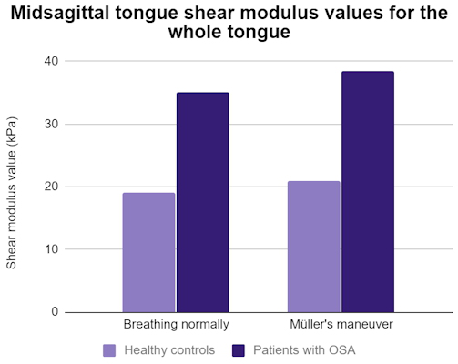
First, the researchers performed conventional B-mode ultrasound using a curvilinear-array transducer to identify the tongue tissues of the patients. Then, they performed SWE while the participants breathed normally and when they performed Müller's maneuver, a technique in which they attempt to inhale while forcing their mouth and nostrils closed.
radiology graph
Patients with sleep apnea showed significant differences from the healthy controls in multiple tongue stiffness values while breathing normally and during Müller's maneuver. Those with OSA also had significantly higher values during Müller's maneuver than when breathing normally for the anterior and middle thirds of the tongue. For comparison, controls had no significant differences for any part of the tongue.
The differences remained statistically significant predictors of OSA after the authors adjusted for age, sex, BMI, and neck circumference, middle tongue shear modulus measured during normal breathing, and whole and posterior tongue and geniohyoid muscle shear modulus measured during Müller's maneuver.
"Quantifying tongue tissue stiffness in a clinical setting is difficult," the authors wrote. "Through [ultrasound] SWE, we found that the stiffness of the tongue was higher in awake OSA patients than in healthy controls during normal breathing and the [Müller's maneuver]."
Based on their findings, the authors recommended cut-off shear-wave modulus values to help detect patients with obstructive sleep apnea. For some values, the accuracy could exceed 80%. The authors cautioned, however, that the findings are preliminary and that more research is needed to validate the results.

"Future application of the technique may involve ultrasonographic measurement of tongue stiffness during sedative or natural sleep, which will help to define the pathophysiology for an individual patient with OSA and to verify treatment results for those procedures targeting the tongue," the authors wrote.

---------

Müller's manoeuvre is a procedure used in medicine to diagnose sleep apnea, some types of tinnitus,[1] and other medical problems.

Procedure

After a forced expiration, an attempt at inspiration is made with closed mouth and nose, whereby the negative pressure in the chest and lungs is made very subatmospheric; the reverse of a Valsalva maneuver.
This technique is designed to look for collapsed sections of airways such as the trachea and upper airways. In this maneuver, the patient attempts to inhale with his mouth closed and his nostrils plugged, which leads to a collapse of the airway. Introducing a flexible fiberoptic scope into the hypopharynx to obtain a view, the examiner may witness the collapse and identify weakened sections of the airway. Müller's maneuver is used to help determine the cause of sleep apnea. A positive test result means the site of upper airway obstruction is likely below the level of the soft palate, and the patient will probably not benefit from a uvulopalatopharyngoplasty alone. This maneuver is very helpful in doing MRI for sleep apnea, when sedation to patient can be avoided.
There is some evidence that the sites of obstruction with Müller's maneuver do not represent reliably the sites of obstruction during normal sleep. Other factors such as the body's position whilst conducting the manoeuvre may well affect this.
Müller's maneuver can also be used to terminate supraventricular tachycardia in an acute primary care setting.
Người đăng: VIETNAMESE MEDIC ULTRASOUND DIAGNOSIS vào lúc 22:03 Không có nhận xét nào :

Thứ Bảy, 16 tháng 5, 2020

ULTRASOUND for LUNG COVID-19


Người đăng: VIETNAMESE MEDIC ULTRASOUND DIAGNOSIS vào lúc 13:19 Không có nhận xét nào :

Thứ Ba, 5 tháng 5, 2020

Course teaches ob/gyns to identify COVID-19 on lung US.

By Theresa Pablos, AuntMinnie staff writer


May 4, 2020 -- A two-and-a-half-hour online course boosted the knowledge of COVID-19 lung ultrasound presentation among ob/gyn clinicians with no prior lung ultrasound experience, according to the findings of a small study published on April 29 in Ultrasound in Obstetrics & Gynaecology.
....
Lung ultrasound scans have proved to be an effective, noninvasive modality for screening patients, including pregnant patients, with confirmed or suspected cases of the novel coronavirus. The latest study adds to existing research demonstrating ob/gyn clinicians may be well-poised to recognize B-lines, subpleural consolidations, a thickened or patchy pleural line, and other hallmark signs of potential infection with the novel coronavirus.
For their study, Buonsenso and Moro recruited two pneumologists and one pediatrician, each with more than 10 years of lung ultrasound experience, to create an online training module.
The module begins with a 15-minute pretest that includes 10 ultrasound video clips showing healthy, normal lungs or ones with pathological patterns. The test asks participants to choose whether the lung video shows the following:
a) Normal findings
b) Clear and distinct vertical artifacts with small pleural alterations
c) Broken pleural line and small consolidations
d) White lung without evidence subpleural consolidations
e) White lung with evidence subpleural consolidations
f) Pattern not compatible, vast consolidations
g) Pattern not compatible, pleural effusions
Following the pretest, participants watch a two-hour theoretical course providing lung ultrasound education. Finally, the course ends with a 15-minute test showing the same 10 ultrasound clips.
.....
Người đăng: VIETNAMESE MEDIC ULTRASOUND DIAGNOSIS vào lúc 20:11 Không có nhận xét nào :

Thứ Bảy, 2 tháng 5, 2020

Adherence to the Standards for Reporting of Diagnostic Accuracy (STARD) 2015 Guidelines in Acute Point-of-Care Ultrasound Research.


Abstract

IMPORTANCE Incomplete reporting of diagnostic accuracy research impairs assessment of risk of bias and limits generalizability. Point-of-care ultrasound has become an important diagnostic tool for acute care physicians, but studies assessing its use are of varying methodological quality.

OBJECTIVE To assess adherence to the Standards for Reporting of Diagnostic Accuracy (STARD) 2015 guidelines in the literature on acute care point-of-care ultrasound.

EVIDENCE REVIEW MEDLINE was searched to identify diagnostic accuracy studies assessing point-of-care ultrasound published in critical care, emergency medicine, or anesthesia journals from 2016 to 2019. Studies were evaluated for adherence to the STARD 2015 guidelines, with the following variables analyzed: journal, country, STARD citation, STARD-adopting journal, impact factor, patient population, use of supplemental material, and body region. Data analysis was performed in November 2019.


Người đăng: VIETNAMESE MEDIC ULTRASOUND DIAGNOSIS vào lúc 19:59 Không có nhận xét nào :

Chủ Nhật, 26 tháng 4, 2020

SIÊU ÂM CƠ VÂN LY GIẢI [DO CHẤN THƯƠNG]

Sonographic appearance of rhabdomyolysis – a systematic review of the literature 

Łukasz Sein Anand, Wojciech Kosiak

Abstract 

Rhabdomyolysis is the process of striated muscle cell lysis, during which proteins and microelements such as myoglobin are released into the bloodstream. It is important to diagnose rhabdomyolysis as soon as possible and start the treatment according to severity, as it is a state that significantly increases the mortality of the patients. The current gold standard of rhabdomyolysis diagnosis is the creatine kinase plasma concentration test, but it can be also diagnosed with imaging techniques, such as ultrasound (US). This review aims to gather previously published information regarding sonographic appearance of rhabdomyolysis. We searched through PubMed and ScienceDirect databases for studies using designed queries. After the selection process we were left with 13 studies containing a description of US appearance of rhabdomyolysis confirmed with a CK plasma level test. Findings described in the majority of the cases were muscle thickening, ground glass opacity, traits of edema and anechoic areas. Other than these, there were several less often reported findings. As a conclusion, rhabdomyolysis seems to have its own US appearance, but for now it cannot be precisely specified and needs further research for clarification.

Keywords: ultrasonography; rhabdomyolysis; point-of-care


Rhabdomyolysis seems to have its own US appearance, mostly in a form of muscle thickening, ground glass opacity, traits of edema, anechoic areas and various less often reported lesions. Due to the small number of published cases the typical aspect of rhabdomyolysis is not clear and needs further research for clarification.
Người đăng: VIETNAMESE MEDIC ULTRASOUND DIAGNOSIS vào lúc 18:46 Không có nhận xét nào :

Thứ Bảy, 25 tháng 4, 2020

FDA relaxes rules on ultrasound in COVID-19


The FDA carved out a special section of the guidance for ultrasound systems, which have become useful in diagnosing patients with COVID-19. The agency said that because ultrasound scanners do not emit ionizing radiation, the FDA believes users can have "additional flexibility," with modifications allowed under the following circumstances:
  • Modifications that would allow an ultrasound scanner to be used outside of the environment for which it is cleared, such as a general practitioner's office or a field hospital
  • Changes that would enable the acquisition of images by healthcare practitioners who are not trained in sonography, under appropriate supervision
  • The addition of lung scanning clinical applications, as long as labeling is included that is based on established practice guidelines, such as from the American Institute of Ultrasound in Medicine (AIUM)
  • The availability of probes and machine settings that are suitable for lung scanning, such as probes at a frequency of 3 MHz and higher, and with linear, curvilinear, and phased-array technologies
  • The use of a mechanical index (MI) < 1.4 as an indicator for potential biological effects on tissues containing gas bodies
The agency would frown on ultrasound modifications as follows:
  • Changes that would result in an increase of the derated maximum acoustic output parameters
  • Modifications that would exceed known safety limits in humans, such as a MI ≥ 1.4 for lung images
  • Modifications that would allow lay users to acquire images without guidance of healthcare practitioners.
Người đăng: VIETNAMESE MEDIC ULTRASOUND DIAGNOSIS vào lúc 05:37 Không có nhận xét nào :

Thứ Tư, 22 tháng 4, 2020

Abdominal Pain: An Unusual -- But Not Rare -- COVID-19 Presentation


  • Whitney J. Palmer
April 20, 2020
Fever. Cough. Shortness of breath. These are what have become known as the classic, tell-tale COVID-19 symptoms. But, there’s a different, less common, one that should alert you to possible infection– abdominal discomfort.


Recent literature has revealed that as many as 20 percent of patients present to the hospital with a digestive symptom, such as diarrhea, vomiting, pain, accompanying their respiratory symptoms. And, roughly 5 percent show up with an abdominal complaint alone.
This is where abdominal radiologists can play an integral role, said industry experts in a recent article published in the American Journal of Roentgenology. A team, led by Abraham Dachman, M.D., professor of radiology and abdominal imaging specialist with UChicago Medicine, shared three cases where patients were referred for abdominal imaging and providers distinguished findings indicative of COVID-19 infection in the lung base.
“It’s important not to be lulled into a false sense of security by a chief complaint of abdominal pain,” Dachman wrote. “The fact that we observed several patients presenting in this way in such a short time period suggests that although abdominal symptoms are an unusual presentation of the infection, they are not rare, and abdominal radiologists must be on the lookout to diagnose COVID-19, even when it is not suspected clinically.”
Because these findings first appeared on abdominal scans, Dachman wrote, it’s important for all radiological sub-specialties to be aware that COVID-19 can produce abdominal complaints.
“Careful evaluation of the lung bases for the typical nodular, peripherally distributed ground-glass opacities may lead to the correct diagnosis before progressive respiratory manifestations,” he wrote. “An early diagnosis is helpful not only in patient care, but also in allowing healthcare workers to be prepared with the appropriate personal protective equipment.”
Người đăng: VIETNAMESE MEDIC ULTRASOUND DIAGNOSIS vào lúc 22:26 Không có nhận xét nào :
Bài đăng mới hơn Bài đăng cũ hơn Trang chủ
Đăng ký: Bài đăng ( Atom )

Giới thiệu về tôi

VIETNAMESE MEDIC ULTRASOUND DIAGNOSIS
Xem hồ sơ hoàn chỉnh của tôi
Được tạo bởi Blogger.