Tổng số lượt xem trang

Thứ Sáu, 24 tháng 9, 2021

CEUS of the Lung Reveals Multiple Areas of Microthrombi in a COVID-19 Patient


A case of a 61-year-old woman (see Fig.  1) with severe COVID-19 and a negative CTPA study. We were able show that these areas of irregularity, labelled as subpleural consolidation by Peng et  al. were avascular and therefore most likely to represent 3–5 mm microinfarcts. Conversely, non-thrombotic consolidation would be seen to have some enhancement; a process not appreciated using other imaging modalities due to the superior spatial resolution of ultrasound. Anecdotally, we have also seen these areas resolve with clinical improvement. It is becoming apparent that severe cases of COVID-19 are characterised by hyperinfammation and a thrombotic phenomenon.

The theory of an underlying thrombotic process also corresponds with studies identifying admission D-dimer, prothrombin time and thrombocytopenia as prognostic markers [3]. Immuno-thrombosis is used to describe the interaction between platelets, coagulation factors and innate immune effector systems that, during an infection, results in secondary thrombus formation. This is not the first time CEUS has been used to evaluate the presence of emboli. Trenker et  al. found that, despite the lack of definite confirmation of PE on CT, peripheral subpleural consolidations with no or inhomogenous enhancement on CEUS to be highly suggestive of embolic consolidations [4].

A follow-up study by the same team used histological examination of six cases, and pulmonary infarction was found in all of them [5].

Thứ Tư, 22 tháng 9, 2021

Lung abnormalties persist in 91% of COVID-19 patients

By Kate Madden Yee, AuntMinnie.com staff writer

September 10, 2021 -- More than 90% of patients who developed COVID-19 showed lung parenchymal abnormalities on chest CT and x-ray three months after recovery in a study published in the September issue of Clinical Infectious Diseases.


The findings highlight the need for further investigation into COVID-19's long-term effects, wrote a team led by Dr. Bram van den Borst of Radboud University Medical Center in Nijmegen, the Netherlands.

"Long-term health consequences of this coronavirus disease ... are yet largely unknown, but many patients are likely to experience long-lasting morbidity," the authors wrote. "Indeed, based on observations from diseases that share COVID-19 characteristics ... it is hypothesized that, in the long-term, a significant number of patients with COVID-19 will suffer from lung function impairment, residual pulmonary parenchymal abnormalities, decreased physical capacity, loss of muscle mass, anxiety, depression, cognitive deficits, post-traumatic stress disorder, fatigue, and poor health status" (Clin Infect Dis, September 2021, Vol. 73:5, pp. e1089-e1098).

The study included 124 patients who had been treated for COVID-19 at Radboud in 2020. Of the 124 patients, 27 had mild, 51 had moderate, 26 had severe, and 20 had critical disease. Each participated in a follow-up examination three months after recovery that consisted of lung function measurement tests, chest CT and x-ray, a six-minute walking test, and a body composition evaluation; each participant also responded to a questionnaire about their mental, cognitive, and health status, as well as their quality of life.

Although the majority of patients showed reduced ground-glass opacities on CT and normal chest x-rays at the three-month follow-up, the researchers also found that 91% had persistent parenchymal lung abnormalities, including ground-glass opacities, pleural lines and parenchymal bands, bronchiolectasis, and fibrosis. Nearly a third of patients had three lung abnormalities on CT at follow-up, and nearly a quarter had four.

CT imaging results in COVID-19 patients 3 months after recovery
Type of abnormality
Ground-glass opacity86%
Lines and bands64%
Bronchiolectasis60%
Fibrosis26%
No. of abnormalities
09%
120%
220%
327%
424%

The team also found the following:

  • 72% of patients reported lower quality of life.
  • 69% showed fatigue.
  • 64% demonstrated functional impairment.
  • 36% showed mental and/or cognitive function difficulties.

"Our results indicate that a substantial proportion of patients still experience severe problems in various health domains three months after COVID-19," the group wrote.

The study suggests that more research on the long-term effects of COVID-19 is needed, according to the authors.

"Longer follow-up studies are warranted to elucidate natural trajectories of COVID-19 recovery, to find predictors of complicated long-term trajectories, and to develop strategies to decrease long-term COVID-19 morbidity," they concluded.

Thứ Bảy, 18 tháng 9, 2021

LUS POST-COVID-19 at MEDIC CENTER


SƠ KẾT SIÊU ÂM PHỔI SAU NHIỄM COVID-19

KHOA SIÊU ÂM MEDIC

ĐẶT VẤN ĐỀ

Sau nhiễm COVID-19 một số bệnh nhân có nhu cầu khám kiểm tra tại MEDIC. Một bài báo của 

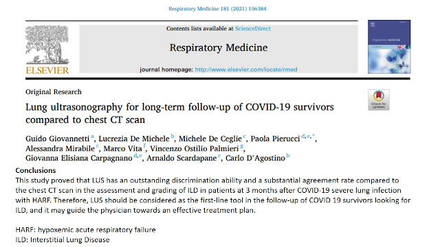
Respiratory Medicine 181, March 2021 đã công bố về vấn đề này với 38 bệnh nhân post COVID-19 infection 03 tháng, dùng siêu âm phổi [LUS] đối chiếu với CT và một số xét nghiệm cận lâm sàng. Nội dung xác nhận tổn thương phổi còn tồn tại ở bệnh nhân từng có suy hô hấp trong thời gian điều trị trong bệnh viện trước đó.

Tại thành phố HCM, hiện chưa có báo cáo nào về khám kiểm tra siêu âm phổi [LUS] sau nhiễm COVID-19. Nhưng nhận thấy có xu hướng cần khám hậu nhiễm COVID-19 tại MEDIC Hòa Hảo nên chúng tôi, được phép của BS Giám đốc, thực hiện việc theo dõi và ghi nhận lại những kết quả ban đầu.

MỤC TIÊU

Ghi nhận tổn thương phổi tồn tại bằng siêu âm phổi (LUS) dựa theo đề nghị của ROUBY, [ROUBY's Protocol] bằng cách khảo sát 12 vùng của 2 phổi P và T và sau đó tính điểm LUS Score total.


PHƯƠNG PHÁP

Khảo sát tiền cứu mô tả và cắt ngang những bệnh nhân và gia đình có nhiễm COVID-19 nay đã có xét nghiệm RT-PCR (-) tự nguyện đến khám tại MEDIC Hòa Hảo.

Thực hiện việc khám LUS và chuẩn hóa (theo y văn tham khảo): tinh chỉnh hệ số mechanical index MI <0,7, tắt tất cả các bộ lọc filters [vốn dùng cho làm đẹp hình ảnh trên màn hình siêu âm] như Harmonic và S-Harmonic, THI… tùy theo hãng máy siêu âm có tại MEDIC.

 
Phối hợp với X-Quang và CT nếu có yêu cầu của bệnh nhân và bác sĩ phòng khám.
 Lưu ý những bệnh nền [phổi, tim, gan, thận, tuyến giáp...] đi kèm. 

Tìm thêm những thương tổn mới ở  tim, gan, thận, tụy, ruột, giáp, mắt, cơ vân...là những cơ quan có thể bị ảnh hưởng do tình trạng nhiễm COVID-19 thúc đẩy, theo hiểu biết hiện nay.


Thống kê nhân thân và bệnh sử. Hồ sơ được lưu trữ điện toán hóa và truy cập qua mã QR code của MEDIC cho từng bệnh nhân.

-------------

mechanical index [MI] is an attempt to measure part of an ultrasound beam's bioeffects. The MI is found on most ultrasound display screens along with the thermal index  

MI of the ultrasound beam is the amount of negative acoustic pressure within  an ultrasonic field

In fact, studies on animal lung tissue have reported effects such as pulmonary capillary haemorrhage for mechanical indices in the diagnostic range.

---------------


KẾT QUẢ

Từ cuối tháng 8 năm 2021 đến 18/9/2021, đợt sơ kết này gồm 06 nam và 05 nữ, tuổi từ 34-60, trung bình sau nhiễm COVID-19 khoảng 30 ngày, trong đó có một nhân viên nữ của khoa siêu âm. Tất cả đều có thời gian cách ly và điều trị trong bệnh viện, đều có khó thở mức độ trung bình, có một trường hợp bệnh nhân nữ hôn mê 3 ngày. Một bệnh nhân nam có khạc tia máu bầm khi đến khám. Một bệnh nhân nam khác, dược sĩ, đã chích ngừa 2 mũi AZ nhưng sau đó lại trở thành F0 và tự điều trị tại nhà trong khi mẹ bị lây nhiễm và chết vì nghi nhiễm COVID-19.


Hình ảnh LUS  post COVID infection của đợt sơ kết này

 gồm còn tồn tại dày màng phổi có gián đoạn [10/11 ca], light beam [8/11 ca], subpleural consolidation nhỏ dưới 20mm [8/11 ca]. Đa số phát hiện tồn tại tổn thương  ở vùng phổi sau bên [posterior lateral] cả 2 bên nhưng thường là phổi trái. Phần lớn thuộc phân loại nhóm C và D, có

 LUS Score total <10, cá biệt có một ca có LUS Score total=15.

Lúc khám LUS các bệnh nhân đều được đo SpO2 =95-99%, bằng oxymeter xung, nhịp thở 10-18 lần/phút, không khó thở khi khám nhưng đều than phiền leo cầu thang hay gắng sức còn thấy khó thở, mệt, chưa trở lại bình thường. 02 bệnh nhân nữ khai báo bị tê vùng lưng và đau cánh tay T (post COVID-19 myositis?]. Tất cả  đều trong quá  trình hồi phục dần [lên cân và ăn ngủ khá tốt].

BÀN LUẬN

Siêu âm phổi LUS  giúp phân biệt các bệnh lý tràn khí màng phổi, viêm phổi vi trùng, phân biệt phù phổi do tim và không do tim... từ hơn 20 năm nay. Khi phát hiện tràn dịch màng phổi, LUS chỉ cần lượng dịch ít hơn X-quang phổi quy ước.

Trong đại dịch COVID-19, LUS được sử dụng nhiều hơn, cơ động hơn, tránh được lây nhiễm với các máy siêu âm cầm tay [handheld] và kỹ thuật PoCUS.Tuy nhiên có khuyến cáo được đồng thuận cao là không dùng LUS để loại trừ nhiễm COVID-19 trong giai đoạn bệnh khởi phát.

Hình ảnh LUS trong nhiễm COVID-19 và sau nhiễm COVID-19 đã được mô tả bởi nhiều tác giả, được hệ thống hóa vùng khám, và định lượng bằng cách cho điểm theo phân loại thống nhất (4 loại A B C D) và thang điểm 0 1 2 3, như đã đề cập. Có tác giả nhận xét rằng hình ảnh siêu âm LUS trong nhiễm COVID-19 rõ nhất khoảng 2 tuần sau khởi phát rồi kém rõ dần. 

Trong nhóm bệnh sau nhiễm COVID-19 tại MEDIC, như các tác giả khác, chúng tôi cũng dựa theo phân loại và thang điểm định lượng trên.


Hình ảnh siêu âm phổi sau nhiễm COVID-19 trong báo cáo sơ kết này gồm những tổn thương tồn tại nhỏ như dày màng phổi, light beam, subpleural consolidation;  thường ở vùng phổi đáy sau bên [posterior lateral] của phổi trái. Đo oxymeter xung lúc khám LUS, kết quả SpO2 dao động từ 95-99% và bệnh nhân không biểu hiện khó thở. LUS Score total <10 trong đa số 10/11 ca cho thấy mức độ các thương tổn tồn tại này không cần can thiệp và không ảnh hưởng nhiều đến chức năng sống sau nhiễm COVID-19.

Những tổn thương này, theo y văn, với 2 trong 4 tiêu chí, trong giai đoạn mới mắc phải COVID-19, đủ để có hướng chẩn đoán nhiễm COVID-19 gây viêm phổi mô kẽ do siêu vi bằng LUS, trong khi chờ đợi kết quả RT-PCR và xét nghiệm vi sinh học khác khi sàng lọc và phân tầng nguy cơ.

 Y văn về tổn thương phổi sau nhiễm COVID-19 trên LUS và CT cũng nêu ý kiến chưa biết rõ nguyên nhân và hướng điều trị tiếp tục ra sao. Do vậy, tác giả đề xuất nên khám LUS định kỳ cho bệnh nhân sau nhiễm COVID-19, nhất là bệnh nhân từng bị khó thở hay suy hô hấp, nếu cần phải chụp CT thì nên dựa theo kết quả [outcomes] LUS trước đó.


KẾT LUẬN


Sơ kết với 11 ca  sau nhiễm COVID-19 đến khám tại MEDIC, chúng tôi ghi nhận có tồn tại tổn thương phổi mức độ nhẹ, đa số có LUS Score total <10,  cần có thời gian theo dõi thêm 3 – 6 tháng, 1 năm hoặc hơn để xem các tổn thương phổi này bao giờ biến mất. Và vì COVID-19 là bệnh đa cơ quan nên cần theo dõi định kỳ tiếp tục cho đến khi nào chức năng sống của bệnh nhân trở lại như trước.


TÀI LIỆU THAM KHẢO


Giovannettia G et al: Lung ultrasonography for long-term follow-up of COVID-19 survivors compared to chest CT scan,  Respiratory Medicine 181, March 2021 https://doi.org/10.1016/j.rmed.2021.106384
 McDermott C et al: Combatting COVID-19: is ultrasound an important piece in the diagnostic puzzle? , Emerg Med J, 2020 Oct;37(10):644-649. doi: 10.1136/emermed-2020-209721. Epub 2020 Sep 9.   

Vetrugno et al : The “pandemic” increase in lung ultrasound use in response to Covid-19: can we complement computed tomography findings? A narrative review, Ultrasound J (2020) 12:39  https://doi.org/10.1186/s13089-020-00185-4


Thứ Tư, 15 tháng 9, 2021

L U S for long-term follow-up of COVID-19 survivors


US COMBATTING COVID-19


PATIENT SELECTION FOR LUS DURING THE COVID-19 PANDEMIC

 The role of LUS during the COVID-19 pandemic is to identify characteristic sonographic abnormalities as well as to support clinical decision making. Not all patients with clinically suspected COVID-19 will warrant LUS, and appropriate patient selection is essential to minimise unnecessary exposure of healthcare workers (HCWs) to this virus. LUS should be performed after the medical history is taken, when a specific clinical question arises and with a pretest probability of COVID-19 diagnosis already in mind.

► The majority of patients who are clinically well and fit for discharge are unlikely to benefit from LUS, as they will be managed based on clinical appearance.

► In clinically well patients with risk factors for severe COVID-19 (such as chronic lung disease, obesity, diabetes mellitus or cardiovascular disease), abnormal LUS findings may identify a patient cohort that would benefit from closer observation such as a home pulse oximeter and remote monitoring.

► Critically ill patients should be resuscitated without delay, and LUS is not useful for the primary diagnosis of COVID-19. Ultrasound is useful in critically unwell patients to examine for other causes of undifferentiated shock, for example, PE, cardiac tamponade or hypovolaemia, thus avoiding anchoring bias in the midst of the current pandemic.

► Goal-directed focused cardiac ultrasound may help identify left ventricular and right ventricular size and function in the case of COVID-19 heart–lung complications, which include myocarditis, right-sided and left-sided heart failure and PE.27 28

► Ultrasound can also be used to assess volume status and guide fluid resuscitation where necessary.29

 ► Ultrasound can be used to assist with emergency central or peripheral venous access.

LUS SCANNING TECHNIQUE

 In general, principles and techniques of LUS are the same for patients with suspected COVID-19 as they were in the preCOVID-19 era. Some modifications necessary for patients with suspected COVID-19 will also be outlined. Transducer selection30

► Linear transducers (5–10 MHz) are better for visualising superficial structures (figure 5). These may be used to view pleural line irregularities, small superficial effusions, skip lesions and B-lines.

► Curvilinear transducers (2–7 MHz) may be better for posterior and deeper or central pathology such as consolidation, hepatisation and air or fluid bronchograms.

Optimising settings

► Optimise the depth of field of view so that the pleural line is in the middle of the screen.

 ► Adjust the transducer focal zone to the level of the pleural line for increased spatial resolution.

► Turn off smoothing algorithms such as compounding and tissue harmonic imaging filters to allow visualisation of lung artefacts. Most lung presets will default to this mode.

► Record cine loop clips rather than still images to visualise subtle pleural changes that may not appear on a single frame. Transducer hold Hold the transducer close to the crystal matrix, between the tips of the index finger and the thumb of the insonating hand (figure 5). Fingers of the insonating hand should be spread out to stabilise the transducer and hand position. Brace the insonating hand against the surface being scanned. These techniques will facilitate small adjustments of the transducer and will allow for greater probe stability and better quality images to be shown on the screen. Scanning protocol Traditional lung scanning protocols suggest evaluation of several anterior, lateral and posterior lung zones. Chinese authors have described COVID-19 scanning using a 12-zone protocol (figure 6).6 Soldati et al30 have proposed a 9-zone protocol and associated scoring system to quantify pulmonary involvement. It is possible to perform a focused study (six chest zones) in less than 2min,31 and the Intensive Care Society has endorsed this approach as part of the Focused Ultrasound in Intensive Care (FUSIC) lung accreditation module (figure 6).32 

Modifications to minimise exposure risk COVID-19 changes are often found in postero-basal zones.6 30 It may be quicker and safer for the point-of-care ultrasound provider to:

► Scan with the patient facing away from the operator to minimise healthcare worker (HCW) exposure to droplets (figure 5). The ultrasound machine may also become less contaminated if placed behind the patient

Start by scanning the patient’s back using the linear transducer in vertical orientation.

► Start medial to the scapula sliding inferior to the lower rib border and moving laterally towards the posterior axillary line.

 ► Evaluate each rib space first with the transducer in a vertical (crossing the ribs) orientation (figure 5) then evaluate each rib space again with the transducer in a horizontal orientation (between the ribs) especially if any abnormalities are seen.

► Finish by scanning lateral zones of the lung in the midaxillary line. Using the curvilinear probe here may be helpful (figure 5).

 Cleaning and disinfection protocols

Strict adherence to decontamination strategies are vital to prevent patient-to-patient COVID-19 transmission as well as patient-to-HCW transmission. What follows are summary points drawn from a number of international best practice standards33 34 and should be considered when using ultrasound with suspected COVID-19 patients:

► Place a dedicated ultrasound machine in the COVID-19 ‘hot zone’ of the ED. 

► Wear standard personal protective equipment when performing LUS and wear gloves when moving the machine between cubicles.

► Strip away ECG leads, gel bottles, extra buckets and straps from the machine. 

► Use a barcode scanner to enter patient details to avoid further contact with the machine.

► Use the machine in battery mode; precharge at all times to avoid use of cables.

► Use a touchscreen device rather than a keyboard, cart-based system.

► Consider using a handheld device, for example, Lumify or ButterflyIQ systems, with the advantage that the whole device can be placed within a probe cover and images are uploaded to the cloud for remote reviewing.

 ► Consider use of a transparent, disposable drape to cover the screen, cradle and cart of the ultrasound machine.

► Use chlorhexidine/alcohol or soap-based wipes to clean transducer heads, as well as the entire length of probe cables, screen and cart after scanning.35 Wait for up to 3min ‘dry time’ after using disinfectant wipes before using the machine again.

► Use a transducer sheath/probe cover for all high-risk patients.

► Use single-use gel packets rather than gel bottles.

CONCLUSION

LUS appears promising as a comprehensive imaging modality in clinically suspected or diagnosed COVID-19, when implemented mindfully and in conjunction with other diagnostic modalities. LUS findings should be interpreted alongside a careful history, physical examination and with pretest probability in mind. Point-of-care ultrasound may help to identify the need for further investigations or may guide the physician towards an alternative diagnosis. Incorporating ultrasound into the evaluation of COVID-19 patients will depend on available resources, expertise of personnel and logistic configurations unique to each situation.


COVID-19: The New Ultrasound Alphabet in SARS-CoV-2 Era


COVID-19: The New Ultrasound Alphabet in SARS-CoV-2 Era

 To the Editor

We applaud the proposal of Piliego et al1 to use lung ultrasound (US) as a bedside test for triage of coronavirus disease 2019 (COVID-19) patients and for subsequent management of clinical workload and level of care in the scenario of a hospital overloaded with severe acute respiratory syndrome coronavirus 2 (SARS-CoV-2) pandemic patients.2 We describe an expanded role for US. Preliminary reports from the Italian outbreak2 prompted us to adopt some variations on our standard clinical protocols and to implement or upgrade techniques we already use in our critical care practice before SARS-CoV-2 pandemic. We propose the following COVID-US alphabet (Figure).

 C: cardiac evaluation 1. Cardiac chambers diameters and kinesis 2. Pericardium (effusion, tamponade) 3. pulmonary artery pressure 4. ejection fraction% 5. inferior vena cava diameter variations differential 

O: outputs 1. renal resistive index 2. velocity-time integral 

V: ventilation 1. B-lines patterns 2. B-lines spatial distribution 3. Hyperinflation and recruitment response 4. Lung score 5. Search for pneumothorax/effusion

 I: intubation 1. Prediction of difficult laryngoscopy/intubation 2. Endotracheal intubation confirmation 

D: Doppler and deep venous thromboembolism/ pulmonary embolism 

The study of cardiac function, diameters and kinesis, including pericardium, is performed bedside at admission and at times when there are significant hemodynamic changes during the intensive care unit (ICU) stay. Cardiac evaluation includes determination of EF%, PAPs, aortic VTI at apical fifth chamber time/velocity integral at apical fifth chamber at aortic efflux as a more comprehensive parameter than sole stroke volume variation (SVV%),3 ΔIVCD%4 with respiratory cycle and RRI.3 Comparative observation of preload parameters (inferior vena cava diameter variations differential %) with contractile (EF%) and ejective function (VTI) and perfusion indexes such as RRI, allow us to tailor hemodynamic and ventilator therapy based on the specific physiopathological picture of the single patient and to exclude pulmonary embolism. Hemodynamic management of COVID-19 patients is particularly challenging because of cardiopulmonary interactions in mechanically ventilated patients. COVID-19 patients show specific lung abnormality patterns, including lesser effect on pulmonary compliance, increase in pulmonary vascular resistance with consequences to the right ventricle, inferior vena cava and renal function, and on left ventricle and systemic perfusion. In this setting, US is useful in decisions regarding pharmacological choices, fluid administration, ventilator adjustments together with metabolic indexes (ie, blood lactate), and the whole clinical picture. In this perspective, RRI, though not as well established as EF or VTI, is added to US hemodynamic evaluation with emphasis on evaluation of “effective” organ perfusion,4 and as added decisional support for administration of vasoactive drugs, diuretics, or renal vasodilators,5 for renal replacement therapy, including Cyto-Sorb (CytoSorbents Corp, Monmouth Junction, NJ) for cytokine storm control. 

Similarly, the choice of best positive end-expiratory pressure (PEEP) is based not only on arterial oxygen partial pressure/fraction of inspired oxygen ratio and driving pressure evaluation but also on its hemodynamic effects and kidney repercussions. In a 22-patient sample, we observed 9% (2 cases) of exnovo kidney failure, compared with 22.2% in New York6 experience. Ventilation was regularly assessed by US between 3 and 4 times in 24 hours to follow evolutional trends of COVID-19–specific US lung findings,7 to score the amount of B-lines and titrate ventilation accordingly. Response to recruitment maneuver with PEEP escalation was evaluated with US, addressing the need for high (recruiters) or low (nonrecruiters) PEEP settings and the decision for early/late/no prone positioning.2 A potential US application that we have not yet adopted is the assessment of respiratory fatigue through respiratory muscle evaluation, with implications for decision to intubate after the noninvasive ventilation trial2 and extubation readiness assessment. 

In our practice, we also use US for preintubation airway evaluation, given the aerosolization risk associated with the performance of conventional tests (measuring interincisor distance, determining Mallampati score),8 intubation confirmation when end-tidal CO2 is not immediately available,2 diagnosis of intubation-related complications (pneumothorax, pneumomediastinum, airway trauma), and for lung and ventilation assessment.7 Finally, evaluation of right cardiac chamber diameters and lung windows, and eventual integration with lower limbs US, is used to monitor thromboembolic phenomena as part of the routine coagulative evaluation (thromboelastography/thromboelastometry), given the high thrombotic risk associated with COVID-19.7 

We believe that our approach has 2 important novelties.

 First of all, it is not only lung US but integrated US, involving cardiac and pulmonary evaluation, fluid repletion status and perfusion, airway evaluation, and thrombosis screening

The second point is that COVID-US approach is not only a diagnostic tool but also an integrated monitoring approach following patient’s evolution and step-by-step clinical and therapeutic decisional support. 

US applications in COVID-19 patients are promising, though they deserve larger studies and robust data to be validated and adopted in clinical practice. We propose a simple, patient-tailored, bedside approach to COVID-19 patients that reflects the multiorgan involvement of SARS-CoV-2.

Antonio Anile, MD Giacomo Castiglione, MD Anesthesia and Intensive Care Policlinico San Marco University Hospital Catania, Italy 

Chiara Zangara, MD Chiara Calabrò, MD Postgraduate School Anesthesia and Intensive Care University of Catania Catania, Italy

 Mauro Vaccaro, MD Postgraduate School Emergency Medicine University of Catania Catania, Italy 

Massimiliano Sorbello, MD Anesthesia and Intensive Care Policlinico San Marco University Hospital Catania, Italy maxsorbello@gmail.com


Thứ Hai, 13 tháng 9, 2021

PoCUS LUNG in ASSESSMENT for RISK STRATIFICATION and THERAPY in COVID-19 PATIENTS


ABSTRACT

Background: Lung ultrasound (LUS) is feasible for assessing lung injury caused by COVID19. However, the prognostic meaning and time-line changes of lung injury assessed by LUS in COVID-19 hospitalized patients, is unknown. 

Methods: Prospective cohort study designed to analyze prognostic value of LUS in COVID-19 patients by using a quantitative scale (LUZ-score) during the first 72 hours after admission. Primary endpoint was in-hospital death and / or admission to the intensive care unit. Total length of hospital stay, increase of oxygen flow or escalate medical treatment during the first 72 hours, were secondary endpoints.

  Results: 130 patients were included in the final analysis; mean age was 56.7 ± 13.5 years. Time since the beginning of symptoms until admission was 6 days (4 - 9). Lung injury assessed by LUZ-score did not differ during the first 72 hours (21 points [16-26] at admission vs 20 points [16-27] at 72 hours; p = 0.183). In univariable logistic regression analysis estimated PaO2/FiO2 (HR 0.99 [0.98 – 0.99]; p=0.027) and LUZ-score > 22 points (5.45 (1.42 – 20.90); p=0.013) were predictors for the primary endpoint. 

Conclusions: LUZ-score is an easy, simple and fast point of care ultrasound tool to identify patients with severe lung injury due to COVID-19, upon admission. Baseline score is predictive of severity along the whole period of hospitalization. The score facilitates early implementation or intensification of treatment for COVID-19 infection. LUZ-score may be combined with clinical variables (as estimated PAFI) to further refine risk stratification. 


Lung ultrasound 

Lung US examinations were performed with the UPROBE-C5PL wireless ultrasound device (Leleman ©), convex probe of 3.5 to 5 MHz, with a gain between 80-100 dB, and a maximum depth of between 160 and 220 mm. Images and videos were stored (Ipad 10.2. Apple ©). Researchers responsible for LUS were Internal Medicine specialists, with extensive experience in clinical ultrasound (more than two years and more than 180 thoracic LUS explorations)[11– 13]. 

In each examination, 12 areas were analyzed according to previous studies[14] (2 anterior, 2 lateral and 2 posterior for each lung). 

Given the progressive nature of ultrasound changes in COVID-19, a score between 0 and 4 points was assigned to each quadrant according to the pattern of observed findings, resulting in a total score between 0 and 48 points

(0 point: A lines and normal pleural line; 

1 point: A lines coexist with isolated and small "B "lines; 

2 points: A lines disappear and multiple "B" lines are seen alternating with preserved lung parenchymal spaces. Pleural line thickens and small "bites" may be seen; 

3 points: "B" lines merge and form a giant "B" line that fills the entire intercostal space. Pleural line is blurred, "bites" appear more frequently

4 points: Pleural line is broken and subpleural consolidations (1 to 1,5 cm deep) are observed. ―Sun rays and ―Waterfall‖ patterns coexists.


(Figure 1,supplementary figure 5 and supplementary multimedia)

 -we called this protocol ―Lung Ultrasound Zaragoza Score‖ (LUZScore).

 In case of multiple patterns coexisting in the same lung quadrant (according to the intercostal space analyzed), the finding with highest score was annotated. Number of affected areas, presence of sub-pleural consolidations and presence of pleural effusion were also recorded.

Limitations The study was carried out in a single center, so their results cannot be generalizable. We did not analyzed correlations between LUS and CT due to the study design. The sample size was designed based on the collection of samples for biomarkers analysis, which could have underestimated power of multivariable logistic regression analysis. Finally, although all physicians who took LUS images had a large previous experience in LUS, this technique is operator-dependent, and could have influenced final results. 


Lung ultrasound and LUZ-Score allow quantifying degree of pulmonary involvement in patients with COVID-19. There are no changes in the score during the first 72 hours of admission, which reinforces the importance of the very first ultrasound assessment, which should be performed soon after admission. 

A baseline admission LUZ-Score > 22 is a predictor of ICU admission or in-hospital death. Despite the improvement in clinical condition, ultrasound lung artifacts remain at discharge in a proportion of patients. This particular finding has not been previously reported and its significance is not clear


Bedside LUS Assessment of PEEP-induced Lung Recruitment


Chủ Nhật, 12 tháng 9, 2021

Siêu âm Phổi MEDIC có LUS SCORING đánh giá và theo dõi tiến triển


Bảng ghi kết quả khám LUS có ghi nhận Oxymeter xung cùng lúc 


Bảng phân loại A B C D tổn thương phổi 


Thang điểm LUS đánh giá tình trạng mất thông khí lúc khám và tái thông khí cho các lần khám sau