


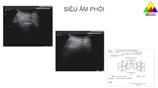
TUYẾN GIÁP có liên quan COVID-19 


ĐÃ GẶP TẠI MEDIC


What does pleural thickness of LUS [or TUS] mean ?


B-lines is a specific term for RDA found in LUS and they must have the following characteristics, in addition to the standard properties of RDA:
• it originates at the pleural line (PL),
• it has a strong light ray-like appearance, obliterating other background LUS artefacts,
• it moves with lung sliding.
Physiologic B-lines
The normal lung is characterized by the absence or presence of very few B-lines; less than three per field of view. These “physiologic” B-lines, seen in only 10 % of young healthy subjects [21], are often a transient phenomenon and change with posture. The dependence of B-lines creation on availability of a medium that could create a resonating vibration (bubble-tetrahderal complexes or equivalent) probably explains why they are more likely found in the better perfused dependent areas of the lung or at the thicker interlobular septa [5,15]. Age related changes in the lung parenchyma such as fibrosis and sub-pleural lesions account for increased B-lines observed in 37% of the elderly [21].
Pathologic B-lines
In pathological processes involving the lung parenchyma, fluid, inflammatory infiltrates or cellular content progressively increase, greatly enhancing the environment for the generation of B-lines (fig 2). As a guide, a finding of a cluster of three or more B-lines per intercostal space may be pathological. However, the number of B-lines and the number of chest areas positive for (multiple) B-lines increases with age [21-23] and therefore one must be cautious of a false positive interpretation in older persons.
The often-used term “interstitial syndrome” [13,24] is a description of ultrasound findings of pathological B-lines. The term is not specific or synonymous to acute interstitial lung disease [6,25]. These are examples of conditions that produce a generalized and often bilateral interstitial syndrome: cardiogenic pulmonary edema [13], acute or chronic interstitial lung disease [26,27] or acute lung injury / acute respiratory distress syndrome [28]. Focal interstitial syndrome may be observed in relation to pneumonia [13,29], pulmonary contusion [30,31], lung tumors [29,32] or other pulmonary consolidating processes [29,33]. As B-lines are sensitive but non-specific for pathological lung parenchyma changes, the findings of interstitial syndrome must be correlated with the following information [34] to make a clinical diagnosis:
• distribution of B-lines (interstitial syndrome) in the lung fields: patchy, uniform, symmetry,
• evaluation of PL morphology [35,36]: lung sliding, thickening, unevenness,
• other LUS features: consolidation, sub-pleural lesions, effusion,
• clinical information of the patient: oxygen saturation, blood gases, other laboratory findings,
• medical history of the patient: e.g. history of pleuritis, thorax injury or surgery, interstitial lung disease or connective-tissue/ rheumatic disease. In the clinical context of acute dyspnea and desaturation, the utility of B-lines in LUS diagnosis is limited in patients with pre-existing interstitial syndrome unless prior LUS reports and images are available for comparison.
Additional US studies such as echocardiography are often helpful in clarifying the cause of the interstitial syndrome.
What kinds of CTA can be seen in LUS?
Lung comets
In the normal lung, a specific type of CTA can be seen. These are short (almost always less than 1cm) vertical artefacts that taper and fade with increasing depth. Similar to B-lines, they arise from the PL and move with lung sliding; these signs point to their origin from a reverberation mechanism occurring at the peripheral lung parenchyma or the inter-pleural layer and the dependence on the apposition of the visceral and parietal pleura for their creation. They are found in all areas of the lung [3,14,18,19], best visualized with higher frequency transducers. It is interesting to note that studies that use low frequency transducers for LUS do not specifically mention them. Numerous CTA seen together sometimes gives the PL a “beads on a string” appearance. As there is currently no specific term to describe this type of CTA, the authors propose the term ‘lung comets’ for the purpose of discussion henceforth.
Z-Lines
Z-lines are observed as vertical artefacts in LUS, especially in thin individuals. Z-lines do not originate at the PL and do not move with lung sliding [37,38]. They are typically weak in appearance, blend with surrounding artefacts (e.g. A-lines) and fade with increasing depth. These characteristics and the varying periodicity and thickness of the horizontal bands that comprises the Zline are testimonial to a reverberation mechanism occurring outside the lung. They have no clinical significance, but it is important not to confuse them with B-lines.
Lung comets versus B-lines in disease diagnosis
The visualization of lung comets or B-lines in LUS
gives proof that the visceral and parietal pleura are in
contact. They are therefore important supporting signs,
besides lung sliding, in excluding a pneumothorax.
Verification of B-lines is often taught as a method
for ruling out pneumothorax. However, this criterion
is limited by low sensitivity because B-lines are rarely
found in the normal lung [17,19], especially in the upper parts where pneumothorax tends to occur. This fact
was clearly expressed in Lichtenstein’s validation of B-lines (then called “CTA”) in ruling out pneumothorax
[38], where none of the subjects with normal lungs had
B-lines. B-lines are more useful if their presence is already demonstrated prior to the pneumothorax event,
e.g. in an intubated patient with pulmonary edema, and
comparisons of scans show the subsequent disappearance.
In contrast, the use of lung comets in this context
is much more effective simply because of their ubiquitous nature. In the challenging situation of determining
the cause of absent lung sliding (e.g. pneumothorax vs
non-ventilation in critical care), one should actively look
for lung comets and be less reliant on B-lines. This approach suggested by the authors has not been validated
in research but it is already well known as evidenced by
images of lung comets used in several discussions on
pneumothorax diagnosis [39,40].
Due to their common occurrence in the normal lung,
lung comets (c.f. B-lines) should not be used to assess for
interstitial syndrome and to do so is to make a diagnosis
when there is none.
RDA other than B-lines
RDA at pathological interfaces
RDA that do not originate from PL are not B-lines. A classic example is seen at the shred line; the interface between a consolidation and the aerated part of the lung [37]. This region has the right conditions for forming bubble tetrahedral complexes and RDA often arise from it. A lung compressed under the weight of a pleural effusion sometimes generates RDA (named sub B-lines by Lichtenstein) from the visceral pleural. In both these scenarios, these RDAs do not necessarily indicate the presence of interstitial syndrome.
I-lines
I-lines [37] share all the same characteristics as B-lines except they are short, about 3-4 cm in length. They are occasionally seen at the sites of the inter-lobar fissures. They could represent a partially visualized B-line, and currently have no clinical significance assigned to them.
Other described vertical artefacts in lung ultrasound
E-lines
Air in the subcutaneous tissues of the chest wall can cause LUS artefacts that simulate the lung. The sonographer may mistake the layer of air as the PL. On closer examination, the pseudo-PL is uneven and varying in thickness; the ribs are not visible and there is no bat sign. Uncommonly, small pockets of air may create RDA or CTA. These vertical artefacts that arise from the subcutaneous layer are collectively called E-lines [37,41]
W-lines
W-lines [37] is just a variation of E-lines where the point of origins of the vertical artefacts are scattered at different planes.


Summary of Coronavirus COVID-19 examination and follow-up
In comparison between non-COVID-19 pneumonia and COVID-19 pneumonia, COVID-19 pneumonia is more likely to have a peripheral distribution [84] (Figure 23). In addition to HR-CT scan and X-ray of the lung, ultrasound can also be used for the diagnosis and follow-up of the disease [85]. By using a curved transducer (5–1 MHz), the morphology and changes of subpleural lesions are clearly displayed. Due to the option to use even the low-frequency of the transducer, changes of air and water contents in consolidated peri-pulmonary tissues and an air bronchogram sign can be depicted (Figure 24).

The blood supply and lesion progression in peripulmonary consolidation can be monitored by using the color or power Doppler technique [86]. Currently, ultrasound of the lung is limited in the diagnosis and treatment of central lung diseases due to the attenuation of sound waves by normal lung and bone tissues. The diagnosis of lung pathologies relies on the artifacts of peri-pulmonary lesions [87–88]. The artifacts exist because of an abnormal ratio of air and water contents in alveoli and interstitial tissues. In order to improve the diagnostic ultrasound lung tool, the use of an abdominal curved array probe (5–1 MHz) seems to be helpful. Typical for the COVID-19 disease are the thickening of the pleural line with pleural line irregularity. The pleural line could be unsmooth, discontinuous and interrupted [85, 89] (Figures 25–26).
The appearance of B-lines artifacts could vary from focal, to multifocal and confluent pattern. The consolidations could vary in different patterns, including multifocal small subpleural consolidations up to non-translobar and translobar with occasional air bronchograms [5]. Pleural effusions are uncommon in coronavirus COVID-19 disease.
An indirect sign for recovering is the appearance of A-lines during the recovery phase [85] (Figure 27).
In summary, in our experience, we consider that lung ultrasound will have a major utility for the management of COVID-19 pneumonia in the ICU due to its safety, repeatability, low cost and point of care use. HR-CT may be reserved in the follow-up if lung ultrasound is not able to answer the clinical question. In our personal experience lung ultrasound could be used for rapid assessment of the severity of SARS-CoV-2 pneumonia, to track the evolution of disease during follow-up and to monitor lung recruitment maneuvers. Additional ultrasound can track the response to prone position and the management of extracorporeal membrane therapy [85]. With increased use of bedside ultrasound in the ICU, patients can be protected from unnecessary radiation and therapy delays. The transport of high-risk patients to X-ray examinations can be avoided.


LU findings presented correlation with HRCT images.
LU can be used in respiratory system propaedeutics as an alternative to the “stethoscope use”. Special clothing and individual protection equipment are indispensable, since the manipulation of the stethoscope in pulmonary evaluation might create contamination risks for the health professionals and patients.
COVID-19 normally induces a bilateral and diffuse interstitial pneumonia with asymmetric lesions and uneven distribution, mainly involving the lung periphery, which makes it particularly suitable for investigation using LU.60
Studies have identified potential correlation between the LU patterns and the patients’ clinical outcome. One of the assays in this study reported that each pulmonary area could be in a different stage of the disease, therefore, the global evaluation of the lungs is fundamental.69
The POCUS allows for hemodynamic, cardiac and vascular evaluations (thromboembolic phenomena — deep venous thrombosis).
LU should be associated to the multisystem point-of-care exam, since the SARS-CoV-2 infection might be linked to myocarditis and a high incidence of thromboembolic events. Thus, multiorgan ultrasound evaluation in early treatment is useful to screening these complications at the bedside.
More studies on LU application in the pediatric population are necessary.
LU in COVID-19 score standardization.
Improvement of reading/automatic identification of B line software, as reported in this study is still needed.74
The advancement of the remote robotic ultrasound scanning technology assisted by the 5G network in real time by the use of big data, cloud storage and artificial intelligence must be improved.73


