Tổng số lượt xem trang

Thứ Ba, 27 tháng 3, 2018

NHÂN 03 CA DIFFUSE LARGE B CELL LYMPHOMA CÓ TRIỆU CHỨNG HÀM MẶT

Trong số 20 ca lymphom lan tỏa dòng tế bào B lớn [diffuse large B cell lymphoma] ở Trung tâm Y khoa Medic từ 2008,  có 03 ca có triệu chứng đau răng và hàm mặt điều trị giảm đau, nhổ răng..không bớt, sau đó xuất hiện nhiều tổn thương khác ngoài hạch. Trong khi đó, tìm được một bài báo về đau xương hàm dưới trong lymphom ác. Sau đây là một hồi cứu về đề tài này.

             Diffuse Large B Cell Lymphoma
and Appearences  in Oral Cavity, and Maxillary Area
Nguyen Thien Hung, Jasmine DCB Thanh Xuan, Le van Tai, Le Thong Nhat, Le Thanh Liem, Phan Thanh Hai
Medic Medical Center, HCMC, Vietnam

INTRODUCTION:
May there had been an initial symptom in oral cavity, maxillary and mandibular area for diffuse infiltration of lymphoma?  We will represent some cases of diffuse lymphoma with appearences  in oral cavity,  and maxillary areas.

MATERIALS and METHODS:
We retrospectively reviewed  20 cases of diffuse lymphoma that were histopathologically diagnosed in our center from 2008 in which presented 03 cases  with oral cavity,  maxillary symptoms and  bone involvement [01 case in male 42 yo and 02 cases in female gender, 33 and 40 year-old].

RESULTS:

CASE 1= A 33 year-old female patient with pain in lower maxillary bone for one month and tension in both  2 breats), hyperpigmented edema of areolar area both 2 sides without pregnancy.  ABVS scanning  detected  multiple nodules infiltrating in 2 breasts.
Biopsy of 2 breasts  reported  microscopic with immunohistochemistry scanning, diffuse large B cell lymphoma.
MRI  full body with gado detected  bone marrow changing, 2 breats  hypercaptured contrast,  ascites and kidney infiltration. In pelvis  2 ovarian tumors and big uterine cervix were detected. Blood tests showed   lower platelets,  EGFR  lower  46, beta2 microglobuline raised  3816,  ferritin raised  911, LDH-l  raised  1360.


CASE 2= A 40 year-old female patient with toothache but pain remained  after removing tooth, and in total body examination detected right pleural effusion  and some nodular tumors in thyroid, left breast and subcutaneous area of right neck, left chest all and  lumbar region. Erosions of left clavicle and right 1st rib. Biopsy of left breast tumor reported diffuse large B cell lymphoma.


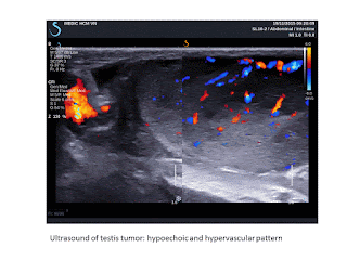
CASE 3= Man 42 year-old, one month ago, pain in oral sinus, difficult eating and 2 days after,  pain appears in left testis. Ultrasound of left big and  hot testis represented  hypoechoic  infiltration,  hypervascular of one part of testis and elastoscan ultrasound value #  10.5 kPa).
 FNAC of this mass of left testis  having abnormal cells. Biopsy of  tumor in oral area represented diffuse large  B cell lymphoma.


DISCUSSION:
There are published reports of pain in mandibular area as initial symptom but symptoms in oral cavity and maxillary bone as onset symptom seems to be not represented in literature. But it exists a case of pain in oral cavity for lymphoma infiltrating in testis and an another case of pain in maxillary area for lymphoma in whole body in this report.Toothache and pain in maxillary region remained with management guided a survey in all body and detected extranodal lesions: bone erosions, perirenal edema, ovarian, uterine cervix, testicular edema, subcutaneous nodules. But in literature, only mandibular lesion with tooth pain were published which not included.

CONCLUSIONS:
Seveval  reports of lymphoma in mandible were published with pain as initial symptom,  but in cases of diffuse infiltration of lymphoma in whole body, the appearences in oral cavity, maxillary and mandibular areas with pain symptom may be added criteria of diagnosis for lymphoma in clinical examination.

REFERENCE

 Mochizuki, Y.,  Harada, H.,  Sakamoto, K.,  Kayamori,  K.,  Nakamura,  S.,  Ikuta, M.,  Kabasawa, Y., Marukawa, E., Shimamoto, H., Tushima, F. and Omura, K. (2015) Malignant Lymphoma with Initial Symptoms in the Mandibular Region. Journal of Cancer Therapy, 6, 554-565


Abstract
Primary intraosseous lymphoma is rare and there are few case reports manifesting with a mass in the mandible. Thus, we retrospectively reviewed and analyzed the clinical characteristics,  treatment, and outcome of extranodal non-Hodgkin’s lymphoma (NHL) with initial mandibular symptoms in our department. At initial treatment of dental clinics, dentists had diagnosed as dental or gingival diseases and had performed dental treatment. Neurological disorder to involvement of the inferior alveolar nerve was present in 80.0% of our cases. On dental or panoramic radiography a specific radiolucent lesion in the mandible was not detected, except for dental lesions. On CT, NHL of the mandible region has no widening and no clear destruction but a slit-like the cortex bone destruction pattern with keeping in shape of the mandibular body  (62.5% of CT-examined cases), and extraosseous soft tissue mass are clearer on MRI  (100.0% of MRI-examined cases).
Histopathologically, 80.0% of our cases were diagnosed as diffuse large B cell lymphoma (DLBCL).
One case as B-cell lymphoblastic lymphoma and one case as B-cell lymphoma unclassifiable with features intermediate between DLBCL and Burkitt lymphoma were Stage IV  (Ann Arbor staging system) and had poor prognosis. The disease-specific survival rate was 77.8% at 5 years. If unexplained non-specific symptoms such as swelling of the jaw, pain, neurological disorder of the inferior alveolar nerve, tooth mobility are observed, oral surgeons and dentists should not perform
dental treatments. CT and MRI show disease specific appearance to be able to give a definitive diasnosis as NHL. PET/CT is useful for scaninng of whole body. A deep bone biopsy is preferred for
suspected malignant lymphoma.

Chủ Nhật, 18 tháng 3, 2018

Acute and Chronic DVT and Ultrasound (US)

SOURCE: Advanced imaging in acute and chronic deep vein thrombosis, Gita Yashwantrao Karande , Sandeep S. Hedgire , Yadiel Sanchez , Vinit Baliyan , Vishala Mishra , Suvranu Ganguli , Anand M. Prabhakar © Cardiovascular Diagnosis and Therapy. Cardiovasc Diagn Ther 2016;6(6):493-507

DVT and Ultrasound (US)[extracted]

US is used in evaluation of both symptomatic and asymptomatic DVT (patients at high risk of DVT). It is useful not only in assessing DVT but can also identify other conditions causing signs and symptoms indistinguishable from DVT.

Compression US
Compression US has been procedure of choice for investigation of suspected upper and lower extremity DVT for decades (14). Other modification to this technique like two-point compression US (15), extended compression US (16) and complete compression US (17) are used in different combinations at different institutions.


Venous duplex US
 Lower extremity venous duplex US combines 2 components to assess for DVT: B-mode or gray-scale imaging with transducer compression maneuvers and Doppler evaluation consisting of color-flow Doppler imaging and spectral Doppler waveform analysis. Respiratory phasicity and cessation of flow with the valsalva maneuver offer indirect evidence of patent abdominal and pelvic veins (18). The primary diagnostic US criteria for acute DVT remains non-compressibility of the vein with secondary diagnostic criteria being echogenic thrombus within the vein lumen, venous distention, complete absence of spectral or color Doppler signal within the vein lumen, loss of flow phasicity, and loss of response to valsalva or augmentation (18). US can also be used to differentiate acute from chronic thrombus. In acute thrombosis, vein is distended by hypoechoic thrombus and shows partial or no compressibility without collaterals (Figure 1). In chronic thrombosis, the vein is incompressible, narrow and irregular and shows echogenic thrombus attached to the venous walls with development of collaterals (Figure 2). According to American College of Radiology (ACR) guidelines and technical standards, lower extremity US should include compression, color and spectral Doppler sonography with assessment of phasicity and venous flow augmentation (19,20). Advantages of lower extremity venous Duplex US are that it is readily available, quick, cost effective, noninvasive, devoid of ionizing radiation, lacks need for intravenous contrast and can be portable for critically ill patients prone for developing DVT.


Limitations include that it is difficult and less sensitive in patients with obesity, edema, tenderness, recent hip or knee arthroplasty, cast, overlying bandages and immobilization devices. It also has limitations in patients who had previous DVT and have new symptoms shortly after the treatment. False-positive results include extrinsic compression of a vein by a pelvic mass or other perivascular pathology (21) and thrombosis in the distal popliteal vein. False-negative studies may occur in the presence of calf DVT, proximal DVT in asymptomatic (even high-risk) patients or in the presence of a thrombosed duplicated venous segment. In a systematic review of accuracy of US in diagnosis of DVT in asymptomatic patients, Kassai et al. suggested that US was accurate in proximal veins for diagnosis of DVT in patients hospitalized for orthopaedic surgery (11) with lower sensitivity in other settings.


Sonographic elasticity imaging (SEI)
As described previously venous duplex US is considered primary noninvasive imaging for DVT. However, this method cannot assess the age and maturity of the thrombus i.e., it cannot distinguish pure post thrombotic syndrome (PTS) (which develops in 20–50% of patients after DVT) from new development of acute DVT with or without PTS (22). It is important to distinguish acute on chronic DVT from PTS as the latter doesn’t require anticoagulant therapy with highly potent fast acting anticoagulants associated with high risk of bleeding (23). Also chronic clots are treated with oral warfarin sodium, which has better safety profile than heparin. SEI is the latest promising technique for estimating age of the thrombus. It uses tissue deformation to assess the tissue hardness and hence clot maturity (24,25). It has shown promising results in animals and in a few studies in humans. As SEI requires tissue to be deformed during imaging, it is consistent with venous duplex US which also requires compression. The degree of compression required for standard strain measurements on SEI is lesser than compression US that is beneficial for patients with swollen painful legs (26-28). Thus, even if evaluation of clot age does not work, just the use of SEI to detect thrombi may be an improvement over the present compression US technique. SEI can hence be incorporated into standard duplex US so that thrombus can be diagnosed and presumably aged simultaneously. SEI is in preliminary stages of investigations and there is limited data on its ability to determine the age of DVT in human subjects (26,29,30). It is highly operator dependent. Another limitation is that comparison between the two clots requires an internal standard with the same hardness in both images, as it is difficult to know the force that was used to deform the tissue in each case. Rubin et al. used the wall of the vein as standard but the hardness difference estimate was conservative due to lack of another standard reference (31). Another limitation is that it is difficult to distinguish between subacute and chronic thrombi that are closer in age (29).
….

Conclusions


The incidence of DVT is increasing, not just in the lower extremity but also in upper extremity, where malignancy and central venous catheters are the major precipitating factors. While US still has advantages, it has various limitations and in such cases advanced imaging techniques such as MRI, should be considered. CTV, while sensitive, can be incorporated into CTPA in suspected PE but has the disadvantage of radiating sensitive pelvic organs especially in young patients. MRI will almost likely feature more commonly in DVT evaluation in the near future with new “blood pool” contrast agents allowing a comprehensive examination for PE and DVT in the same scan. Although advanced imaging techniques in nanotechnology/ biotechnology, molecular imaging and PET are also being investigated, these may not replace the established first line modalities in diagnosis, but may be useful as adjuncts in patients who are not good candidates for structural imaging like renal disease patients with contraindication for intravenous contrast. Most of these agents are under trial and will hopefully come into routine use in diagnosing this potentially fatal disease.

Thứ Tư, 14 tháng 3, 2018

PoCUS MEDIC:Foreign Body [Thorn # 02mm]

Thôn Dakrong.
Bé trai 10 tuổi . Mủ dưới da bàn chân. Siêu âm thấy có dị vật d# 2mm.
Xử trí: rạch da lấy dị vật tại chỗ, gai.

PoCUS MEDIC detected a 2mm foreign body in an abscess of left subcutaneous forefoot of a 10 year-old young boy.
Management: Removed right away a thorn in place.


Why does it need an abscess evaluation by ultrasound?
- to differentiate abscess from cellulitis.
- to find out a foreign body in an abscess.
- to use appropriate antibiotics in case of cellulitis.