Tổng số lượt xem trang

Thứ Ba, 4 tháng 9, 2018

Ultrasound in Chronic Liver Disease, a Review


Ultrasound in Chronic Liver Disease, a Review

J. F. Gerstenmaier and R. N. Gibson

Insights Imaging. 2014 Aug; 5(4): 441–455.
Published online 2014 May 24. doi:  10.1007/s13244-014-0336-2

Abstract
Background 
With the high prevalence of diffuse liver disease there is a strong clinical need for noninvasive detection and grading of fibrosis and steatosis as well as detection of complications.

Methods
B-mode ultrasound supplemented by portal system Doppler and contrast-enhanced ultrasound are the principal techniques in the assessment of liver parenchyma and portal venous hypertension and in hepatocellular carcinoma surveillance.

Results
Fibrosis can be detected and staged with reasonable accuracy using Transient Elastography and Acoustic Radiation Force Imaging. Newer elastography techniques are emerging that are undergoing validation and may further improve accuracy. Ultrasound grading of hepatic steatosis
currently is predominantly qualitative.

Conclusion
A summary of methods including B-mode, Doppler, contrast-enhanced ultrasound and various elastography techniques, and their current performance in assessing the liver, is provided.

Teaching Points

• Diffuse liver disease is becoming more prevalent and there is a strong clinical need for noninvasive detection.
• Portal hypertension can be best diagnosed by demonstrating portosystemic collateral venous flow.
• B-mode US is the principal US technique supplemented by portal system Doppler.
• B-mode US is relied upon in HCC surveillance, and CEUS is useful in the evaluation of possible HCC.
• Fibrosis can be detected and staged with reasonable accuracy using TE and ARFI.
• US detection of steatosis is currently reasonably accurate but grading of severity is of limited accuracy.

Future perspective

The increasing prevalence of chronic liver disease, including fatty liver disease, together with the need to minimise invasive liver biopsies will continue to provide substantial drive for ultrasound research and development.

Conventional B-mode ultrasound =
 + Quantification of echointensity in the diagnosis of fatty liver disease is an area of recent interest. Standardisation of technique and interobserver eproducibility will need to be addressed.
  + Quantitative and semi-quantitative assessment of echotexture in the diagnosis of fibrosis and fatty liver disease is being developed

Doppler ultrasound =
 The role of Doppler ultrasound in chronic liver disease is likely to remain principally in the diagnosis of portal venous hypertension

Contrast-enhanced ultrasound =
 Kupffer-phase contrast agents can be used in the grading of fibrosis as well as in the detection of focal lesions,and it is hoped that these agents will become more widely available.

Elastography
 More widespread use of the two most extensively validated and established techniques of TE and ARFI is expected, and further shear wave elastography techniques are likely to become more widely adopted and evaluated.


Thứ Hai, 3 tháng 9, 2018

PoCUS for WRIST TRAUMA


Abstract

Objective

We evaluated the effectiveness of point-of-care wrist ultrasonography compared with 3T-magnetic resonance imaging (MRI) for diagnosing triangular fibrocartilage complex (TFCC) injuries in trauma patients with ulnar-sided pain and instability. Moreover, we assessed the inter-observer variability between an emergency physician and a musculoskeletal radiology fellow.

Material and methods

A prospective cross-sectional study was conducted in an emergency department; patients with ulnar-sided sprain and instability were recruited. An emergency physician and a musculoskeletal radiology fellow independently evaluated the TFC, meniscal homologue, volar and dorsal distal radioulnar ligaments, and extensor carpi ulnaris using point-of-care ultrasonography. Findings were classified as normal, partial rupture, or complete rupture. Wrist 3T-MRI was used as the reference standard. We compared the diagnostic values for point-of-care ultrasonography obtained by both reviewers using DeLong's test. Intra-class correlation coefficients (ICCs) were calculated for agreement between each reviewer and the reference standard, and directly between the two reviewers.

Results

Sixty-five patients were enrolled. Point-of-care wrist ultrasonography showed acceptable sensitivity (97.2–99.1%), specificity (96.8–97.3%), and accuracy (96.9–97.9%); these diagnostic performance values did not differ significantly between reviewers (p = 0.58–0.98). Agreement between each reviewer and the reference standard was excellent (emergency physician, ICC = 0.964; musculoskeletal radiology fellow, ICC = 0.976), as was the inter-observer agreement (ICC = 0.968).

Conclusion

Point-of-care wrist ultrasonography is as precise as MRI for detecting TFCC injuries, and can be used for immediate diagnosis and further preoperative imaging. Moreover, it may shorten the interval from emergency department admission to surgical intervention while reducing costs.

Chủ Nhật, 2 tháng 9, 2018

PTOSIS and DIABETES

From this case, we suggest that when evaluating patients with acute onset ptosis as the only manifestation, diabetic-vasculopathic neuropathy should be considered. 
This case also implies that the most interior portion of the third cranial nerve may consist of nerve fibers mainly innervating the levator palpebrae superioris.


Handheld ultrasound + CADx helps triage breast masses


By Kate Madden Yee, AuntMinnie.com staff writer
August 31, 2018 -- Researchers successfully used a combination of handheld ultrasound and computer-aided diagnosis (CADx) software to triage women in Mexico who had palpable breast masses. They believe the combination is good news for women who may not have access to screening mammography, according to an article published online August 29 in the Journal of Global Oncology.
"[Our] results open up the possibility of implementing practical, cost-effective triage of palpable breast lumps, ensuring that scarce resources can be dedicated to suspicious lesions requiring further workup," wrote a team led by Dr. Susan Love of the Dr. Susan Love Research Foundation in Encino, CA.
In low- to middle-income countries, 23% of new breast cancer cases occur among women ages 15 to 49 years, compared with 10% of new breast cancer cases in high-income countries. Because screening mammography is not necessarily available, women often present with palpable breast lumps; although most of these masses are benign, it's important to determine which of them need further evaluation, Love and colleagues wrote.
In these situations, portable ultrasound is often available, but image interpretation has traditionally been performed by radiologists or breast surgeons -- who may be in short supply.
"At [our study] site, the average wait for a diagnostic ultrasound is nine months because of limited available trained radiologists," the researchers wrote. "Many of the participants traveled up to six hours by bus to participate in this study, corroborating the need for a simple accessible local means of triage."
For the study, Love's group trained three nonradiologist healthcare workers in Jalisco, Mexico, to determine whether they could use ultrasound to acquire images of sufficient quality for accurate analysis by CADx software. The workers were a first-year medical student, a surgical nurse, and a gynecologic intern who used a portable ultrasound scanner (Vscan, GE Healthcare) to acquire orthogonal views of 32 breast masses in 32 women.
The images were analyzed using what the researchers call "triage CADx," an artificial intelligence-based CADx program for ultrasound that is based on convolutional neural networks. CAD results were then compared with radiologist readings.
Among the 32 masses, two were malignant. Nonradiologist healthcare staff was able to acquire adequate images, Love and colleagues found. In addition, triage by the software was as accurate as radiologist assessment of the masses, with 100% sensitivity and specificity.
The results suggest a way to make use of limited breast care resources in low- to middle-income countries.
"This study demonstrates that first-level healthcare workers are capable of acquiring images with a portable ultrasound machine comparable to those of a trained radiologist," the group concluded.

Thứ Bảy, 1 tháng 9, 2018

ULTRASOUND and FETAL NERVOUS SYSTEM LESIONS

https://www.e-ultrasonography.org/journal/view.php?number=214

Abstract

Central nervous system (CNS) malformations play a role in all fetal malformations. Ultrasonography (US) is the best screening method for identifying fetal CNS malformations. A good echographic study depends on several factors, such as positioning, fetal mobility and growth, the volume of amniotic fluid, the position of the placenta, the maternal wall, the quality of the apparatus, and the sonographer’s experience. Although US is the modality of choice for routine prenatal follow-up because of its low cost, wide availability, safety, good sensitivity, and real-time capability, magnetic resonance imaging (MRI) is promising for the morphological evaluation of fetuses that otherwise would not be appropriately evaluated using US. The aim of this article is to present correlations of fetal MRI findings with US findings for the major CNS malformations.


Introduction

Fetal imaging evaluation has improved over the years. Faced with more complex diagnoses, magnetic resonance imaging (MRI) has become widely used as an important complement to prenatal ultrasonography (US). Due to its higher contrast resolution than US, fetal MRI allows normal versus abnormal tissue to be better differentiated, providing detailed imaging information on fetal structures, particularly the brain. To date, fetal MRI has been shown to play an important role in the evaluation of structural brain development and in the assessment of abnormalities suspected on US [1,2]. Moreover, the use of fetal MRI has been shown to help in counseling parents during pregnancy and in discussions about treatment [1].
As the indications for fetal brain MRI are mainly based on abnormal US findings, fetal MRI is usually performed during the second half of gestation, from 18 to 20 weeks onward. After that point, the utility of prenatal US is limited due to decreased amniotic fluid volume, fetal positioning, and acoustic shadowing from the ossifying calvaria. For these reasons, MRI represents an important modality for morphologic evaluation of the fetal brain in the second half of gestation [1,3].

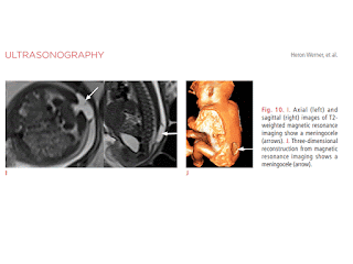
The most common indications for imaging the fetal brain are briefly discussed below, and include anencephaly, ventriculomegaly, corpus callosum agenesis, holoprosencephaly, hydranencephaly, schizencephaly, porencephaly, microcephaly, Chiari malformation, iniencephaly, the Dandy-Walker malformation, vein of Galen malformations, tuberous sclerosis, and encephalocele.
Nota:

anencephaly= vô não
ventriculomegaly= phì đại não thất
corpus callosum agenesis = teo thể chai
holoprosencephaly= tiền não hoàn toàn do không có phân cách vỏ não, (gồm alobar, semilobar và lobar forms).
hydranencephaly = não úng thủy toàn bộ
schizencephaly= não chẻ (nứt)
porencephaly= rỗ não, thông não thất bên ra bề mặt não
microcephaly= não nhỏ
Chiari malformation
iniencephaly = dị dạng não củ hành
dị dạng Dandy-Walker
dị dạng tĩnh  mạch Galen
xơ cứng củ
encephalocele= thoát vị não