Tổng số lượt xem trang
Thứ Bảy, 11 tháng 4, 2015
Chủ Nhật, 5 tháng 4, 2015
CHOROIDAL THICKNESS and RETINAL VASCULAR CALIBER and I C A
ABSTRACT
Purpose
Decreased retinal arteriolar caliber and increased retinal venular caliber have been associated with increased cardiovascular mortality. This study aimed to evaluate correlations of choroidal thickness and retinal vascular caliber measurements with internal carotid artery (ICA) Doppler ultrasound variables.
Methods
In this cross-sectional and observational study, 43 eyes and ICAs of 43 healthy volunteers were examined. Spectral domain optical coherence tomography was used to measure subfoveal choroidal thickness (SFCT) and retinal vascular caliber. The ICA Doppler ultrasonographic parameters were diameter, flow volume, peak-systolic velocity, end-diastolic velocity, resistance index (RI), and pulsatility index (PI).
Results
Negative correlations emerged between ICA RI and SFCT (p = 0.017, r = −0.36) as well as between ICA PI and retinal arteriolar caliber (p = 0.015, r = −0.37). A negative linear correlation appeared between ICA diameter and SFCT (p = 0.005, r = −0.42), although ICA diameter and flow volume showed no association with retinal vessel caliber (p over 0.05).
Conclusions
Choroidal thickness is negatively correlated with ICA diameter and ICA RI, while retinal arteriolar caliber is inversely related with ICA PI in normal volunteers.
© 2015 Wiley Periodicals, Inc. J Clin Ultrasound, 2015;
© 2015 Wiley Periodicals, Inc. J Clin Ultrasound, 2015;
Thứ Tư, 1 tháng 4, 2015
Ultrasound should be 1st choice for female pelvic imaging
by Erik L. Ridley
Thanks to technological advances such as volume imaging, real-time transvaginal ultrasound (TVUS), and 3D Doppler techniques, ultrasound is ideally suited for evaluating the female pelvis and is an effective first-line imaging modality for most gynecologic patients, concluded a multi-institutional research team led by Dr. Beryl Benacerraf of Harvard Medical School. "Consistent use of ultrasound imaging first in women with pelvic symptoms, especially with the adjunct of 3D and Doppler if necessary, would likely render further imaging unnecessary and at the same time be cost-effective and safer," the authors wrote in their Clinical Opinion article (Am J Obstet Gynecol, March 31, 2015).
Ultrasound First initiative
The American Institute of Ultrasound in Medicine's (AIUM) Ultrasound First initiative is particularly relevant for obstetric and gynecologic patients, "for whom a skillfully performed and well-interpreted ultrasound image usually obviates the need to proceed to additional more costly and complex cross-sectional imaging techniques," according to the authors. "Yet still today, many women with pelvic pain, masses, or flank pain first undergo CT scans and those with Müllerian duct anomalies typically have MRIs," they wrote. "Not uncommonly, CT or MRI of the pelvis often yields indeterminate and confusing findings that then require clarification by ultrasound imaging." 3D/4D ultrasound is among the key technical advances that have sparked the modality's improved utility in gynecological imaging. While ultrasound used to require filling a woman's bladder and obtaining a series of 2D images one at a time, that's no longer the case. "Today, 3D volume ultrasound imaging allows the automated acquisition of an entire volume that, in turn, can generate hundreds of images and be used to reconstruct any view in any orientation," the authors wrote. "Furthermore, 3D ultrasound imaging is less expensive and less time-consuming than MRI." Reconstructed views of the pelvis -- such as the coronal view of the uterus -- have greatly improved the modality's ability to answer the vast majority of clinical questions in gynecology, according to the authors. For example, 3D volume sonography has been shown to be just as effective as MRI for demonstrating Müllerian duct anomalies. 3D ultrasound has also "emerged as the ideal imaging modality not only when examining patients with infertility but also for examining patients with pelvic pain associated with embedded intrauterine devices, fibroid tumors, adenomyosis, adnexal masses, torsion, [and] endometriosis," they wrote. "Ultrasound volume imaging makes it possible to localize fibroid tumors, polyps, and hydrosalpinges with high precision, as well as other uterine abnormalities." As a result, the medical community must be educated to consider using 3D ultrasound for the initial assessment of specific gynecologic indications, such as evaluating the uterus for Müllerian abnormalities and localizing intrauterine devices or other intracavitary lesions, according to Benacerraf and colleagues. "In this setting, it is likely that fewer women would require a costly workup that involves multiple advanced imaging studies if 3D ultrasound images were performed first," they wrote.
Real-time TVUS
The transvaginal transducer -- one of the most important innovations in pelvic imaging over the past few decades -- along with the real-time nature of ultrasound imaging enables close probing of pelvic organs to elicit patient symptoms and to correlate them with specific pelvic anatomic locations, according to the authors. "The practitioner therefore can gain crucial information about the degree and area of pain and mobility of organs in the pelvis and correlate the ultrasound findings with the physical examination," they wrote. "The ability to examine and image the patient at the same time offers considerable and too often neglected value, which is unique to ultrasound imaging as a cross-sectional imaging technique." For example, tenderness-guided ultrasound imaging is now the most effective way to detect implants of painful, deep-penetrating endometriosis throughout the pelvis, according to the group. "Ultrasound imaging has proved to be accurate for the evaluation of deep infiltrating endometriosis and for patients with pain because of extensive pelvic adhesions," they wrote. "Not only can we identify abnormalities on the images, but also simultaneous gentle pushing can show whether organs slide past each other, thus providing crucial information about the origin of a mass (adnexal mass versus broad ligament fibroid tumor) and the connections and adhesions between the organs." Real-time ultrasound also facilitates sonohysterography, in which a small catheter is used to inject saline solution into the uterine cavity. Clinicians can then evaluate the endometrium for polyps, submucosal fibroid tumors, synechiae, and uterine shape, according to Benacerraf and colleagues. "Adjunctive to sonohysterography, the installation of microbubbles is useful for the determination of tubal patency and is critical to those patients with contrast allergies," they wrote.
3D Doppler for blood flow
Ultrasound combines morphologic and vascular imaging, an advantage for characterizing pelvic masses. Without requiring the injection of contrast, Doppler yields valuable information about the location and degree of blood flow in and around pelvic lesions, according to the authors. "Not only is the characteristic grayscale image of pelvic abnormalities key in making a diagnosis, but also 3D Doppler ultrasound imaging can evaluate the mapping and density of blood flow and even provide a quantitative measure of the amount of blood flow in a lesion," they wrote. Color Doppler mapping also provides important information for evaluating adnexal masses and differentiating an endometrioma from an ovarian tumor or an ovarian fibroma. "For example, the unique Doppler pattern of a hemorrhagic corpus luteum permits this definitive diagnosis as a cause of acute pelvic pain," they wrote.
An education gap?
The authors noted that it would be best if all practitioners could be comfortable with high-resolution 3D ultrasound imaging, tenderness-guided TVUS, and pelvic Doppler imaging. "It is unfortunate that ultrasound users have such a wide range of experience, such that not everyone uses the modality to its full potential," they wrote. "Inexperience should not justify ordering an MRI or CT scan." Although ultrasound technology has advanced very quickly, many practitioners still provide only basic 2D ultrasound imaging. This dynamic highlights the need for education and dissemination of information on the newer techniques that ultrasound offers, according to the authors. "In this era of cost concerns, it is very important to recognize that ultrasound technology now offers multiple applications such as 3D volume imaging (similar to CT and MRI), real-time evaluation of pelvic organs along [with] the physical examination, and Doppler blood flow mapping (without contrast)," they wrote. "Collectively, these applications make ultrasound imaging a unique imaging modality that ideally is suited to evaluate the female pelvis.
Copyright © 2015 AuntMinnie.com
Thứ Bảy, 28 tháng 3, 2015
Thứ Sáu, 27 tháng 3, 2015
BREAST DENSITY ULTRASOUND BEST
AIUM: Ultrasound bests MRI, mammo in assessing breast tumor size
By Erik L. Ridley, AuntMinnie staff writer
March 25, 2015 -- ORLANDO, FL - Regardless of breast density, ultrasound outperformed MRI and digital mammography for preoperatively assessing breast tumor size, according to research presented on Tuesday at the American Institute of Ultrasound in Medicine (AIUM) meeting
In a retrospective study, a team from the Medical University of South Carolina (MUSC) found that both ultrasound and mammography yielded better performance than MRI, which significantly overestimated tumor sizes. However, ultrasound was the highest overall performer, according to lead author Dr. Rebecca Leddy.
Accurate clinical and pathological tumor size assessment is essential for breast cancer staging and treatment planning, said Leddy, who presented the group's research during a scientific session at AIUM 2015. Preoperative cancer staging could be improved if the accuracy of various imaging modalities could be determined, she said.
Because past studies have provided different results with regards to the imaging techniques as well as inclusion/exclusion criteria, the researchers set out to reassess the accuracy of digital mammography, high-resolution ultrasound, and high-resolution MRI using more specific inclusion criteria. They also wanted to determine whether or not the accuracy of these modalities for assessing tumor size varied based on breast density or type of tumor.
In the retrospective review, the researchers evaluated 93 patients who were newly diagnosed with in situ or invasive breast cancer and seen at MUSC between January 2008 and March 2012. All of the patients had received preoperative mammography, ultrasound, and MRI; had undergone needle localization; and did not receive neoadjuvant chemotherapy. Thirty patients who had positive margins were excluded from the study, as were six patients with mammographically occult lesions.
The remaining 57 patients had a mean age of 63, and 43.9% had dense breasts. There were 48 ductal cancers (84.2%) and nine lobular carcinomas (15.8%).
Modality showdown
Board-certified breast imaging radiologists with fellowship training provided maximum tumor size on mammography, ultrasound, and MRI. The researchers documented breast density for each of the cases, and they reviewed pathology results for maximum tumor size and type of cancer. Pathology results served as the gold standard for the study.
On the digital mammograms, the maximum tumor size was measured using standard, nonmagnified craniocaudal (CC) and mediolateral-oblique (MLO) views. Standard MRI sequences were obtained using the American College of Radiology (ACR) breast MR accreditation guidelines on a dedicated phased-array breast coil. The maximum tumor size was recorded using the first postcontrast axial and sagittal T1-weighted images, or in cases of larger tumors, on a maximum intensity projection (MIP) image.
Images were acquired by radiologists or by dedicated breast imaging technologists trained in screening and diagnostic mammography and breast sonography. They used handheld techniques with a linear transducer in the 7-16 MHz frequency range. Scanning and images were obtained in the sagittal and transverse planes and three measurements of the tumor were documented in the sagittal, transverse, and anteroposterior planes. The transducer was rotated to find and record the largest tumor dimension in any plane, Leddy said.
Mean tumor measurement sizes were as follows:
- Pathology: 1.401 mm ± 0.084
- MRI: 1.768 mm ± 0.019
- Ultrasound: 1.481 mm ± 0.090
- Mammography: 1.305 mm ± 0.077
The mean tumor size with MRI was significantly larger (p < 0.001) than the pathology results; however, the differences between pathology and ultrasound and mammography were not statistically significant. MRI tumor size measurements were also significantly higher than both ultrasound and mammography (p = 0.003 and p < 0.001, respectively).
Lin's concordance correlation coefficient (CCC) analysis -- performed by the researchers to assess reproducibility -- showed that ultrasound moderately agreed with pathology (CCC = 0.71, 95% confidence interval [CI]: 0.56-0.82). Meanwhile, mammography (CCC = 0.58, 95% CI: 0.38-0.72) and MRI (CCC = 0.50, 95% CI: 0.31-0.65) had poorer levels of agreement with pathology.
No effect from density
Breast density did not affect the measurement results: There was no significant difference in measurement performance between women with dense breasts and those who had nondense breasts. A significant difference was seen, however, by type of cancer.
MRI significantly overestimated tumor size in ductal cancers (p < 0.001), while slightly underestimating tumor size in lobular cancers, according to Leddy. In addition, measurements of lobular cancers on mammography were significantly smaller than the ultrasound and MRI measurements (p < 0.05).
The higher accuracy of ultrasound when compared with MRI and mammography differed from other studies reported in the literature, according to Leddy.
"We feel this was because we obtained the maximum tumor measurement irrespective of the plane of scanning," she said.
Leddy acknowledged a number of limitations of the study, including its retrospective nature and small sample size. In addition, it did not account for mammographically occult cases, she said.
Thứ Sáu, 20 tháng 3, 2015
SITUS INVERSUS and Ultrasound
Practice of Ultrasound: Part 21 -- Situs inversus
By Dr. Jason Birnholz, AuntMinnie.com contributing writer
March 3, 2015 -- AuntMinnie.com presents the 21st in a series of columns on the practice of ultrasound from Dr. Jason Birnholz, one of the pioneers of the modality.
Dr. Jason Birnholz.
However, I started to wonder about that surmise when I heard of a recent case at a local academic institution where situs inversus was missed on multiple fetal exams. It was discovered in early infancy because of malrotation and volvulus.
It is not easy to determine how often situs inversus is found or missed in routine prenatal exams, especially with an incidence of about one in 4,000 and a frequent absence of overt health issues in the afflicted. One paper from Children's Heart Center Nevada (Evans WN et al, Pediatric Cardiology, January 14, 2015) reported a detection rate of 68% in 2002, which later improved to the expected 100% in 2013.
How might we explain the baseline rate of 68%, which may very well be lower in less-expert scanning units?
The radiologist brain
When I applied for a diagnostic radiology residency, there was radiography or video fluoroscopy, each with its variations, techniques, and applications. The interviewers, whether they knew it or not, and the apprentice-like rotations during the residency, all sought to identify a particular visual-spatial talent of effortlessly forming a mental 3D spatial model of a body part, rotating it as needed about any axis.
One of the principal maneuvers in fluoroscopy is to locate a lesion or structure by rotating the patient or some body part about an axis and seeing which way the object shifts. In chest films, the equivalent is to identify the pulmonary subsegment from appearances in frontal and lateral (and sometimes oblique) views.
My teachers all had this ability, probably innately, so they just did it and assumed everyone else could too. Perhaps this is like a child with the much rarer talent of perfect pitch who identifies individual or multiple simultaneous notes automatically and with perfect accuracy, and only later learns this is not universal.
In a study that was published in Radiology in 2005, Swiss researchers performed functional MRI scans on both radiologists and nonradiologists while showing them various kinds of films. They found several distinct differences for radiologists, including selective activation of some left-sided parietal lobules -- identified as being strongly involved in mental rotation of complex stimuli and for spatial working memory.
Finding situs inversus
There is fluid plainly visible in the fetal stomach, pretty much anytime after the middle of the first trimester, and the head and spine are obvious even earlier. The way I would describe identifying situs for beginners is to form a mental image of a baby or a doll floating horizontally in space before them. Locate the head as toward you or away from you and note the position of the spine like compass points or on a clock face.
If the fetus is in breech position and the spine is at 3 o'clock, the left side is up and the stomach should be in the top of the abdominal field in a transverse view. With vertex presenting, you get the same positioning with the spine at 9 o'clock. In real scanning, start with a four-chamber view of the heart with the transducer in front of the chest. The line of the interventricular septum toward the front of the chest defines the axis of the heart.
Angle the probe toward the fetus' feet: The stomach and spleen should be evident. Go back to the heart: The axis of the heart should align with the side of the stomach. Continuing the cephalad angulation reveals, first, the aortic root angled to the opposite side, and then the pulmonic outflow tract with an isolateral direction. All it takes is one transverse plane sweep of the transducer array.
Handshaking and x-ray viewing conventions
Chest x-rays are always positioned on light boxes or monitors as if you are looking directly at a patient, who is looking back at you. The standard chest film is performed with the x-ray source behind the back and the chest against the sensor or film cassette, i.e., posterior to anterior. The image is reversed to maintain the convention. Lateral views are oriented with the patient's front toward the viewer's left. Most x-ray departments require that technologists affix a lead marker to the cassette identifying the laterality of the film relative to the patient.
Chest x-rays can be oriented up or down, back or front. That's four different ways. The random chance of getting it "right" is therefore one in four, which probably explains why chest films in the background of so many TV shows set in morgues, doctors' offices, or ORs are more often wrong than right.
The persistence of right-handed handshaking. Funerary stele of Thrasea and Euandria image by Marcus Cyron. Licensed under CC BY-SA 3.0 via Wikimedia Commons.
I am pretty sure that medical students, worldwide, and for the past few centuries, have all been taught to take a patient's history face to face wherever possible and to do a physical exam from the patient's right side, independent of their handedness. Could it be that there are basic perceptual reasons for this and other ultrasonic and physical exams and other historically persistent right-handed activities such as handshaking?
Feng shui
Have you ever arranged or rearranged an ultrasound room? You may not have a lot of choice for windows or lighting or the placement of electrical outlets. You will want to think of the position of the patient relative to the door to preserve modesty and confidentiality.
In radiology ultrasound suites, rooms are configured with the examiner and equipment on the patient's right. The probe is usually held in the right hand, with the left free for manipulating controls or acquiring permanent images or video clips. You need to know the arrangement and display convention to decipher the laterality of imaging findings unambiguously.
I have had a chance to visit a lot of ultrasound facilities, including exam rooms in other specialties with innovative arrangements, especially in fields like obstetrics, where laterality, per se, is not a major concern. This emphasis may also have passed its primacy in radiology, as CT and MRI with their rigid patient positioning requirements differ from the free-form scanning of ultrasound.
I couldn't come up with anything directly pertinent to ultrasound when writing this article, but I did find a U.K. study of about 100 senior fiber-optic bronchoscopists. Of these, 97% were right-handed and about 60% operated facing the patient on the patient's right (Mathew JJ, Howes T, Respiratory Medicine, June 2010, Vol. 104:6, pp. 921-922). The right-handedness is interesting in view of the tidbit above about the left parietal lobe being important for conceptual spatial orienting.
I have always been in favor of -- and tried to promote -- the diffusion of ultrasound from radiology into primary care venues. I have also thought that exams should be by physicians and that the process should be with full support from radiology.
On one hand, knowledge of the pathology that one may encounter in a particular situation is essential for structuring the exam, and on the other, there are a host of mainly unrecognized or unapparent factors, such as the perceptual ones noted herein, that can profoundly influence the accuracy of ultrasound imaging.
Global and local metrics
Because the fetal skull, spine, and stomach are gross structures, visible with any equipment that is or has been approved for clinical use, it is evident that missed prenatal recognition of situs inversus has to be operator error. Or, if operators assume this is a "diagnosis," which may not be in their job description, then it is an interpreter error when these functions are separated. Given fetal lie and knowing the local practice for image orientation, one can always determine fetal situs from a transverse abdominal view.
One of the major issues that has plagued ultrasound as long as I can remember is the impossibility of determining the accuracy of the method itself for a specific diagnostic task. The best we can do is to report the statistics of the experience of a facility under the conditions and timing of the "study."
There are legal, medical, and medical system consequences for performance metrics that are only local and not global. Standardization will be possible when we are at a technical plateau. I hope no one doing ultrasound would ever ask: Is it OK to miss situs inversus in a routine exam because it is rare and because many people with the condition seem to be normal?
Despite its apparent rarity, dextrocardia has become a trope in the media community used to "explain" survival with a major left chest injury or death with a lesser insult to the right-side chest in print, movie, and TV productions. Dextrocardia is the thoracic component of situs inversus, which means mirror imaging around the right-left axis of the body of the organs and vessels in the chest and abdomen.
The more correct term for complete mirror imaging is situs inversus totalis. Situs solitus is the term for "normal," and situs ambiguous is for the murky intermediate where some things are switched and others are not. Heterotaxy is a more general term, basically meaning "strange arrangement," but it is commonly applied to situs ambiguous. Situs ambiguous has a very high association with other congenital abnormalities, particularly the heart -- an area of notoriously poor prenatal diagnostic performance.
Situs inversus is an example of an observational condition with an old terminology, followed by a relatively recent phase of understanding of its nature and our current technology for unraveling its molecular genetics. As visceral laterality occurs in the first few weeks of embryogenesis, it is safe to assume that it has a genetic basis.
Trying to fit situs inversus into a Mendelian framework, it was thought that this was an autosomal recessive condition. However, now it's known that there are multiple recessive and dominant forms of transmission, so that the foundations are polygenic with the added complexity that they only affect thoracic and/or abdominal parts of the body. There are also reports of prenatal environmental promoters of the condition, including diabetes and cocaine.
Situs inversus was first reported by Fabricius at the start of the 17th century. Early in the 20th century, there was a syndromic association of situs inversus, sinusitis, and bronchiectasis, a triad referred to as Kartagener's syndrome from observations published in 1933. Situs inversus totalis occurs in about 50% of cases; most of the time there is limited or absent olfactory acuity, frequent inner ear infections, and male infertility from immotile sperm.
Cilia
Multiple, exquisite ultrastructural studies in the 1970s unmasked the common denominator, which involves the number, formation, and/or beat sequence of cilia on specialized cells throughout the respiratory tract and auditory system, and which comprise the motor for sperm. Kartagener's syndrome has been replaced by the broader and more exact designation of primary ciliary dyskinesia.
Even more impressive and technologically astounding discoveries during the next 25 years, and continuing now, are unraveling the gene expressions involved in ciliary structure and function. The prevailing notion from these elegant investigations is that visceral rotation during early development follows flow patterns established by ciliary motion at critical organizing patches of early mammalian embryos. At least 24 genes are operant in the formation, energy transport, and signaling pathways for cilia, some of which have also been implicated causally in polycystic kidney disease!
Congenital heart defects are common with all the heterotaxias, going from maybe 10% of cases with situs inversus to nearly all cases with situs ambiguous. Partial or focal rotation may be difficult to identify ultrasonically when there are no gross findings such as a missing or misplaced spleen. The basic concept is that there are temporally and spatially critical genetic events early in the first trimester that will result in situs inversus totalis; small variations in timing or spatial localization disrupt normal development in a region or individual organ. The heart has a well-known pattern of looping as it goes from a tube to a four-chamber pattern around six weeks after conception, which makes it particularly vulnerable.
For more on this topic, see the excellent and well-illustrated review article by Francis et al in the American Journal of Physiology - Heart and Circulatory Pathology (May 15, 2012, Vol. 302:10, pp. H2102-H2111).
Appreciation
Mathematical physicists have been mulling over the classic three-body problem for a few hundred years and have managed with great difficulty to identify about a dozen families of soluble movement patterns. Molecular genetics may be dealing with a playing field of 30,000 moving bodies. It is astoundingly complex, but there have already been awesome glimpses of how some parts of normal development happen and how distinct forms of pathology may arise.
Those of us who perform ultrasound can rightly take pride in our work and its consequences for patients, but we need to recognize that we use a relatively simple form of technology at a naked-eye level of organization. The heroes we need to emulate and the works we need to appreciate to progress in our craft are in the molecular realm. It is through their super high-tech, painstaking, diligent, frequently frustrating, and endlessly rechecked work that our own progress will be illuminated.
In other words, the sooner we replace the traditional observational, "syndromic" model of disease with goal-oriented exams based on principles established by molecular biology, the better we will all do.
Chủ Nhật, 1 tháng 3, 2015
Thứ Năm, 26 tháng 2, 2015
Thứ Tư, 25 tháng 2, 2015
Ultrasound +Lab Tests can reliably spot Pediatric Appendicitis
February 24, 2015 -- Combining blood tests with sonography findings significantly outperformed ultrasound alone for diagnosing or ruling out pediatric appendicitis, and the combination could also reduce the number of CT scans in children, according to research published online in the Journal of the American College of Surgeons.
In a retrospective study of 845 children seen in the emergency department between 2010 and 2012 for suspected appendicitis, researchers from Boston Children's Hospital found that 51% of ultrasound studies were considered to be equivocal. However, the use of blood test data -- specifically, white blood cell (WBC) count and polymorphonuclear leukocyte differential (PMN%) -- increased negative predictive value (NPV) from 41.9% to 95.8% in these patients, a statistically significant difference (p under 0.001).
In the 18% of patients who had only primary signs of appendicitis on ultrasound, such as increased blood flow or thickening of the appendix wall, the risk of appendicitis increased from 79.1% to 91.3% when the lab studies indicated a bacterial infection. In the 24% of patients who had only secondary signs of appendicitis, such as fat near the appendix, the appendicitis risk climbed from 89.1% to 96.8% when laboratory results were abnormal (JACS, January 30, 2015).
Notably, the researchers said that using the tests could obviate the need for CT. Following guidelines recommending against the use of CT for very high-risk and low-risk categories based on combined ultrasound and laboratory data would have avoided 101 (27.1%) of the 373 CT exams performed during the study period.
"Although our results reflect the experience of a single freestanding children's hospital, the conceptual approach described in this study for characterizing predictive profiles on the basis of laboratory and sonographic data is likely to be generalizable to many, if not all, hospitals that treat appendicitis in the pediatric population," said senior author and pediatric surgeon Dr. Shawn Rangel.
However, instead of suggesting that other institutions adopt their sonographic categories or lab value thresholds for pediatric appendicitis, the researchers advocate that hospitals work with their own radiologists and emergency department physicians to develop a customized approach for categorizing sonographic findings. They should then "develop risk profiles that are tailor-made for their patients after incorporation of their institution's laboratory data," he said in a statement.
"Institutions can use the risk profiles as educational vehicles and clinical guideline decision tools to help emergency department physicians and surgeons avoid unnecessary [CT] scans and admissions for observation for very low-risk patients, and avoid treatment delays in very high-risk patients," Rangel said.
Related Reading
Copyright © 2015 AuntMinnie.com
Thứ Hai, 23 tháng 2, 2015
Superb Micro-Vascular Imaging (SMI)
Ultrasound Doppler mode detects low-grade MSK inflammation
By Erik L. Ridley, AuntMinnie staff writer
February 17, 2015 -- A new Doppler ultrasound technique can provide better visualization of low-grade inflammation in joints and tendons than power Doppler ultrasound, yielding clinical information that often influences patient management, a team of U.K. researchers recently found.
The technique, called Superb Micro-Vascular Imaging (SMI, Toshiba Medical Systems), offered better visualization of microvasculature, enabling the detection of low-grade inflammation not previously identified with power Doppler, concluded Dr. Adrian Lim, from Imperial College Healthcare National Health Service (NHS) Trust, and colleagues.
"This has significant clinical impact, leading to a change in management in 25% of our patients in this study population," said Lim, who presented the research at the RSNA 2014 meeting in Chicago.
Chủ Nhật, 22 tháng 2, 2015
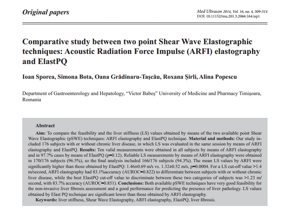
ElastPQ and LIVER FIBROSIS, [PQ=point quantification]
Liver stiffness values obtained by Elast PQ technique are
significantly lower than those obtained by ARFI elastography and future studies
are needed to establish the best LS cut-offs assessed by ElastPQ for predicting
different liver fibrosis stages.
--------------------------------------------------------------------------------------------------
ElastPQ
(Philips)
from JSUM
Ultrasound Elastography Practice Guideline Liver
Masatoshi Kudo1*,
Tsuyoshi Shiina2, Fuminori Moriyasu3, Hiroko Iijima4, Ryosuke Tateishi5,
Norihisa Yada1, Kenji Fujimoto6, 7, Hiroyasu Morikawa8, Masashi Hirooka9,
Yasukiyo Sumino10, Takashi Kumada11
A) Introduction
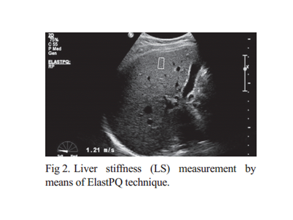
Name: ElastPQ (PQ:
point quantification)
Equipment: The iU22
xMATRIX ultrasound system (iU: Intelligent Ultrasound)
ElastPQ is a
non-invasive diagnostic tool to measure tissue stiffness using an ARFI-based
technology. Immediately after image acquisition, the screen displays the image
and measurement results, including the mean and median values and the
deviations in kPa or m/s (Fig. 33).
*If measurement reliability is low, 0.00 kPa will be
displayed as the result.
Elastic value E [kPa] is calculated using the equation
E = 3ρVsଶ where Vs
[m/s] is defined as the shear wave propagation velocity and ρ as tissue density
(whose approximated value in the human body is 1).
An ROI can be placed
anywhere but at a depth of uder 8 cm.
B) Indication
1. Quantitative
assessment of liver fibrosis in diffuse liver diseases
2. Neoplastic lesions of the liver
C) Procedures (including Tips and Tricks)
1. Perform right intercostal scanning to visualize the liver
2. Steadily place the
probe with minimum compression
3. Set the ROI with a
depth of under 8 cm.
4. Ask the patient to breath hold (if not possible, ask the patient to breathe as shallowly as possible).
5. Push the” Update” button for quantification
5. Push the” Update” button for quantification
6. The use of a mean value from more than 10 measurements is recommended.
Approach the right hepatic lobe from the right intercostal space. Avoid the left hepatic lobe because the measurement is affected by cardiac movement. Breath hold without exerting abdominal pressure. The most appropriate ROI is the center of the image, namely, immediately below the probe, and 3–5 cm from the probe surface. Avoid blood vessels, any necrotic areas, the boundary between organs, and areas influenced by cardiac movement (ex. left hepatic lobe). Three frequencies (R1/RP/P1) are available. The measurement sensitivity of areas deep inside the body can be improved by using a lower frequency.
D) Results (What does the value mean?)
・Healthy liver: 4 kPa (2.5–4.7 kPa, 1–1.5 m/s)
・Mild fibrosis: 7 kPa (4.7–12.0 kPa, 1.5–2.0 m/s)
・Moderate–severe fibrosis: 12 kPa (12.0–21.0 kPa, 2.0–2.5 m/s)
・Severe fibrosis: over 21
kPa (over 2.5 m/s)
E) Limitations
・There is a limit to
measurable depth.
・ElastPQ is affected by respiratory and body movement.
・Cardiac movement also affects the system.
・Accuracy of measurement depends on the skills of the
examiner.
・Measurement accuracy is generally low at the sides of an
image.
・Ribs may cast lateral acoustic shadows.
F) Recommendations
At present, the number of studies using ElastPQ is not large
enough to reach a definitive conclusion. We look forward to having more study
results in the near future.
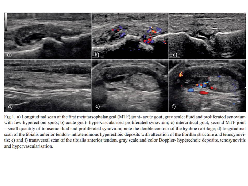
The place of musculoskeletal ultrasonography in gout diagnosis
Daniela Fodor et al
Investigation questions
One of the
problems in US investigation of gouty patients is which sites to be
investigated (only symptomatic joints or structures or more extensive
examination) and what findings to be search (MSU deposits, effusion, synovial
hypertrophy, vascularisation) in order to have good sensibility and specificity
of the method. In a recent published paper by Naredo et al [17] the authors
found that US examination of 12 anatomical sites (bilateral radio-carpal joint,
first metatarsal head cartilage, talar cartilage, second metacarpal head, knee
femoral condyle cartilage, patellar and triceps tendons) for DC sign and
hyperechoic aggregates gives the best results for sensitivity and specificity
(84.6% and 83.3%, respectively). This approach had high positive and good
negative predictive values (92% and 71%, respectively) for diagnosing gout.
Peiteado et al [57] studied the presence of six types of lesions in gouty
patients: hyperechoic spots in the synovial fluid, HCA, bright stippled
aggregates, DC sign, erosions, and the Doppler signal in 17 joints and 8
tendons. They found that the knees and MTP joints are the most frequent
affected sites (in 93% of patients) and a six-minute US examination of four
joints (knees and the 1st MTPs) allowed the detection of HCA or DC sign in 97%
of cases. Clinical diagnosis of gout is sometimes difficult, even if the
manifestations are in MTF-1 (classical podagra). Kienhorst et al [58] recently
demonstrated that in patients with MTP-1 arthritis the gout was the correct
diagnosed only in 77% of cases but the general practitioner supposed gout in
98% of cases. In these cases US examination of the joint could simplify the
diagnosis process. This fact was demonstrated by Lamers-Karnebeek et al [59] in
patients with monoarthrithis in which 48% of patients had MSU crystals-proven
gout. In this cases sensitivity of DC sign and any US abnormality (DC sign,
tophi, or snowstorm appearance of the joint effusion) was 77 and 96 %,
respectively. The authors considered that the DC sign is an important
informative finding for clinician, with positive predictive value of 74 % and
negative predictive value of 78 %.
The new US
techniques probably will increase the US capacity to detect the crystals
aggregates. MicroPure, a new US image processing function designed to improve
the visualization of microcalcifications, was used by Yin et al [60] in
patients with gout. Significantly more microcalcifications were seen with
MicroPure compared to gray scale US and the level of agreement between
investigators was consistently improved. The method seems to be useful
especially for the cases where the presence of gray scale US artifacts or
unspecific findings make difficult the interpretation. The use of different
imaging methods (dual energy CT, MRI, positron emission tomography) may improve
the description of lesions [61].
The main question is if there is a really need in clinical practice for such a complex approach. The need for good education and training for identification and interpretation of the US findings encountered in gout was underline by Filippucci et al [62].The authors highlight the necessity for dedicated training-programme to avoid the false positive and false negative results. After 7 days of training the rheumatologists with limited US experience gain satisfactory skills to identify MSU aggregates in different tissues. Due to the lower diagnostic utility of the clinical gouty features (exception tophi and response to colchicine) [63], there is an incremental need for imaging techniques to confirm the clinicians’ suppositions. The place of the US in patients suspected of gout must be clearly defined [59] due to the high numbers of patients presented with monoarthritis. The research agenda concerning the use of US in gouty patients is still busy despite of the recent achievements in this pathology. In conclusion in the assessment of the gouty patients US is an important and valuable tool. For a complete examination and a correct interpretation of the imagines the examiner need to have solid US knowledge about normal and pathological aspects of the musculoskeletal structures. Knowing the specific US aspects of MSU depositions will permit a rapid diagnosis. Moreover, when combining the clinical examination with US scan in patients with suspicion of gout a proper and rapid decisions for the patient management can be taken.
The main question is if there is a really need in clinical practice for such a complex approach. The need for good education and training for identification and interpretation of the US findings encountered in gout was underline by Filippucci et al [62].The authors highlight the necessity for dedicated training-programme to avoid the false positive and false negative results. After 7 days of training the rheumatologists with limited US experience gain satisfactory skills to identify MSU aggregates in different tissues. Due to the lower diagnostic utility of the clinical gouty features (exception tophi and response to colchicine) [63], there is an incremental need for imaging techniques to confirm the clinicians’ suppositions. The place of the US in patients suspected of gout must be clearly defined [59] due to the high numbers of patients presented with monoarthritis. The research agenda concerning the use of US in gouty patients is still busy despite of the recent achievements in this pathology. In conclusion in the assessment of the gouty patients US is an important and valuable tool. For a complete examination and a correct interpretation of the imagines the examiner need to have solid US knowledge about normal and pathological aspects of the musculoskeletal structures. Knowing the specific US aspects of MSU depositions will permit a rapid diagnosis. Moreover, when combining the clinical examination with US scan in patients with suspicion of gout a proper and rapid decisions for the patient management can be taken.
PLEURAL ULTRASOUND
Conclusions
US is a very important diagnostic method for pleural pathology, providing high resolution images, useful in all kinds of clinical scenarios – emergency, diagnostic and interventional ultrasonography. It is superior to chest radiography for the diagnosis of pneumothorax and characterization of pleural effusions. US represents the best method for guiding fluid aspiration and transthoracic biopsy of pleural-based lesions. It is also a reliable method for evaluating the parietal infiltration of the lung tumors, but CT is superior providing more information concerning all parts of the pleura. The limit of this method is the inability to visualize the mediastinal pleura, and the lesions obscured by subcutaneous emphysema and the bony structures of the thorax – scapulae, sternum and the spine. US can provide useful information for almost all pleural pathology and it should be use more extensively for all these reasons, being also accessible, cheap and widely available for many medical specialties.
Đăng ký:
Nhận xét
(
Atom
)


