Tổng số lượt xem trang

Thứ Sáu, 18 tháng 6, 2021

UltraVisions debuts breast microcalcifications software

By AuntMinnie.com staff writers


June 17, 2021 -- UltraVision recently announced it has developed a patent-pending software to visualize microcalcifications for breast cancer diagnosis and biopsy guidance

The software, called MC-mode, can be implemented on ultrasound scanners and used for visualizing microcalcifications, with the initial application being Ultravision's point-of-care ultrasound scanner, UltraVision-XS.

Microcalcifications in a blood vessel in the breast
Microcalcifications in a blood vessel in the breast. Image courtesy of UltraVision.

Company leaders say the software aims to expand breast cancer detection with ultrasound into physician's offices, mammography centers, and underserved international markets.

The company has filed for a patent on MC-mode, which detects microcalcifications and their locations from their vibration. The company also filed a patent on differentiating between type-1 and type-2 microcalcifications.

Thứ Năm, 10 tháng 6, 2021

U S can determine lung congestion in kidney patients

 By Amerigo Allegretto, AuntMinnie.com staff writer


June 10, 2021 -- Ultrasound is a useful guide in determining lung congestion for hemodialysis patients at high cardiovascular risk, according to research presented on June 6 at the European Renal Association -- European Dialysis and Transplant Association virtual congress.

A team of researchers led by Dr. Claudia Torino from the National Research Council of Italy found that patients with end-stage kidney disease who underwent lung ultrasound showed significantly less frequent episodes of decompensated heart failure and major cardiovascular events.

"Ultrasound examinations are available virtually everywhere in the hospital environment and do not take long to perform, so they can be deployed to diagnose and treat an ominous complication like lung congestion in hemodialysis patients," Torino said.

Lung congestion is common for hemodialysis patients, including those at high cardiovascular risk. While x-ray imaging can detect heart alterations, they cannot be heard easily through a stethoscope.

For hemodialysis patients, lung congestion is a strong risk factor for mortality. Between dialysis sessions, all fluid that patients introduce is retained. Severe overhydration may follow, which can lead to heart decompensation.

Ultrasound has been shown to determine just how much lung congestion there is. This in turn can help clinicians adjust fluid removal during hemodialysis and drug therapies.

"Extravascular lung water can be easily measured by lung ultrasound, being quantified on the basis of the number of ultrasound B-lines," Torino said.

The team studied 363 patients from several hospital systems in Europe and Asia to see if ultrasound can improve patient outcomes over standard care, with 183 patients being tested with ultrasound and the remaining 180 patients in the control group. Out of those, 152 patients completed the ultrasound-guided study and 155 completed the standard care study.

The primary endpoints the researchers studied were mortality, heart attack, and decompensated heart failure. This strategy was compared with standard care in hemodialysis patients with a high cardiovascular risk.

Sonographers measured B-lines in images, with the target for hemodialysis management being less than 15 sonographic B-lines, a threshold that signified less fluid accumulation in the lungs.

Torino said the team used a 28-point lung ultrasound technique. Depending on the user's experience, scans took between three and 15 minutes.

Impact of ultrasound on treatment of hemodialysis patients
 Control groupUltrasound group
Change in number of B‑lines from start to end of study16 B‑lines30 B‑lines15 B‑lines9 B‑lines
Number of patients reaching target of less than 15 B‑lines85 patients117 patients
Number of patients reaching primary endpoint71 patients62 patients*
*Difference is not statistically significant

Secondary analyses showed significantly less frequent episodes of decompensated heart failure and major cardiovascular events for the sonography group. For heart failure, lung ultrasound showed an incidence reduction rate of 63% while for cardiovascular events, that rate decreased by 37%.

"Because decompensated heart failure was not the primary end point of the study, new trials are still needed to confirm this finding," Torino noted.

Thứ Tư, 9 tháng 6, 2021

Education leads to confidence for new POCUS users

By Amerigo Allegretto, AuntMinnie.com staff writer


June 9, 2021 -- Instructional training increases confidence among family medicine residents using point-of-care ultrasound (POCUS), according to a study published June 4 in Family Medicine.

team of researchers from the University of New Mexico led by Dr. Jerica Johnson found residents felt "significantly more confident" in their ability to perform and interpret a POCUS exam after a curriculum was implemented and that baseline attitudes toward POCUS were "very" favorable in both the pre- and postintervention surveys.

"Our residents and faculty have all been very excited to use POCUS. There are of course barriers to adoption of any new technology," Johnson told AuntMinnie.com. "The most significant barrier has been residents and faculty feeling that they are trained well enough to use POCUS and that there is not enough time to obtain this training."

The use of POCUS in family medicine residency training is increasing. However, researchers said attitudes about the technology, including confidence levels with performing and interpreting POCUS exams, are unknown.

The team wanted to find out attitudes and confidence levels of family medicine residents before and after the implementation of a new POCUS curriculum.

Residents in their postgraduate years at the University of New Mexico Family Medicine Residency Program were trained with POCUS in a yearlong program. The university's current POCUS curriculum was implemented in 2017.

"We had a loose curriculum with limited POCUS experiences prior to this when we made intentional changes to create a robust POCUS curriculum," Johnson said.

Attitudes and confidence levels with various POCUS exams were assessed through pre and postintervention surveys. On a 5-point confidence scale, a score of 1 meant strong disagreement while 5 indicated strong agreement.

The research team held a three-hour, hands-on workshop for residents, an 18-hour workshop for faculty and residents who participated as volunteer models, and 30-minute sessions every other week for residents who were rotating on the program's inpatient service.

Twenty-one residents responded to the preintervention survey. The average confidence scale score for education on ultrasound principles for performing and interpreting POCUS exams was 4.38. For the postintervention survey, which included 25 residents, that score was 3.96 (p = 0.05).

While agreement was high among residents when it came to ready access to ultrasound equipment and hands-on training, the results did not achieve statistical significance.

The team wrote that future research opportunities include investigating the effectiveness of different POCUS training technologies among family medicine residents and effects on patient-oriented outcomes.

Johnson told AuntMinnie.com that the team is hoping to implement an outpatient-based curriculum for the university's family medicine residents that is geared toward POCUS applications that are useful in the outpatient clinic.

"We are hoping to demonstrate that minimal training interventions can lead to improved revenue generation from the POCUS exams and essentially provide evidence that POCUS training for family medicine residents 'pays for itself,'" she said. "We hope that this outpatient-based curriculum can then be shared and applied by other family medicine residency programs.

Thứ Sáu, 4 tháng 6, 2021

SHORTER LUNG U S exams can help kidney disease patients


By Amerigo Allegretto, AuntMinnie.com staff writer

June 4, 2021 -- Lung ultrasound with an abbreviated scanning protocol can efficiently diagnose pulmonary congestion caused by fluid overload in hemodialysis patients, according to research published June 3 in the American Journal of Kidney Diseases.


team of researchers led by Dr. Nathaniel Reisinger from the University of Pennsylvania found that several abbreviated lung ultrasound protocols that focus on a limited number of lung zones (4, 6, or 8-zone) performed similarly to comprehensive 28-zone studies among patients with kidney failure on hemodialysis seeking care in an emergency department.

The researchers also did not find any differences in mortality between patients with no-to-mild and moderate-to-severe pulmonary congestion.

"Abbreviated lung ultrasound protocols take less time, are less bothersome to patients, and are nearly as accurate," Reisinger told AuntMinnie.com.

Pulmonary congestion from fluid overload is common among patients with kidney failure on hemodialysis and contributes to excess morbidity and mortality in patients. The team said that physical examination is an insensitive approach to detecting pulmonary congestion.

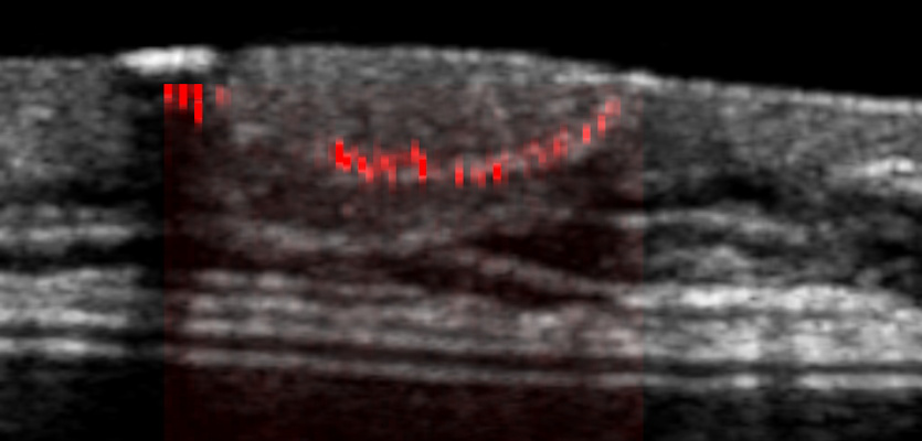
Lung ultrasound with a comprehensive 28-zone protocol has been shown to be sensitive for detecting pulmonary congestion, according to previous research. However, the 28-zone technique requires complete disrobing of the patient and longer scanning times for operators.

"In the constrained environment of the emergency department, this commitment is prohibitive to both patients and providers," the authors wrote.

Reisinger and his team wanted to see if abbreviated forms of lung ultrasound could produce similar results while saving time for operators and providing a more comfortable experience for patients.

The group started with research by Buessler and colleagues that studied the use of four-, six-, and eight-zone scanning for heart failure patients, as well as mapped the specific lung zones that would be most equivalent to a 28-zone study. The researchers noted that previous research has found that the regional distribution of pulmonary edema on imaging based on clinical condition has been well-demonstrated.

They then tested the protocol in 98 patients in the U.S. with kidney failure on hemodialysis participated, with a follow-up time of 30 days. Out of those, 84 were African American and 97 were non-Hispanic or Latino in ethnicity.

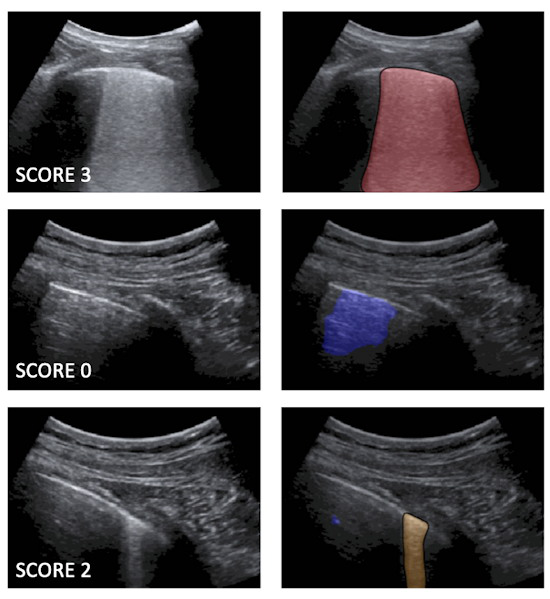
The team found high correlation and good agreement between 28-zone and abbreviated lung ultrasound studies. Each of the short-form studies was able to discriminate between no-to-mild pulmonary congestion versus moderate-to-severe pulmonary congestion when compared with the long-form 28-zone study, the authors wrote.

For short-form studies, the highest sensitivities were seen in 6C, 8C, and 8D zone ultrasound studies at 93.0%, 95.8%, and 98.6% respectively. Meanwhile, 6C and 8A studies had the highest specificities, at 96.3% and 100% respectively. Finally, 6C, 8C, and 8D studies had the highest area under the curve (AUC), at 0.95, 0.94, and 0.94 respectively.

Of the four-zone studies, lung zone 4C had the highest sensitivity at 90.1%, specificity at 92.6%, and AUC at 0.91.

"With AUC values of greater than 0.88, any of the 8-zone studies clearly seems sufficient for clinical use," the study authors wrote. "Similarly, the 4C pattern can easily be performed without removing a patient's shirt, while still achieving a point estimate of AUC greater than 0.90, demonstrating accuracy better than the current standard-of-care assessment of pulmonary congestion."

However, Reisinger said that despite the promise, abbreviated lung ultrasound protocols are "slightly less accurate" than more comprehensive tests and may miss focal pathology such as pneumonia.

He told AuntMinnie.com that the research team is looking at randomized controlled trials as the next step in investigating whether lung ultrasound-guided ultrafiltration therapy improves outcomes in patients with end-stage kidney disease chronically on hemodialysis.

Thứ Tư, 2 tháng 6, 2021

A I can help stratify COVID-19 risk on lung ultrasound

By Erik L. Ridley, AuntMinnie.com staff writer


June 1, 2021 -- Deep-learning algorithms can be used to automatically provide risk scores on lung ultrasound exams in COVID-19 patients, researchers from Italy reported in an article published online May 27 in the Journal of the Acoustical Society of America.


 A multi-institutional team of researchers trained deep-learning models to automatically score and provide semantic segmentations of lung ultrasound exams in COVID-19 patients. In testing, the deep-learning algorithms yielded a high level of agreement with lung ultrasound experts for stratifying patients as having either low- or high risk of clinical worsening, according to the group led by corresponding author Libertario Demi, PhD, of the University of Trento.

"These encouraging results demonstrate the potential of [deep-learning] models for the automatic scoring of [lung ultrasound] data, when applied to high-quality data acquired according to a standardized imaging protocol," the authors wrote.

Although lung ultrasound has been shown to be useful for evaluating COVID-19 patients, the modality is often limited to the visual interpretation of ultrasound data. As a result, there are concerns over reliability and reproducibility, according to the researchers.

Following up on prior work to develop standard lung ultrasound protocols and training deep-learning algorithms for COVID-19 patients, the researchers wanted to compare the performance of their models with that of expert clinicians. The first algorithm labels each video frame with a score, while the second provides semantic segmentation -- assigning one or more scores to each frame.

They used their algorithms to evaluate 314,879 ultrasound video frames from 1,488 lung ultrasound videos on 82 COVID-19 patients. The ultrasound exams were acquired using multiple ultrasound scanner types at the Agostino Gemelli University Hospital Foundation in Rome and the San Matteo Polyclinic Foundation in Pavia, Italy. None of the images were used in training the models.

Examples of lung ultrasound images corresponding to score 3, score 0, and score 2
Left: Examples of lung ultrasound images corresponding to score 3 (top), score 0 (middle), and score 2 (bottom). Right: the corresponding semantic segmentations obtained with the described deep-learning algorithms. Color maps are informative of the score level (red for score 3, blue for score 0, orange for score 2), as well as indicate the relevant part of the image that determines the score. These maps explain the decision progress of the algorithms. Images and caption courtesy of Libertario Demi, PhD.

The algorithm's scores for each frame are then used to generate an aggregate score for each video. The researchers tested two methods for producing an aggregate score: one based only on the labeled video frames and another that combined the labeled frames with the segmented frames.

These video-level scores were then compared with the risk scores provided by expert clinicians.

AI algorithm performance for risk stratifying COVID-19 patients
 AI risk scores based on only labeled framesAI risk scores based on combination of labeled and segmented frames
Agreement with expert clinicians83.3%86%

Although the combined approach generally outperforms the first method, the improvement isn't significant. What's more, the first method seems more stable to threshold variations, according to the researchers.

"Therefore, the first approach can be considered as a self-standing strategy to classify [lung ultrasound] videos starting from a frame-based classification," the authors wrote. "Nevertheless, the semantic segmentation remains essential for clinicians, as it provides the explainability of the decision by highlighting the specific [lung ultrasound] patterns."

In future work, the researchers plan to expand their existing database and train deep-learning algorithms on video-labeled data instead of just frame-based labeled data. That approach would be more consistent with the process used by clinicians when evaluating lung ultrasound videos, they said.

How to implement POCUS at your hospital

By Amerigo Allegretto, AuntMinnie.com staff writer


June 1, 2021 -- So, you want to integrate point-of-care ultrasound (POCUS) into your hospital enterprise image network. But how? Breaking down what goes into a successful POCUS implementation was the subject of a May 26 talk at the Society for Imaging Informatics in Medicine meeting.


Successfully implementing POCUS requires swift strategy and teamwork, including taking advantage of existing imaging technology, according to registered nurse Laurie Perry from Cincinnati Children's Hospital in Ohio.

"Point-of-care ultrasound is only going to grow, and there will be divisions that continually need to be implemented. You'll need to do that quickly," she said.

POCUS has been looked at in recent years as a convenient clinical tool that can bring imaging to a patient's bedside. Advantages include portability and cost-effectiveness.

However, there are some limitations to POCUS, Perry said. One is that images remain on the ultrasound device and are not clinically available to anyone else taking care of the patient. POCUS images also can't be compared with other specialty images, and there are challenges to getting reimbursed.

Although many divisions at Cincinnati Children's Hospital wanted to use POCUS, Perry said they were unable to bill for the procedure until providers had been credentialed and images stored in the enterprise archive.

"If you're not storing images, you cannot bill for the ultrasound," Perry said.

To integrate POCUS with the rest of the enterprise, the implementation team needed to create a standard POCUS workflow within the electronic health record. Then, it needed to work with business managers to help them to understand the process for billing imaging studies.

The team that was assembled for this included clinical and business leaders from clinical divisions, imaging informatics professionals, electronic medical records analysts, a project manager, a trainer, and an enterprise imaging physician leader.

The group solved the existing challenges by working with clinical leaders and business managers in creating a process that fits into the existing workflow. Team members utilized ad-hoc order creation and automated as many steps as possible. They also worked with the radiology business director to identify and create appropriate procedure codes.

"All radiology and cardiology, including ultrasound, is sent to the hospital's enterprise archive. We've acquired and sent over 5,000 POCUS studies to the archive," Perry said. "We have billed more than $2.25 million of new services for our organization."

Children's Hospital began using POCUS for anesthesia in 2015 and has expanded its use in other care units and departments, such as emergency, pediatric intensive care, and cardiac intensive care among others. The hospital is currently working on implementing POCUS for physical medicine and rehab and its gastrointestinal division.

Perry said the hospital has used the following guiding principles over the years to successfully implement POCUS into its departments:

  • Take advantage of DICOM to power workflow.
  • Automate workflow for providers as much as possible.
  • Provide a link to the images in the electronic medical record, preferably within the appropriate context for the division.
  • The goal should be to create a standard, reproducible process.

Perry also said implementation teams should evaluate a division's current ultrasound machines for such things as WiFi capability, DICOM capability, and security requirements. Credentialing providers, as well as determining procedures and billing, developing the electronic medical record, and training users are also needed.

Thứ Bảy, 29 tháng 5, 2021

Special training not needed for COVID lung ultrasound scoring

By Amerigo Allegretto, AuntMinnie.com staff writer


May 26, 2021 -- Although there were some variations in performance, it may not be necessary for users of ultrasound scanners to receive special training before scoring lung exams of patients with COVID-19, according to research published May 21 in Nature.

group of researchers led by Dr. Markus Lerchbaumer from Charité Institute of Radiology in Berlin, Germany found that while different types of observers had "fair to moderate" interobserver agreement in interpreting specific lung findings in patients with COVID-19, a little background in lung ultrasound goes a long way.

"As long as observers have some experience in lung ultrasound, no specific clinical background is needed for scoring the findings, even though specific expertise is often reported as a requirement," the authors wrote.

Examining the lungs in patients is usually performed with nonenhanced CT scans, but point-of-care ultrasound (POCUS) is being looked at as a safer method since patients infected with the SARS-CoV-2 virus would not need to be transferred, and risk of exposure for medical staff would decrease.

The researchers said lung ultrasound may have a big advantage for COVID-19 due to its widespread availability and cost-effectiveness.

"Additionally, lung ultrasound has emerged over the last two decades as a noninvasive tool for the fast differential diagnosis of pulmonary diseases and is now used in different settings in intensive care," they wrote.

In the current study, 10 observers from three different medical specialties participated in rating 100 lung ultrasound images from 13 patients. These included observers specializing in intensive care medicine, emergency medicine, and physiology.

Images were acquired by a radiologist using a hand-held POCUS system (Viamo sv7, Canon Medical Systems) performed at the bedside. Ultrasound presets were optimized for lung ultrasound.

Through an online tool, observers could use multiple-choice options with predefined answers for rating the scans. Options included typical COVID-19-associated lung ultrasound findings; these included the following:

  • Pleural thickening and fragmentation
  • Presence of B-lines subclassified in single or confluent, subpleural consolidations
  • Positive air bronchogram

Selecting none of these pathologies was also an option.

The team found that observers in the intensive care unit tended to interpret B-lines more accurately, while physiology researchers and emergency physicians more often categorized B-lines as confluent rather than single. This tendency became even stronger over the course of viewing instances, probably explaining the poorer than expected overall inter- and intraobserver agreement.

"We assume that ICU observers have greater clinical experience with patients with severe ARDS or cardiogenic edema and their corresponding lung ultrasound findings, especially compared to scientists whose experience relies on lung ultrasound in rodents," they wrote.

ICU observers, on the other hand, differed from the latter two groups regarding the identification of pleural thickening.

Meanwhile, agreement was highest for more distinct lung ultrasound findings such as air bronchograms and subpleural consolidations, as well as more severe lung ultrasound scores.

The researchers wrote that training lung ultrasound users may improve agreement and clinical feasibility. They also suggest that training material used for lung ultrasound in POCUS should pay better attention to areas such as B-line quantification and differentiation of intermediate scores, which revealed only "mediocre" agreement in the study.

Thứ Tư, 26 tháng 5, 2021

US AI model can help evaluate chronic kidney disease


By Erik L. Ridley, AuntMinnie.com staff writer

May 24, 2021 -- Artificial intelligence (AI)-based analysis of kidney ultrasound studies could serve as a first-line method for evaluating patients with chronic kidney disease, according to research published online May 24 in JAMA Network Open.


team of researchers led by Dr. Ambarish Athavale of Cook County Health in Chicago developed a deep-learning algorithm that yielded approximately 90% accuracy on a test set for quantifying interstitial fibrosis and tubular atrophy (IFTA).

"This article provides proof-of-principle that a [deep-learning] system can be used to noninvasively, accurately, and independently predict IFTA grade in patients with kidney disease," the authors wrote. "Although the system in its current form may not be an alternative to kidney biopsy, after robust external validation, a [deep learning]-based, noninvasive assessment of IFTA has the potential to significantly enhance clinical decision-making and prognostication in patients with CKD."

A strong indicator for decline in kidney function, interstitial fibrosis and tubular atrophy is currently measured using histopathological assessment of a kidney biopsy core. There currently isn't a noninvasive test for IFTA, according to the researchers.

The authors utilized AI to test their hypothesis that subtle signs of IFTA are ingrained within kidney ultrasound images and could be quantitatively extracted and analyzed. A deep-learning algorithm was trained and tested to segment the kidney and classify IFTA using 6,135 consecutive Crimmins-filtered kidney ultrasound images acquired at their institution between January 1, 2014, and December 31, 2018. The longitudinal images were obtained from both kidneys and were acquired between six months before and two weeks after kidney biopsy.

Of the total image dataset, 5,122 were used for training and 401 were used for validation. The researchers then tested the model on 612 images. The algorithm was 91% accurate for segmenting the kidney ultrasound images.

Performance of AI algorithm for quantifying IFTA on kidney ultrasound
 Image levelPatient level
Precision0.8930.900
Recall0.8040.842
Accuracy0.8680.896
F1 score0.8390.864

In other results, the researchers noted that the algorithm's accuracy remained high irrespective of the timing of the ultrasound studies and the biopsy diagnosis. Also, adding baseline clinical characteristics into the model's analysis didn't significantly improve its performance.

"From a clinical standpoint, it is foreseeable that a [deep-learning] system such as the one developed in this study has the potential to act as a gatekeeper for rationalizing the decision to conduct a kidney biopsy in patients with CKD," the authors wrote. "We anticipate that because of the ability of this system to provide [a] probabilistic estimate of IFTA in real-time, the system is likely to be acceptable (because it is unlikely to put any time burden on the technicians) and can also reduce the costs associated with kidney biopsy."

The researchers acknowledged that more work is needed to improve the accuracy of the model before it's ready for clinical use. Furthermore, the algorithm needs to be validated on external datasets to assess its performance across varying clinical settings.